

| 产品编号 | bs-1540R |
| 英文名称 | IL7R Rabbit pAb |
| 中文名称 | 白细胞介素-7受体a(CD127)抗体 |
| 别 名 | CD127; CDW127; IL-7R-alpha; IL-7Ralpha; IL7RA; IL7Ralpha; ILRA; IMD104; lnc-IL7R; sIL-7R; IL7RA_HUMAN; IL7R; IL-7 receptor subunit alpha; IL-7R subunit alpha; IL-7RA; IL7RA_MOUSE; |
 |
Specific References (1) | bs-1540R has been referenced in 1 publications.
[IF=13.081] Xu Jun Yan. et al. Interleukin-5-induced eosinophil population improves cardiac function after myocardial infarction. CARDIOVASC RES. 2022 Jul;118(9):2165-2178 IF ; Mouse.
|
| 研究领域 | 细胞生物 免疫学 发育生物学 干细胞 淋巴细胞 t-淋巴细胞 b-淋巴细胞 |
| 抗体来源 | Rabbit |
| 克隆类型 | Polyclonal |
| 克 隆 号 | |
| 交叉反应 | Human,Mouse,Rat (predicted: Pig,Sheep,Cow,Dog,Horse) |
| 产品应用 | WB=1:500-2000
not yet tested in other applications. optimal dilutions/concentrations should be determined by the end user. |
| 理论分子量 | 50 kDa |
| 检测分子量 | 70 |
| 细胞定位 | 细胞膜 分泌型蛋白 |
| 性 状 | Liquid |
| 浓 度 | 1mg/ml |
| 免 疫 原 | KLH conjugated synthetic peptide derived from human IL-7Ra: 251-350/459 |
| 亚 型 | IgG |
| 纯化方法 | affinity purified by Protein A |
| 缓 冲 液 | 0.01M TBS (pH7.4) with 1% BSA, 0.02% Proclin300 and 50% Glycerol. |
| 保存条件 | Shipped at 4℃. Store at -20℃ for one year. Avoid repeated freeze/thaw cycles. |
| 注意事项 | This product as supplied is intended for research use only, not for use in human, therapeutic or diagnostic applications. |
| PubMed | PubMed |
| 产品介绍 |
The protein encoded by this gene is a receptor for interleukine 7 (IL7). The function of this receptor requires the interleukin 2 receptor, gamma chain (IL2RG), which is a common gamma chain shared by the receptors of various cytokines, including interleukine 2, 4, 7, 9, and 15. This protein has been shown to play a critical role in the V(D)J recombination during lymphocyte development. This protein is also found to control the accessibility of the TCR gamma locus by STAT5 and histone acetylation. Knockout studies in mice suggested that blocking apoptosis is an essential function of this protein during differentiation and activation of T lymphocytes. The functional defects in this protein may be associated with the pathogenesis of the severe combined immunodeficiency (SCID). Function: Receptor for interleukin-7. Also acts as a receptor for thymic stromal lymphopoietin (TSLP). Subcellular Location: Secreted and Cell membrane. Post-translational modifications: N-glycosylated IL-7Ralpha binds IL7 300-fold more tightly than the unglycosylated form. DISEASE: Defects in IL7R are a cause of severe combined immunodeficiency autosomal recessive T-cell-negative/B-cell-positive/NK-cell-positive (T(-)B(+)NK(+) SCID) [MIM:608971]. A form of severe combined immunodeficiency (SCID), a genetically and clinically heterogeneous group of rare congenital disorders characterized by impairment of both humoral and cell-mediated immunity, leukopenia, and low or absent antibody levels. Patients present in infancy recurrent, persistent infections by opportunistic organisms. The common characteristic of all types of SCID is absence of T-cell-mediated cellular immunity due to a defect in T-cell development. Genetic variations in IL7R are a cause of susceptibility to multiple sclerosis type 3 (MS3) [MIM:612595]. A multifactorial, inflammatory, demyelinating disease of the central nervous system. Sclerotic lesions are characterized by perivascular infiltration of monocytes and lymphocytes and appear as indurated areas in pathologic specimens (sclerosis in plaques). The pathological mechanism is regarded as an autoimmune attack of the myelin sheat, mediated by both cellular and humoral immunity. Clinical manifestations include visual loss, extra-ocular movement disorders, paresthesias, loss of sensation, weakness, dysarthria, spasticity, ataxia and bladder dysfunction. Genetic and environmental factors influence susceptibility to the disease. Note=A polymorphism at position 244 strongly influences susceptibility to multiple sclerosis. Overtransmission of the major 'C' allele coding for Thr-244 is detected in offspring affected with multiple sclerosis. In vitro analysis of transcripts from minigenes containing either 'C' allele (Thr-244) or 'T' allele (Ile-244) shows that the 'C' allele results in an approximately two-fold increase in the skipping of exon 6, leading to increased production of a soluble form of IL7R. Thus, the multiple sclerosis associated 'C' risk allele of IL7R would probably decrease membrane-bound expression of IL7R. As this risk allele is common in the general population, some additional triggers are probably required for the development and progression of MS. Similarity: Belongs to the type I cytokine receptor family. Type 4 subfamily. Contains 1 fibronectin type-III domain. SWISS: P16871 Gene ID: 3575 Database links: Entrez Gene: 3575 Human Entrez Gene: 16197 Mouse Omim: 146661 Human SwissProt: P16871 Human SwissProt: P16872 Mouse Unigene: 591742 Human Unigene: 635723 Human Unigene: 389 Mouse |
| 产品图片 |
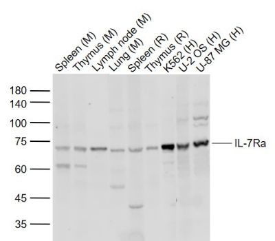
Sample:
Lane 1: Mouse Spleen tissue lysates
Lane 2: Mouse Thymus tissue lysates
Lane 3: Mouse Lymph node tissue lysates
Lane 4: Mouse Lung tissue lysates
Lane 5: Rat Spleen tissue lysates
Lane 6: Rat Thymus tissue lysates
Lane 7: Human K562 cell lysates
Lane 8: Human U-2 OS cell lysates
Lane 9: Human U-87 MG cell lysates
Primary: Anti-IL-7Ra (bs-1540R) at 1/1000 dilution
Secondary: IRDye800CW Goat Anti-Rabbit IgG at 1/20000 dilution
Predicted band size: 50 kD
Observed band size: 70 kD
|
| 1、抗体溶解方法 | |
| 2、抗体修复方式 | |
| 3、常用试剂的配制 | |
| 4、免疫组化操作步骤 | |
| 5、免疫组化问题解答 | |
| 6、Western Blotting 操作步骤 | |
| 7、Western Blotting 问题解答 | |
| 8、关于肽链的设计 | |
| 9、多肽的溶解与保存 | |
| 10、酶标抗体效价测定程序 | |


