

| 产品编号 | bs-1635R |
| 英文名称 | B3GAT1 Rabbit pAb |
| 中文名称 | 髓鞘相关糖蛋白(CD57)抗体 |
| 别 名 | CD57; GLCATP; GLCUATP; HNK1; LEU7; NK-1; NK1; B3GA1_HUMAN; B3GAT1; Beta-1,3-glucuronyltransferase 1; Glucuronosyltransferase P (GlcAT-P); UDP-GlcUA:glycoprotein beta-1,3-glucuronyltransferase (GlcUAT-P); 2.4.1.135; |
 |
Specific References (2) | bs-1635R has been referenced in 2 publications.
[IF=5.29] Kaneko, Yuji, et al. "Kainic Acid-Induced Golgi Complex Fragmentation/Dispersal Shifts the Proteolysis of Reelin in Primary Rat Neuronal Cells: An In Vitro Model of Early Stage Epilepsy." Molecular Neurobiology (2015): 1-10. WB ; Rat.
[IF=3.6] Salisbury, Elizabeth A., et al. "Transient Brown Adipocyte-Like Cells Derive from Peripheral Nerve Progenitors in Response to Bone Morphogenetic Protein 2." Stem cells translational medicine 1.12 (2012): 874-885.
|
| 研究领域 | 细胞生物 免疫学 |
| 抗体来源 | Rabbit |
| 克隆类型 | Polyclonal |
| 克 隆 号 | |
| 交叉反应 | Human (predicted: Mouse,Rat,Chicken) |
| 产品应用 | Flow-Cyt=2ug/Test,ICC/IF=1:100-500
not yet tested in other applications. optimal dilutions/concentrations should be determined by the end user. |
| 理论分子量 | 38 kDa |
| 检测分子量 | 45 |
| 细胞定位 | 细胞浆 细胞膜 |
| 性 状 | Liquid |
| 浓 度 | 1mg/ml |
| 免 疫 原 | KLH conjugated synthetic peptide derived from human B3GAT1: 21-120/334 |
| 亚 型 | IgG |
| 纯化方法 | affinity purified by Protein A |
| 缓 冲 液 | 0.01M TBS (pH7.4) with 1% BSA, 0.02% Proclin300 and 50% Glycerol. |
| 保存条件 | Shipped at 4℃. Store at -20℃ for one year. Avoid repeated freeze/thaw cycles. |
| 注意事项 | This product as supplied is intended for research use only, not for use in human, therapeutic or diagnostic applications. |
| PubMed | PubMed |
| 产品介绍 |
The protein encoded by this gene is a member of the glucuronyltransferase gene family. These enzymes exhibit strict acceptor specificity, recognizing nonreducing terminal sugars and their anomeric linkages. This gene product functions as the key enzyme in a glucuronyl transfer reaction during the biosynthesis of the carbohydrate epitope HNK-1 (human natural killer-1, also known as CD57 and LEU7). Alternate transcriptional splice variants have been characterized. [provided by RefSeq, Jul 2008] Function: Involved in the biosynthesis of L2/HNK-1 carbohydrate epitope on glycoproteins. Can also play a role in glycosaminoglycan biosynthesis. Substrates include asialo-orosomucoid (ASOR), asialo-fetuin, and asialo-neural cell adhesion molecule. Requires sphingomyelin for activity: stearoyl-sphingomyelin was the most effective, followed by palmitoyl-sphingomyelin and lignoceroyl-sphingomyelin. Activity was demonstrated only for sphingomyelin with a saturated fatty acid and not for that with an unsaturated fatty acid, regardless of the length of the acyl group. Subunit: Homodimer (Potential). Subcellular Location: Golgi apparatus membrane; Single-pass type II membrane protein. Tissue Specificity: Mainly expressed in the brain. Similarity: Belongs to the glycosyltransferase 43 family. SWISS: Q9P2W7 Gene ID: 27087 Database links: Entrez Gene: 27087 Human Omim: 151290 Human SwissProt: Q9P2W7 Human Unigene: 381050 Human CD57是一种分子量为110KD的糖蛋白,它是自然杀伤细胞(NK)和杀伤细胞的特异性表面抗原。CD57表达于15-20%的外周血单核细胞,60%的NK活化细胞以及T细胞亚群。该抗体可以标记NK细胞,也可识别非淋巴组织的一些细胞。可用于NK细胞介导的细胞毒、NK细胞及T细胞亚群的功能等方面的研究,也可标记神经内分泌细胞及其肿瘤。 |
| 产品图片 |
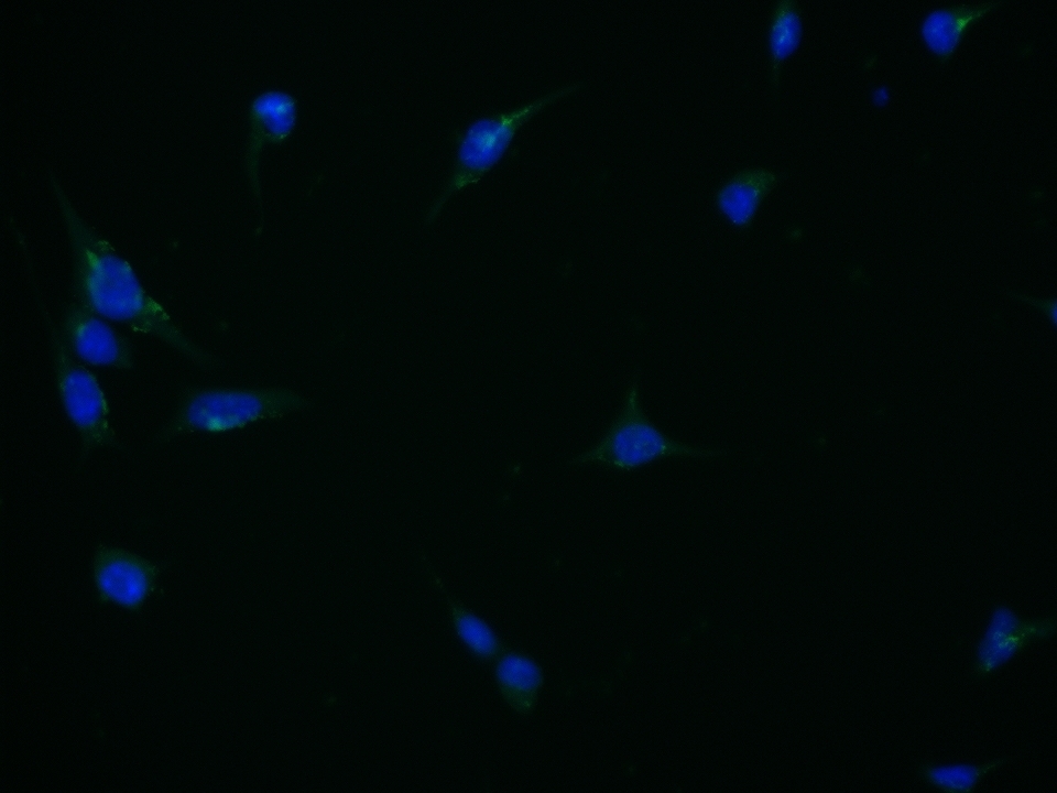
SH-SY5Y cell; 4% Paraformaldehyde-fixed; Triton X-100 at room temperature for 20 min; Blocking buffer (normal goat serum, C-0005) at 37°C for 20 min; Antibody incubation with (B3GAT1) polyclonal Antibody, Unconjugated (bs-1635R) 1:100, 90 minutes at 37°C; followed by a conjugated Goat Anti-Rabbit IgG antibody at 37°C for 90 minutes, DAPI (blue, C02-04002) was used to stain the cell nuclei.
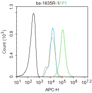
Blank control: U2OS.
Primary Antibody (green line): Rabbit Anti-CD57 antibody (bs-1635R)
Dilution: 2μg /10^6 cells;
Isotype Control Antibody (orange line): Rabbit IgG .
Secondary Antibody : Goat anti-rabbit IgG-AF647
Dilution: 1μg /test.
Protocol
The cells were fixed with 4% PFA (10min at room temperature)and then permeabilized with PBST for 20 min at room temperature. The cells were then incubated in 5%BSA to block non-specific protein-protein interactions for 30 min at at room temperature .Cells stained with Primary Antibody for 30 min at room temperature.The secondary antibody used for 40 min at room temperature.Acquisition of 20,000 events was performed.
|
| 1、抗体溶解方法 | |
| 2、抗体修复方式 | |
| 3、常用试剂的配制 | |
| 4、免疫组化操作步骤 | |
| 5、免疫组化问题解答 | |
| 6、Western Blotting 操作步骤 | |
| 7、Western Blotting 问题解答 | |
| 8、关于肽链的设计 | |
| 9、多肽的溶解与保存 | |
| 10、酶标抗体效价测定程序 | |


