

| 产品编号 | bs-2067R |
| 英文名称 | PIK3CA Rabbit pAb |
| 中文名称 | 磷脂酰肌醇激酶催化亚单位A抗体 |
| 别 名 | CCM4; CLAPO; CLOVE; CWS5; HMH; MCAP; MCM; MCMTC; PI3K; PI3K-alpha; p110-alpha; PK3CA_HUMAN; PIK3CA; PI3-kinase subunit alpha; PI3Kalpha; PtdIns-3-kinase subunit alpha; Phosphatidylinositol 4,5-bisphosphate 3-kinase 110 kDa catalytic subunit alpha (PtdIns- |
 |
Specific References (22) | bs-2067R has been referenced in 22 publications.
|
| 研究领域 | 肿瘤 细胞生物 免疫学 神经生物学 信号转导 细胞凋亡 激酶和磷酸酶 |
| 抗体来源 | Rabbit |
| 克隆类型 | Polyclonal |
| 克 隆 号 | |
| 交叉反应 | Human,Mouse,Rat (predicted: Rabbit,Cow,Chicken) |
| 产品应用 | WB=1:500-2000,IHC-P=1:100-500,IHC-F=1:100-500,IF=1:100-500
not yet tested in other applications. optimal dilutions/concentrations should be determined by the end user. |
| 理论分子量 | 124 kDa |
| 检测分子量 | 124 |
| 细胞定位 | 细胞浆 |
| 性 状 | Liquid |
| 浓 度 | 1mg/ml |
| 免 疫 原 | KLH conjugated synthetic peptide derived from human PI3KCA: 961-1068/1068 |
| 亚 型 | IgG |
| 纯化方法 | affinity purified by Protein A |
| 缓 冲 液 | 0.01M TBS (pH7.4) with 1% BSA, 0.02% Proclin300 and 50% Glycerol. |
| 保存条件 | Shipped at 4℃. Store at -20℃ for one year. Avoid repeated freeze/thaw cycles. |
| 注意事项 | This product as supplied is intended for research use only, not for use in human, therapeutic or diagnostic applications. |
| PubMed | PubMed |
| 产品介绍 |
PI3-Kinases (PI3-Ks) are a family of lipid kinases that are implicated in signal transduction. Phosphatidylinositol 3-kinase is composed of an 85 kDa regulatory subunit and a 110 kDa catalytic subunit. The p85 subunit localize PI3-K activity to the plasma membrane while the p110 subunit contains the catalytic domain of PI3-K which uses ATP to phosphorylate PtdIns, PtdIns4P and PtdInsP2. Four isoforms of p110 has been found; alpha, beta, gamma, and the delta subunit. The alpha isoform, also known as PI3KCA, is a transforming oncogene that was shown to have activating mutations in nine types of cancers such as colon, brain, breast and stomach. Function: PI3K that phosphorylates PtdIns, PtdIns4P and PtdIns(4,5)P2 to generate PIP3. PIP3 plays a key role by recruiting PH domain-containing proteins to the membrane, including AKT1 and PDPK1, activating signaling cascades involved in cell growth, survival, proliferation, motility and morphology. Participates in cellular signaling in response to various growth factors. Involved in the activation of AKT1 upon stimulation by receptor tyrosine kinases ligands such as EGF, insulin, IGF1, VEGFA and PDGF. Involved in signaling via insulin-receptor substrate proteins. Essential in endothelial cell migration during vascular development through VEGFA signaling, possibly by regulating RhoA activity. Required for lymphatic vasculature development, possibly by binding to RAS and by activation by EGF and FGF2, but not by PDGF. Regulates invadopodia formation in breast cancer cells through the PDPK1-AKT1 pathway. Participates in cardiomyogenesis in embryonic stem cells through a AKT1 pathway. Participates in vasculogenesis in embryonic stem cells through PDK1 and protein kinase C pathway. Has also serine-protein kinase activity: phosphorylates PIK3R1, EIF4EBP1 and HRAS. Subunit: Heterodimer of a catalytic subunit PIK3CA and a p85 regulatory subunit (PIK3R1, PIK3R2 or PIK3R3). Interacts with IRS1 in nuclear extracts. Interacts with RUFY3. Interacts with RASD2. Interacts with APPL1. Interacts with HRAS1 and KRAS. Interaction with HRAS1/KRAS is required for PI3K pathway signaling and cell proliferation stimulated by EGF and FGF2. DISEASE: Defects in PIK3CA are associated with colorectal cancer (CRC) [MIM:114500]. Defects in PIK3CA are a cause of susceptibility to breast cancer (BC) [MIM:114480]. A common malignancy originating from breast epithelial tissue. Breast neoplasms can be distinguished by their histologic pattern. Invasive ductal carcinoma is by far the most common type. Breast cancer is etiologically and genetically heterogeneous. Important genetic factors have been indicated by familial occurrence and bilateral involvement. Mutations at more than one locus can be involved in different families or even in the same case. Defects in PIK3CA are a cause of susceptibility to ovarian cancer (OC) [MIM:167000]. Ovarian cancer common malignancy originating from ovarian tissue. Although many histologic types of ovarian neoplasms have been described, epithelial ovarian carcinoma is the most common form. Ovarian cancers are often asymptomatic and the recognized signs and symptoms, even of late-stage disease, are vague. Consequently, most patients are diagnosed with advanced disease. Defects in PIK3CA may underlie hepatocellular carcinoma (HCC) [MIM:114550]. Defects in PIK3CA are a cause of keratosis seborrheic (KERSEB) [MIM:182000]. A common benign skin tumor. Seborrheic keratoses usually begin with the appearance of one or more sharply defined, light brown, flat macules. The lesions may be sparse or numerous. As they initially grow, they develop a velvety to finely verrucous surface, followed by an uneven warty surface with multiple plugged follicles and a dull or lackluster appearance. Similarity: Belongs to the PI3/PI4-kinase family. Contains 1 C2 PI3K-type domain. Contains 1 PI3K-ABD domain. Contains 1 PI3K-RBD domain. Contains 1 PI3K/PI4K domain. Contains 1 PIK helical domain. SWISS: P42336 Gene ID: 5290 Database links: Entrez Gene: 5290 Human Omim: 171834 Human SwissProt: P42336 Human Unigene: 553498 Human Unigene: 715194 Human Unigene: 85701 Human 磷脂酰肌醇激酶(P110α亚单位) |
| 产品图片 |
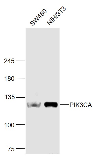
Sample:
SW480 (Human) Cell Lysate at 30 ug
NIH/3T3(Mouse) Cell Lysate at 30 ug
Primary: Anti- PIK3CA (bs-2067R) at 1/1000 dilution
Secondary: IRDye800CW Goat Anti-Rabbit IgG at 1/20000 dilution
Predicted band size: 124 kD
Observed band size: 124 kD
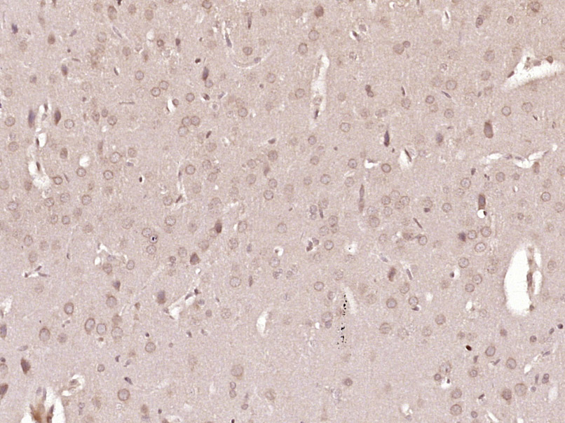
Paraformaldehyde-fixed, paraffin embedded (Rat brain); Antigen retrieval by boiling in sodium citrate buffer (pH6.0) for 15min; Block endogenous peroxidase by 3% hydrogen peroxide for 20 minutes; Blocking buffer (normal goat serum) at 37°C for 30min; Antibody incubation with (PIK3CA) Polyclonal Antibody, Unconjugated (bs-2067R) at 1:400 overnight at 4°C, followed by operating according to SP Kit(Rabbit) (sp-0023) instructionsand DAB staining.
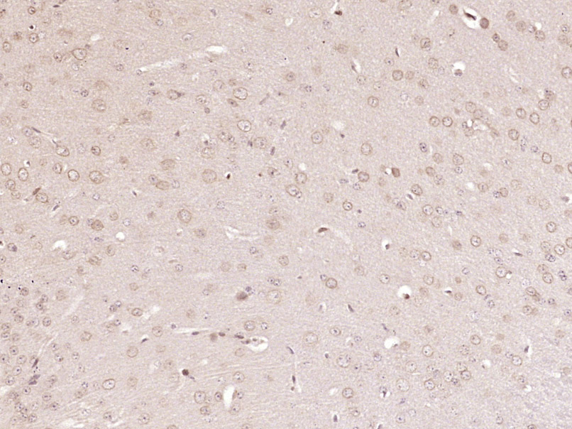
Paraformaldehyde-fixed, paraffin embedded (Mouse brain); Antigen retrieval by boiling in sodium citrate buffer (pH6.0) for 15min; Block endogenous peroxidase by 3% hydrogen peroxide for 20 minutes; Blocking buffer (normal goat serum) at 37°C for 30min; Antibody incubation with (PIK3CA) Polyclonal Antibody, Unconjugated (bs-2067R) at 1:400 overnight at 4°C, followed by operating according to SP Kit(Rabbit) (sp-0023) instructionsand DAB staining.
|
| 1、抗体溶解方法 | |
| 2、抗体修复方式 | |
| 3、常用试剂的配制 | |
| 4、免疫组化操作步骤 | |
| 5、免疫组化问题解答 | |
| 6、Western Blotting 操作步骤 | |
| 7、Western Blotting 问题解答 | |
| 8、关于肽链的设计 | |
| 9、多肽的溶解与保存 | |
| 10、酶标抗体效价测定程序 | |


