

| 产品编号 | bs-2041R |
| 英文名称 | Yellow fever virus envelope glycoprotein E Rabbit pAb |
| 中文名称 | 黄热病毒包膜糖蛋白抗体 |
| 别 名 | YFVgp1; |
| 研究领域 | 免疫学 细菌及病毒 |
| 抗体来源 | Rabbit |
| 克隆类型 | Polyclonal |
| 克 隆 号 | |
| 交叉反应 | Yellow fever virus |
| 产品应用 | WB=1:500-2000
not yet tested in other applications. optimal dilutions/concentrations should be determined by the end user. |
| 理论分子量 | 54/375 kDa |
| 检测分子量 | 30 |
| 细胞定位 | 细胞浆 细胞膜 分泌型蛋白 |
| 性 状 | Liquid |
| 浓 度 | 1mg/ml |
| 免 疫 原 | KLH conjugated synthetic peptide derived from Yellow fever virus envelope glycoprotein E: 601-700/3411 |
| 亚 型 | IgG |
| 纯化方法 | affinity purified by Protein A |
| 缓 冲 液 | 0.01M TBS (pH7.4) with 1% BSA, 0.02% Proclin300 and 50% Glycerol. |
| 保存条件 | Shipped at 4℃. Store at -20℃ for one year. Avoid repeated freeze/thaw cycles. |
| 注意事项 | This product as supplied is intended for research use only, not for use in human, therapeutic or diagnostic applications. |
| PubMed | PubMed |
| 产品介绍 |
Envelope protein E binding to host cell surface receptor is followed by virus internalization through clathrin-mediated endocytosis. Envelope protein E is subsequently involved in membrane fusion between virion and host late endosomes. Synthesized as a homodimer with prM which acts as a chaperone for envelope protein E. After cleavage of prM, envelope protein E dissociate from small envelope protein M and homodimerizes. Function: Capsid protein C self-assembles to form an icosahedral capsid about 30 nm in diameter. The capsid encapsulates the genomic RNA. prM acts as a chaperone for envelope protein E during intracellular virion assembly by masking and inactivating envelope protein E fusion peptide. prM is matured in the last step of virion assembly, presumably to avoid catastrophic activation of the viral fusion peptide induced by the acidic pH of the trans-Golgi network. After cleavage by host furin, the pr peptide is released in the extracellular medium and small envelope protein M and envelope protein E homodimers are dissociated. Envelope protein E binding to host cell surface receptor is followed by virus internalization through clathrin-mediated endocytosis. Envelope protein E is subsequently involved in membrane fusion between virion and host late endosomes. Synthesized as a homodimer with prM which acts as a chaperone for envelope protein E. After cleavage of prM, envelope protein E dissociate from small envelope protein M and homodimerizes. Non-structural protein 1 is involved in virus replication and regulation of the innate immune response. Non-structural protein 2A may be involved viral RNA replication and capsid assembly (Potential). Non-structural protein 2B is a required cofactor for the serine protease function of NS3. Serine protease NS3 displays three enzymatic activities: serine protease, NTPase and RNA helicase. NS3 serine protease, in association with NS2B, performs its autocleavage and cleaves the polyprotein at dibasic sites in the cytoplasm: C-prM, NS2A-NS2B, NS2B-NS3, NS3-NS4A, NS4A-2K and NS4B-NS5. NS3 RNA helicase binds RNA and unwinds dsRNA in the 3' to 5' direction (By similarity). Non-structural protein 4A induces host endoplasmic reticulum membrane rearrangements leading to the formation of virus-induced membranous vesicles hosting the dsRNA and polymerase, functioning as a replication complex. NS4A might also regulate the ATPase activity of the NS3 helicase (By similarity). Peptide 2k functions as a signal peptide for NS4B and is required for the interferon antagonism activity of the latter. Non-structural protein 4B inhibits interferon (IFN)-induced host STAT1 phosphorylation and nuclear translocation, thereby preventing the establishment of cellular antiviral state by blocking the IFN-alpha/beta pathway (By similarity). RNA-directed RNA polymerase NS5 replicates the viral (+) and (-) genome, and performs the capping of genomes in the cytoplasm. NS5 methylates viral RNA cap at guanine N-7 and ribose 2'-O positions. Besides its role in genome replication, also prevents the establishment of cellular antiviral state by blocking the interferon-alpha/beta (IFN-alpha/beta) signaling pathway Subunit: Capsid protein C forms homodimers. prM and envelope protein E form heterodimers in the endoplasmic reticulum and Golgi. In immature particles, there are 60 icosaedrally organized trimeric spikes on the surface. Each spike consists of three heterodimers of envelope protein M precursor (prM) and envelope protein E. NS1 forms homodimers as well as homohexamers when secreted. NS1 may interact with NS4A. NS3 and NS2B form a heterodimer. NS3 is the catalytic subunit, whereas NS2B strongly stimulates the latter, acting as a cofactor. In the absence of the NS2B, NS3 protease is unfolded and inactive. NS3 interacts with unphosphorylated NS5; this interaction stimulates NS5 guanylyltransferase activity. Subcellular Location: Capsid protein C: Virion (Potential). Peptide pr: Secreted. Small envelope protein M: Virion membrane; Multi-pass membrane protein (Potential). Host endoplasmic reticulum membrane; Multi-pass membrane protein (Potential). Envelope protein E: Virion membrane; Multi-pass membrane protein (Potential). Host endoplasmic reticulum membrane; Multi-pass membrane protein (Potential). Non-structural protein 1: Secreted. Host endoplasmic reticulum membrane; Peripheral membrane protein; Lumenal side. Non-structural protein 2A-alpha: Host endoplasmic reticulum membrane; Multi-pass membrane protein (Potential). Non-structural protein 2A: Host endoplasmic reticulum membrane; Multi-pass membrane protein (Potential). Serine protease subunit NS2B: Host endoplasmic reticulum membrane; Peripheral membrane protein; Cytoplasmic side. Serine protease NS3: Host endoplasmic reticulum membrane; Peripheral membrane protein; Cytoplasmic side. Note=Remains non-covalently associated to NS3 protease. Non-structural protein 4A: Host endoplasmic reticulum membrane; Multi-pass membrane protein. Note=Located in RE-associated vesicles hosting the replication complex. Non-structural protein 4B: Host endoplasmic reticulum membrane; Multi-pass membrane protein. RNA-directed RNA polymerase NS5: Host endoplasmic reticulum membrane; Peripheral membrane protein; Cytoplasmic side. Host nucleus. Note=Located in RE-associated vesicles hosting the replication complex. Post-translational modifications: Specific enzymatic cleavages in vivo yield mature proteins. The nascent protein C contains a C-terminal hydrophobic domain that act as a signal sequence for translocation of prM into the lumen of the ER. Mature protein C is cleaved at a site upstream of this hydrophobic domain by NS3. prM is cleaved in post-Golgi vesicles by a host furin, releasing the mature small envelope protein M, and peptide pr. Non-structural protein 2A-alpha, a C-terminally truncated form of non-structural protein 2A, results from partial cleavage by NS3. Specific enzymatic cleavages in vivo yield mature proteins Peptide 2K acts as a signal sequence and is removed from the N-terminus of NS4B by the host signal peptidase in the ER lumen. Signal cleavage at the 2K-4B site requires a prior NS3 protease-mediated cleavage at the 4A-2K site (By similarity). RNA-directed RNA polymerase NS5 is phosphorylated on serines residues. This phosphorylation may trigger NS5 nuclear localization. Envelope protein E and non-structural protein 1 are N-glycosylated. Similarity: In the N-terminal section; belongs to the class I-like SAM-binding methyltransferase superfamily. mRNA cap 0-1 NS5-type methyltransferase family. Contains 1 helicase ATP-binding domain. Contains 1 helicase C-terminal domain. Contains 1 mRNA cap 0-1 NS5-type MT domain. Contains 1 peptidase S7 domain. Contains 1 RdRp catalytic domain. |
| 产品图片 |
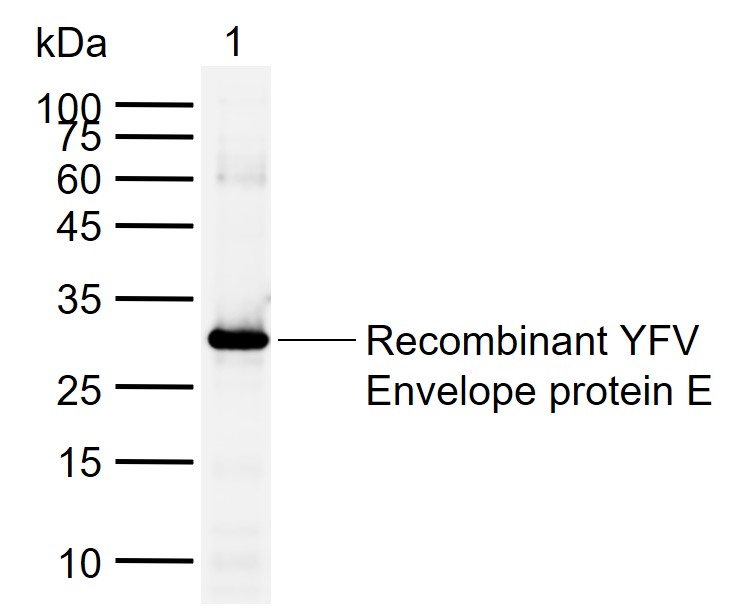
Sample:
Lane 1: Recombinant YFV Envelope protein E, Trx & His(bs-41285P-10ng)
Primary: Anti-Yellow fever virus envelope glycoprotein E (bs-2041R) at 1/1000 dilution
Secondary: IRDye800CW Goat Anti-Rabbit IgG at 1/20000 dilution
Predicted band size: 54/375 kDa
Observed band size: 30 kDa
|
| 1、抗体溶解方法 | |
| 2、抗体修复方式 | |
| 3、常用试剂的配制 | |
| 4、免疫组化操作步骤 | |
| 5、免疫组化问题解答 | |
| 6、Western Blotting 操作步骤 | |
| 7、Western Blotting 问题解答 | |
| 8、关于肽链的设计 | |
| 9、多肽的溶解与保存 | |
| 10、酶标抗体效价测定程序 | |


