

| 产品编号 | bs-1812R |
| 英文名称 | Notch3 Rabbit pAb |
| 中文名称 | 跨膜受体蛋白Notch-3抗体 |
| 别 名 | CADASIL; CADASIL1; CARASIL1; CASIL; FPLD1; IMF2; LMNS; N3; hpbk; NOTC3_HUMAN; NOTCH3; Notch 3; NOTC3_MOUSE; NOTC3_RAT; |
 |
Specific References (2) | bs-1812R has been referenced in 2 publications.
[IF=17.521] Hao Sun. et al. Silencing of NOTCH3 Signaling in Meniscus Smooth Muscle Cells Inhibits Fibrosis and Exacerbates Degeneration in a HEYL-Dependent Manner. Advanced Science. 2023 Apr;:2207020 IHC,IF ; Human.
[IF=1.984] Zhan J et al. In Vivo Study on the Effects of Xiaoaiping on the Stemness of Hepatocellular Carcinoma Cells. Evid Based Complement Alternat Med. 2019 Jun 23;2019:4738243. WB ; Human.
|
| 研究领域 | 染色质和核信号 神经生物学 干细胞 |
| 抗体来源 | Rabbit |
| 克隆类型 | Polyclonal |
| 克 隆 号 | |
| 交叉反应 | Human,Mouse,Rat |
| 产品应用 | IHC-P=1:100-500,IHC-F=1:100-500,IF=1:100-500,Flow-Cyt=1ug/Test
not yet tested in other applications. optimal dilutions/concentrations should be determined by the end user. |
| 理论分子量 | 255 kDa |
| 细胞定位 | 细胞核 细胞膜 |
| 性 状 | Liquid |
| 浓 度 | 1mg/ml |
| 免 疫 原 | KLH conjugated synthetic peptide derived from mouse Notch3: 2001-2100/2318 |
| 亚 型 | IgG |
| 纯化方法 | affinity purified by Protein A |
| 缓 冲 液 | 0.01M TBS (pH7.4) with 1% BSA, 0.02% Proclin300 and 50% Glycerol. |
| 保存条件 | Shipped at 4℃. Store at -20℃ for one year. Avoid repeated freeze/thaw cycles. |
| 注意事项 | This product as supplied is intended for research use only, not for use in human, therapeutic or diagnostic applications. |
| PubMed | PubMed |
| 产品介绍 |
This gene encodes the third discovered human homologue of the Drosophilia melanogaster type I membrane protein notch. In Drosophilia, notch interaction with its cell-bound ligands (delta, serrate) establishes an intercellular signalling pathway that plays a key role in neural development. Homologues of the notch-ligands have also been identified in human, but precise interactions between these ligands and the human notch homologues remains to be determined. Mutations in NOTCH3 have been identified as the underlying cause of cerebral autosomal dominant arteriopathy with subcortical infarcts and leukoencephalopathy (CADASIL). [provided by RefSeq, Jul 2008] Function: Functions as a receptor for membrane-bound ligands Jagged1, Jagged2 and Delta1 to regulate cell-fate determination. Upon ligand activation through the released notch intracellular domain (NICD) it forms a transcriptional activator complex with RBPJ/RBPSUH and activates genes of the enhancer of split locus. Affects the implementation of differentiation, proliferation and apoptotic programs. Subunit: Heterodimer of a C-terminal fragment N(TM) and a N-terminal fragment N(EC) which are probably linked by disulfide bonds. Interacts with MAML1, MAML2 and MAML3 which act as transcriptional coactivators for NOTCH3. Interacts with PSMA1. Interacts with HIF1AN. Subcellular Location: Cell membrane; Single-pass type I membrane protein. Notch 3 intracellular domain: Nucleus. Note=Following proteolytical processing NICD is translocated to the nucleus. Tissue Specificity: Ubiquitously expressed in fetal and adult tissues. Post-translational modifications: Synthesized in the endoplasmic reticulum as an inactive form which is proteolytically cleaved by a furin-like convertase in the trans-Golgi network before it reaches the plasma membrane to yield an active, ligand-accessible form. Cleavage results in a C-terminal fragment N(TM) and a N-terminal fragment N(EC). Following ligand binding, it is cleaved by TNF-alpha converting enzyme (TACE) to yield a membrane-associated intermediate fragment called notch extracellular truncation (NEXT). This fragment is then cleaved by presenilin dependent gamma-secretase to release a notch-derived peptide containing the intracellular domain (NICD) from the membrane. Phosphorylated. Hydroxylated by HIF1AN. DISEASE: Cerebral arteriopathy with subcortical infarcts and leukoencephalopathy, autosomal dominant (CADASIL) [MIM:125310]: A cerebrovascular disease characterized by multiple subcortical infarcts, pseudobulbar palsy, dementia, and the presence of granular deposits in small cerebral arteries producing ischemic stroke. Note=The disease is caused by mutations affecting the gene represented in this entry. Myofibromatosis, infantile 2 (IMF2) [MIM:615293]: A rare mesenchymal disorder characterized by the development of benign tumors in the skin, striated muscles, bones, and, more rarely, visceral organs. Subcutaneous or soft tissue nodules commonly involve the skin of the head, neck, and trunk. Skeletal and muscular lesions occur in about half of the patients. Lesions may be solitary or multicentric, and they may be present at birth or become apparent in early infancy or occasionally in adult life. Visceral lesions are associated with high morbidity and mortality. Note=The disease is caused by mutations affecting the gene represented in this entry. Similarity: Belongs to the NOTCH family. Contains 5 ANK repeats. Contains 34 EGF-like domains. Contains 3 LNR (Lin/Notch) repeats. SWISS: Q61982 Gene ID: 18131 Database links: Entrez Gene: 4854 Human Entrez Gene: 18131 Mouse Omim: 600276 Human SwissProt: Q9UM47 Human SwissProt: Q61982 Mouse Unigene: 8546 Human Unigene: 439741 Mouse Unigene: 53876 Rat Notch3是保守的Ⅰ型跨膜受体,Notch3信号通路在机体发育过程中调控细胞生长、分化和凋亡等多种重要生物学过程。 |
| 产品图片 |
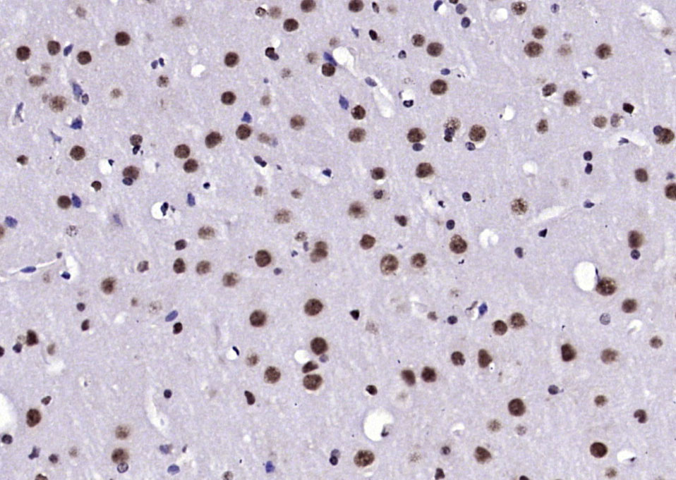
Paraformaldehyde-fixed, paraffin embedded (mouse brain); Antigen retrieval by boiling in sodium citrate buffer (pH6.0) for 15min; Block endogenous peroxidase by 3% hydrogen peroxide for 20 minutes; Blocking buffer (normal goat serum) at 37°C for 30min; Antibody incubation with (Notch3) Polyclonal Antibody, Unconjugated (bs-1812R) at 1:200 overnight at 4°C, followed by operating according to SP Kit(Rabbit) (sp-0023) instructionsand DAB staining.
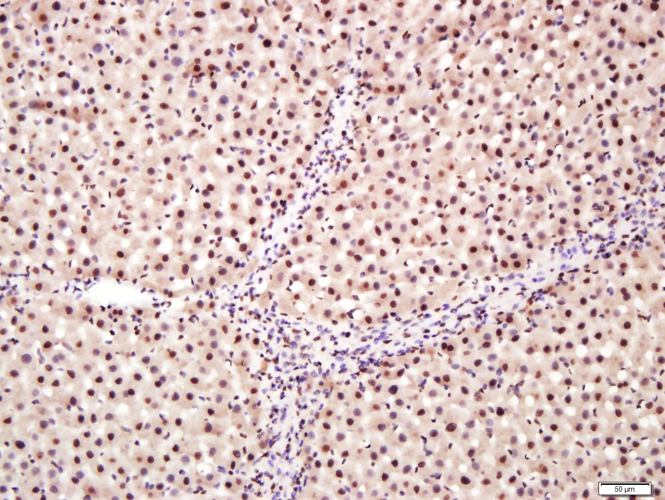
Paraformaldehyde-fixed, paraffin embedded (rat liver tissue); Antigen retrieval by boiling in sodium citrate buffer (pH6.0) for 15min; Block endogenous peroxidase by 3% hydrogen peroxide for 20 minutes; Blocking buffer (normal goat serum) at 37°C for 30min; Antibody incubation with (Notch3) Polyclonal Antibody, Unconjugated (bs-1812R) at 1:400 overnight at 4°C, followed by a conjugated secondary (sp-0023) for 20 minutes and DAB staining.

Paraformaldehyde-fixed, paraffin embedded (mouse brain); Antigen retrieval by boiling in sodium citrate buffer (pH6.0) for 15min; Block endogenous peroxidase by 3% hydrogen peroxide for 20 minutes; Blocking buffer (normal goat serum) at 37°C for 30min; Antibody incubation with (Notch3) Polyclonal Antibody, Unconjugated (bs-1812R) at 1:200 overnight at 4°C, followed by operating according to SP Kit(Rabbit) (sp-0023) instructionsand DAB staining.
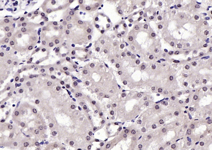
Paraformaldehyde-fixed, paraffin embedded (mouse kidney); Antigen retrieval by boiling in sodium citrate buffer (pH6.0) for 15min; Block endogenous peroxidase by 3% hydrogen peroxide for 20 minutes; Blocking buffer (normal goat serum) at 37°C for 30min; Antibody incubation with (Notch3) Polyclonal Antibody, Unconjugated (bs-1812R) at 1:200 overnight at 4°C, followed by operating according to SP Kit(Rabbit) (sp-0023) instructionsand DAB staining.
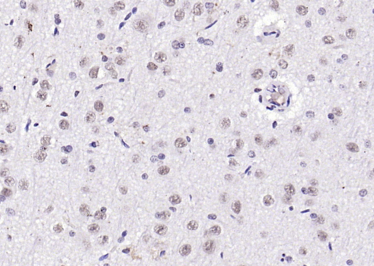
Paraformaldehyde-fixed, paraffin embedded (rat brain); Antigen retrieval by boiling in sodium citrate buffer (pH6.0) for 15min; Block endogenous peroxidase by 3% hydrogen peroxide for 20 minutes; Blocking buffer (normal goat serum) at 37°C for 30min; Antibody incubation with (Notch3) Polyclonal Antibody, Unconjugated (bs-1812R) at 1:200 overnight at 4°C, followed by operating according to SP Kit(Rabbit) (sp-0023) instructionsand DAB staining.
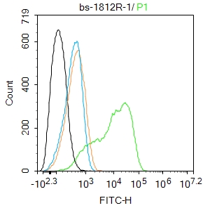
Blank control: SH-SY5Y.
Primary Antibody (green line): Rabbit Anti-Notch3 antibody (bs-1812R)
Dilution: 1ug/Test;
Secondary Antibody : Goat anti-rabbit IgG-FITC
Dilution: 0.5ug/Test.
Protocol
The cells were fixed with 4% PFA (10min at room temperature)and then permeabilized with 90% ice-cold methanol for 20 min at -20℃.The cells were then incubated in 5%BSA to block non-specific protein-protein interactions for 30 min at room temperature .Cells stained with Primary Antibody for 30 min at room temperature. The secondary antibody used for 40 min at room temperature. Acquisition of 20,000 events was performed.
|
| 1、抗体溶解方法 | |
| 2、抗体修复方式 | |
| 3、常用试剂的配制 | |
| 4、免疫组化操作步骤 | |
| 5、免疫组化问题解答 | |
| 6、Western Blotting 操作步骤 | |
| 7、Western Blotting 问题解答 | |
| 8、关于肽链的设计 | |
| 9、多肽的溶解与保存 | |
| 10、酶标抗体效价测定程序 | |


