

| 产品编号 | bs-1867R |
| 英文名称 | PD-1 Rabbit pAb |
| 中文名称 | 程序性死亡1(CD279)抗体 |
| 别 名 | ADMIO4; AIMTBS; CD279; PD-1; PD1; SLEB2; hPD-1; hPD-l; hSLE1; Ly101; Pdc1; PDCD1_HUMAN; PDCD1; Protein PD-1; PDCD1_MOUSE; mPD-1; programmed cell death 1; systemic lupus erythematosus susceptibility 2 |
 |
Specific References (13) | bs-1867R has been referenced in 13 publications.
|
| 研究领域 | 肿瘤 细胞生物 免疫学 细胞凋亡 |
| 抗体来源 | Rabbit |
| 克隆类型 | Polyclonal |
| 交叉反应 | Mouse (predicted: Human,Rat) |
| 产品应用 | Flow-Cyt=1μg /test
not yet tested in other applications. optimal dilutions/concentrations should be determined by the end user. |
| 理论分子量 | 32kDa |
| 检测分子量 | 55-60 |
| 细胞定位 | 细胞膜 |
| 性 状 | Liquid |
| 浓 度 | 1mg/ml |
| 免 疫 原 | KLH conjugated synthetic peptide derived from human PD-1: 201-288/288 |
| 亚 型 | IgG |
| 纯化方法 | affinity purified by Protein A |
| 缓 冲 液 | 0.01M TBS (pH7.4) with 1% BSA, 0.02% Proclin300 and 50% Glycerol. |
| 保存条件 | Shipped at 4℃. Store at -20℃ for one year. Avoid repeated freeze/thaw cycles. |
| 注意事项 | This product as supplied is intended for research use only, not for use in human, therapeutic or diagnostic applications. |
| PubMed | PubMed |
| 产品介绍 |
Programmed cell death protein 1 (PDCD1) is an immune-inhibitory receptor expressed in activated T cells; it is involved in the regulation of T-cell functions, including those of effector CD8+ T cells. In addition, this protein can also promote the differentiation of CD4+ T cells into T regulatory cells. PDCD1 is expressed in many types of tumors including melanomas, and has demonstrated to play a role in anti-tumor immunity. Moreover, this protein has been shown to be involved in safeguarding against autoimmunity, however, it can also contribute to the inhibition of effective anti-tumor and anti-microbial immunity. [provided by RefSeq, Aug 2020] Function: Inhibitory cell surface receptor involved in the regulation of T-cell function during immunity and tolerance. Upon ligand binding, inhibits T-cell effector functions in an antigen-specific manner. Possible cell death inducer, in association with other factors. Subunit: Monomer. Subcellular Location: Membrane; Single-pass type I membrane protein. Tissue Specificity: Ta,Ba,Ma,Thy DISEASE: Systemic lupus erythematosus 2 (SLEB2) [MIM:605218]: A chronic, relapsing, inflammatory, and often febrile multisystemic disorder of connective tissue, characterized principally by involvement of the skin, joints, kidneys and serosal membranes. It is of unknown etiology, but is thought to represent a failure of the regulatory mechanisms of the autoimmune system. The disease is marked by a wide range of system dysfunctions, an elevated erythrocyte sedimentation rate, and the formation of LE cells in the blood or bone marrow. {ECO:0000269|PubMed:12402038}. Note=Disease susceptibility is associated with variations affecting the gene represented in this entry. Similarity: Contains 1 Ig-like V-type (immunoglobulin-like) domain. SWISS: Q15116 Gene ID: 5133 Database links: Entrez Gene: 5133 Human Entrez Gene: 18566 Mouse Omim: 600244 Human SwissProt: Q15116 Human SwissProt: Q02242 Mouse Unigene: 158297 Human Unigene: 5024 Mouse Unigene: 105023 Rat |
| 产品图片 |
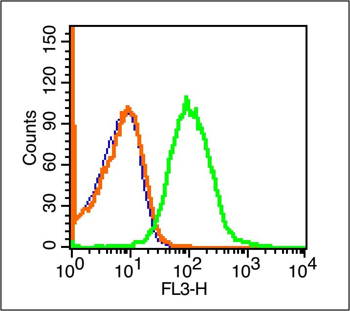
Blank control (blue line): Mouse spleen cells(blue).
Primary Antibody (green line): Rabbit Anti-PD-1/PE-CY7 Conjugated antibody (bs-1867R-PE-CY7)
Dilution: 1μg /10^6 cells;
Isotype Control Antibody (orange line): Rabbit IgG-PE-CY7 .
Protocol
The cells were fixed with 70% ice-cold methanol overnight at 4℃ . The cells were then incubated in 1 X PBS/2%BSA/10% goat serum to block non-specific protein-protein interactions followed by the antibody for 15 min at room temperature. Cells stained with Primary Antibody for 30 min at room temperature.Acquisition of 20,000 events was performed.
|
| 1、抗体溶解方法 | |
| 2、抗体修复方式 | |
| 3、常用试剂的配制 | |
| 4、免疫组化操作步骤 | |
| 5、免疫组化问题解答 | |
| 6、Western Blotting 操作步骤 | |
| 7、Western Blotting 问题解答 | |
| 8、关于肽链的设计 | |
| 9、多肽的溶解与保存 | |
| 10、酶标抗体效价测定程序 | |


