

| 产品编号 | bs-1990R |
| 英文名称 | MITF Rabbit pAb |
| 中文名称 | 微小细胞血症相关转录因子抗体 |
| 别 名 | CMM8; COMMAD; MI; MITF-A; WS2; WS2A; bHLHe32; BCC2; Gsfbcc2; Vitiligo; Wh; bw; vit; MITF_HUMAN; MITF; Class E basic helix-loop-helix protein 32 (bHLHe32); MITF_MOUSE; MITF_RAT; melanocyte inducing transcription factor; Waardenburg syndrome, type 2A; microphthalmia-associated transcription factor; melanogenesis associated transcription factor; homolog of mouse microphthalmia |
 |
Specific References (5) | bs-1990R has been referenced in 5 publications.
[IF=14.976] Qinyu Ma. et al. Small extracellular vesicles deliver osteolytic effectors and mediate cancer‐induced osteolysis in bone metastatic niche. J Extracell Vesicles. 2021 Feb;10(4):e12068 WB ; Mouse.
[IF=4.26] Rice et al. Melanoblast development coincides with the late emerging cells from the dorsal neural tube in turtle Trachemys scripta. (2017) Sci.Rep. 7:12063 IHC-P ; Turtle.
[IF=4.21] Kito, Yusuke, Chiemi Saigo, and Tamotsu Takeuchi. "Novel Transgenic Mouse Model of Polycystic Kidney Disease." The American Journal of Pathology (2017). WB ; Mouse.
[IF=2.766] Egawa et al. Therapeutic potential of CPI-613 for targeting tumorous mitochondrial energy metabolism and inhibiting autophagy in clear cell sarcoma. (2018) PLoS.One. 13:e0198940 IHC ; Mice.
[IF=1.65] Han, Joon‐Seung, Jong Hwan Sung, and Seung Kwon Lee. "Antimelanogenesis Activity of Hydrolyzed Ginseng Extract (GINST) via Inhibition of JNK Mitogen‐activated Protein Kinase in B16F10 Cells." Journal of Food Science (2016). WB ; Mouse.
|
| 研究领域 | 细胞生物 免疫学 染色质和核信号 转录调节因子 表观遗传学 |
| 抗体来源 | Rabbit |
| 克隆类型 | Polyclonal |
| 交叉反应 | Human,Mouse (predicted: Rat,Rabbit,Cow,Chicken,Dog,Horse) |
| 产品应用 | WB=1:500-2000,ICC/IF=1:50-200
not yet tested in other applications. optimal dilutions/concentrations should be determined by the end user. |
| 理论分子量 | 59kDa |
| 检测分子量 | 57 |
| 细胞定位 | 细胞核 |
| 性 状 | Liquid |
| 浓 度 | 1mg/ml |
| 免 疫 原 | KLH conjugated synthetic peptide derived from human MITF: 351-450/526 |
| 亚 型 | IgG |
| 纯化方法 | affinity purified by Protein A |
| 缓 冲 液 | 0.01M TBS (pH7.4) with 1% BSA, 0.02% Proclin300 and 50% Glycerol. |
| 保存条件 | Shipped at 4℃. Store at -20℃ for one year. Avoid repeated freeze/thaw cycles. |
| 注意事项 | This product as supplied is intended for research use only, not for use in human, therapeutic or diagnostic applications. |
| PubMed | PubMed |
| 产品介绍 |
The protein encoded by this gene is a transcription factor that contains both basic helix-loop-helix and leucine zipper structural features. The encoded protein regulates melanocyte development and is responsible for pigment cell-specific transcription of the melanogenesis enzyme genes. Heterozygous mutations in the this gene cause auditory-pigmentary syndromes, such as Waardenburg syndrome type 2 and Tietz syndrome. [provided by RefSeq, Aug 2017] Function: Transcription factor for tyrosinase (TYR) and tyrosinase-related protein 1 (TYRP1) that plays a key role in melanocyte development. Binds to a symmetrical DNA sequence (E-boxes) (5'-CACGTG-3') found in the tyrosinase promoter. Plays a critical role in the differentiation of various cell types as neural crest-derived melanocytes, mast cells, osteoclasts and optic cup-derived retinal pigment epithelium. Subunit: Efficient DNA binding requires dimerization with another bHLH protein. Binds DNA in the form of homodimer or heterodimer with either TFE3, TFEB or TFEC. Interacts with KARS. Subcellular Location: Nucleus. Tissue Specificity: Isoform M is exclusively expressed in melanocytes and melanoma cells. Isoform A and isoform H are widely expressed in many cell types including melanocytes and retinal pigment epithelium (RPE). Isoform C is expressed in many cell types including RPE but not in melanocyte-lineage cells. Post-translational modifications: Phosphorylation at Ser-405 significantly enhances the ability to bind the tyrosinase promoter. Phosphorylated at Ser-180 and Ser-516 following KIT signaling, trigerring a short live activation: Phosphorylation at Ser-180 and Ser-516 by MAPK and RPS6KA1, respectively, activate the transcription factor activity but also promote ubiquitination and subsequent degradation by the proteasome. Ubiquitinated following phosphorylation at Ser-180, leading to subsequent degradation by the proteasome. Deubiquitinated by USP13, preventing its degradation. DISEASE: Defects in MITF are the cause of Waardenburg syndrome type 2A (WS2A) [MIM:193510]. It is a dominant inherited disorder characterized by sensorineural hearing loss and patches of depigmentation. The features show variable expression and penetrance. [DISEASE] Defects in MITF are a cause of Waardenburg syndrome type 2 with ocular albinism (WS2-OA) [MIM:103470]. It is an ocular albinism with sensorineural deafness. [DISEASE] Defects in MITF are the cause of Tietz syndrome (TIETZS) [MIM:103500]. It is an autosomal dominant disorder characterized by generalized hypopigmentation and profound, congenital, bilateral deafness. Penetrance is complete. [DISEASE] Defects in MITF are a cause of susceptibility to cutaneous malignant melanoma type 8 (CMM8) [MIM:614456]. A malignant neoplasm of melanocytes, arising de novo or from a pre-existing benign nevus, which occurs most often in the skin but also may involve other sites. Similarity: Belongs to the MiT/TFE family. Contains 1 basic helix-loop-helix (bHLH) domain. SWISS: O75030 Gene ID: 4286 Database links: Entrez Gene: 4286 Human Entrez Gene: 17342 Mouse SwissProt: O75030 Human SwissProt: Q08874 Mouse MITF微小转录因子是一个黑色素细胞的核蛋白,对黑色素细胞的生成和活性起着关键作用,MITF也是控制细胞外信号的一项调节因子。MITF高度表达于原发和转移的恶性黑色素瘤,也可视为高敏感和高特异的黑色素细胞标记。 |
| 产品图片 |
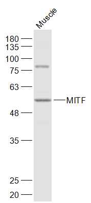
Sample:
Muscle (Mouse) Lysate at 40 ug
Primary: Anti-MITF (bs-1990R) at 1/1000 dilution
Secondary: IRDye800CW Goat Anti-Rabbit IgG at 1/20000 dilution
Predicted band size: 59 kD
Observed band size: 57 kD
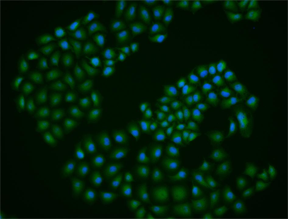
Hela cell; 4% Paraformaldehyde-fixed; Triton X-100 at room temperature for 20 min; Blocking buffer (normal goat serum, C-0005) at 37°C for 20 min; Antibody incubation with (MITF) polyclonal Antibody, Unconjugated (bs-1990R) 1:25, 90 minutes at 37°C; followed by a conjugated Goat Anti-Rabbit IgG antibody at 37°C for 90 minutes, DAPI (blue, C02-04002) was used to stain the cell nuclei.
|
| 1、抗体溶解方法 | |
| 2、抗体修复方式 | |
| 3、常用试剂的配制 | |
| 4、免疫组化操作步骤 | |
| 5、免疫组化问题解答 | |
| 6、Western Blotting 操作步骤 | |
| 7、Western Blotting 问题解答 | |
| 8、关于肽链的设计 | |
| 9、多肽的溶解与保存 | |
| 10、酶标抗体效价测定程序 | |


