

| 产品编号 | bs-2439R |
| 英文名称 | Axin1 Rabbit pAb |
| 中文名称 | 轴蛋白1抗体 |
| 别 名 | AXIN; CMDOH; PPP1R49; AXIN1-RGS; axin-1; Fu; Kb; Ki; fused; kinky; knobbly; AXIN1_CHICK; AXIN1; Axis inhibition protein 1; AXN; AXIN1_HUMAN; Axis inhibition protein 1 (hAxin); AXIN1_MOUSE; Protein Fused; AXIN1_RAT; Axis inhibition protein 1 (rAxin); |
 |
Specific References (1) | bs-2439R has been referenced in 1 publications.
[IF=6.183] Zou et al. Hydroxylase Activity of ASPH Promotes Hepatocellular Carcinoma Metastasis Through Epithelial-to-Mesenchymal Transition Pathway. (2018) EBioMedicine. 31:287-298 WB ; Human.
|
| 研究领域 | 细胞生物 信号转导 干细胞 细胞周期蛋白 |
| 抗体来源 | Rabbit |
| 克隆类型 | Polyclonal |
| 克 隆 号 | |
| 交叉反应 | Human,Mouse,Rat (predicted: Pig,Sheep,Cow,Dog,GuineaPig) |
| 产品应用 | IHC-P=1:100-500,IHC-F=1:100-500,IF=1:100-500,ICC/IF=1:100-500
not yet tested in other applications. optimal dilutions/concentrations should be determined by the end user. |
| 理论分子量 | 99 kDa |
| 细胞定位 | 细胞核 细胞浆 细胞膜 |
| 性 状 | Liquid |
| 浓 度 | 1mg/ml |
| 免 疫 原 | KLH conjugated synthetic peptide derived from human Axin1: 761-862/862 |
| 亚 型 | IgG |
| 纯化方法 | affinity purified by Protein A |
| 缓 冲 液 | 0.01M TBS (pH7.4) with 1% BSA, 0.02% Proclin300 and 50% Glycerol. |
| 保存条件 | Shipped at 4℃. Store at -20℃ for one year. Avoid repeated freeze/thaw cycles. |
| 注意事项 | This product as supplied is intended for research use only, not for use in human, therapeutic or diagnostic applications. |
| PubMed | PubMed |
| 产品介绍 |
This gene encodes a cytoplasmic protein which contains a regulation of G-protein signaling (RGS) domain and a dishevelled and axin (DIX) domain. The encoded protein interacts with adenomatosis polyposis coli, catenin (cadherin-associated protein), beta 1, 88kDa, glycogen synthase kinase 3 beta, protein phosphate 2, and itself. This protein functions as a negative regulator of the wingless-type MMTV integration site family, member 1 (WNT) signaling pathway and can induce apoptosis. The crystal structure of a portion of this protein, alone and in a complex with other proteins, has been resolved. Mutations in this gene have been associated with hepatocellular carcinoma, hepatoblastomas, ovarian endometriod adenocarcinomas, and medullablastomas. Two transcript variants encoding distinct isoforms have been identified for this gene. [provided by RefSeq] Function: Component of the beta-catenin destruction complex required for regulating CTNNB1 levels through phosphorylation and ubiquitination, and modulating Wnt-signaling. Controls dorsoventral patterning via two opposing effects; down-regulates CTNNB1 to inhibit the Wnt signaling pathway and ventralize embryos, but also dorsalizes embryos by activating a Wnt-independent JNK signaling pathway. In Wnt signaling, probably facilitates the phosphorylation of CTNNB1 and APC by GSK3B. Likely to function as a tumor suppressor. Facilitates the phosphorylation of TP53 by HIPK2 upon ultraviolet irradiation. Enhances TGF-beta signaling by recruiting the RNF111 E3 ubiquitin ligase and promoting the degradation of inhibitory SMAD7. Also component of the AXIN1-HIPK2-TP53 complex which controls cell growth, apoptosis and development. Subcellular Location: Cytoplasm. Nucleus. Cell membrane. MACF1 is required for its translocation to cell membrane. On UV irradiation, translocates to the nucleus and colocalizes with DAAX. Tissue Specificity: Ubiquitously expressed. Post-translational modifications: Phosphorylation and dephosphorylation of AXIN1 regulates assembly and function of the beta-catenin complex. Phosphorylated by CK1 and GSK3B. Dephosphorylated by PPP1CA and PPP2CA. Phosphorylation by CK1 enhances binding of GSK3B to AXIN1. ADP-ribosylated by tankyrase TNKS and TNKS2. Poly-ADP-ribosylated protein is recognized by RNF146, followed by ubiquitination and subsequent activation of the Wnt signaling pathway. Ubiquitinated by RNF146 when poly-ADP-ribosylated, leading to its degradation and subsequent activation of the Wnt signaling pathway. Sumoylation at Lys-857 and Lys-860 prevents ubiquitination and degradation. Sumoylation is required for AXIN1-mediated JNK activation. Deubiquitinated by USP34, deubiquitinated downstream of beta-catenin stabilization step: deubiquitination is important for nuclear accumulation during Wnt signaling to positively regulate beta-catenin (CTNBB1)-mediated transcription. DISEASE: Defects in AXIN1 are involved in hepatocellular carcinoma (HCC) [MIM:114550]. Defects in AXIN1 are a cause of caudal duplication anomaly (CADUA) [MIM:607864]. Caudal duplication anomaly is characterized by the occurrence of duplications of different organs in the caudal region. Note=Caudal duplication anomaly is associated with hypermethylation of the AXIN1 promoter. Similarity: Contains 1 DIX domain. Contains 1 RGS domain. SWISS: O15169 Gene ID: 8312 Database links: Entrez Gene: 8312 Human Entrez Gene: 395786 Chicken Entrez Gene: 100065116 Horse Entrez Gene: 12005 Mouse Omim: 603816 Human SwissProt: O42400 Chicken SwissProt: O15169 Human SwissProt: O35625 Mouse Unigene: 592082 Human Unigene: 23684 Mouse Unigene: 31781 Rat |
| 产品图片 |
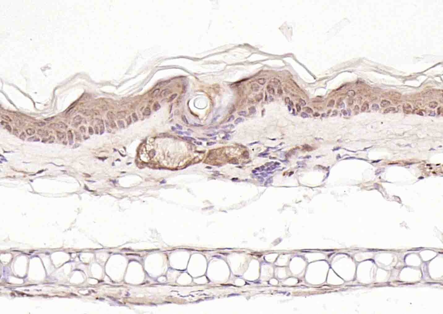
Paraformaldehyde-fixed, paraffin embedded (mouse skin); Antigen retrieval by boiling in sodium citrate buffer (pH6.0) for 15min; Block endogenous peroxidase by 3% hydrogen peroxide for 20 minutes; Blocking buffer (normal goat serum) at 37°C for 30min; Incubation with (Axin1) Polyclonal Antibody, Unconjugated (bs-2439R) at 1:200 overnight at 4°C, followed by operating according to SP Kit(Rabbit) (sp-0023) instructionsand DAB staining.
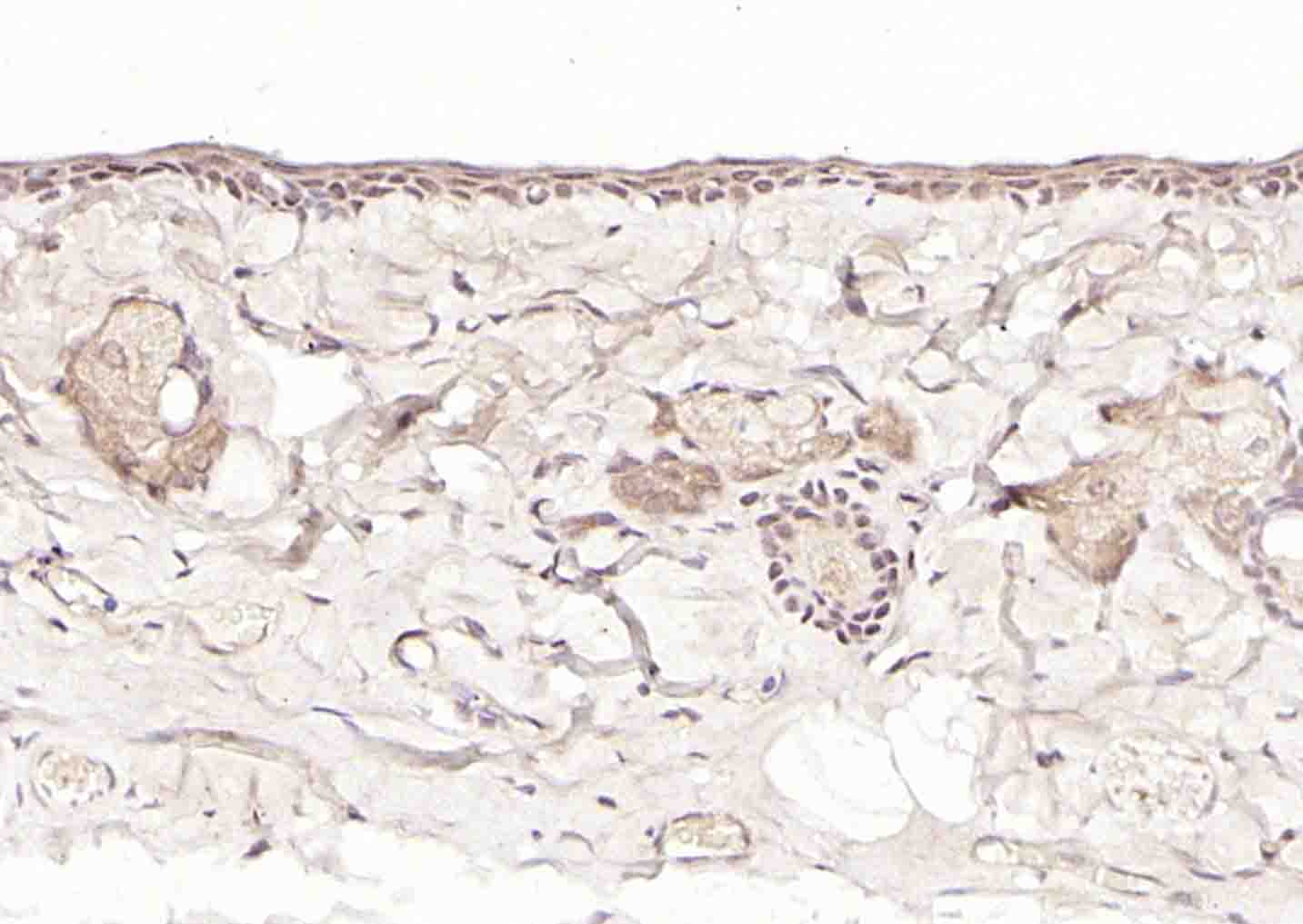
Paraformaldehyde-fixed, paraffin embedded (rat skin); Antigen retrieval by boiling in sodium citrate buffer (pH6.0) for 15min; Block endogenous peroxidase by 3% hydrogen peroxide for 20 minutes; Blocking buffer (normal goat serum) at 37°C for 30min; Incubation with (Axin1) Polyclonal Antibody, Unconjugated (bs-2439R) at 1:200 overnight at 4°C, followed by operating according to SP Kit(Rabbit) (sp-0023) instructionsand DAB staining.
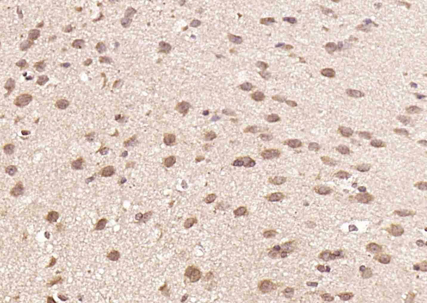
Paraformaldehyde-fixed, paraffin embedded (mouse brain); Antigen retrieval by boiling in sodium citrate buffer (pH6.0) for 15min; Block endogenous peroxidase by 3% hydrogen peroxide for 20 minutes; Blocking buffer (normal goat serum) at 37°C for 30min; Incubation with (Axin1) Polyclonal Antibody, Unconjugated (bs-2439R) at 1:200 overnight at 4°C, followed by operating according to SP Kit(Rabbit) (sp-0023) instructionsand DAB staining.
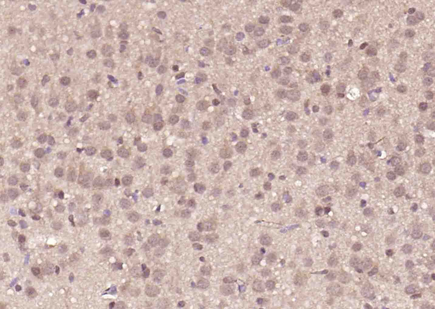
Paraformaldehyde-fixed, paraffin embedded (rat brain); Antigen retrieval by boiling in sodium citrate buffer (pH6.0) for 15min; Block endogenous peroxidase by 3% hydrogen peroxide for 20 minutes; Blocking buffer (normal goat serum) at 37°C for 30min; Incubation with (Axin1) Polyclonal Antibody, Unconjugated (bs-2439R) at 1:200 overnight at 4°C, followed by operating according to SP Kit(Rabbit) (sp-0023) instructionsand DAB staining.
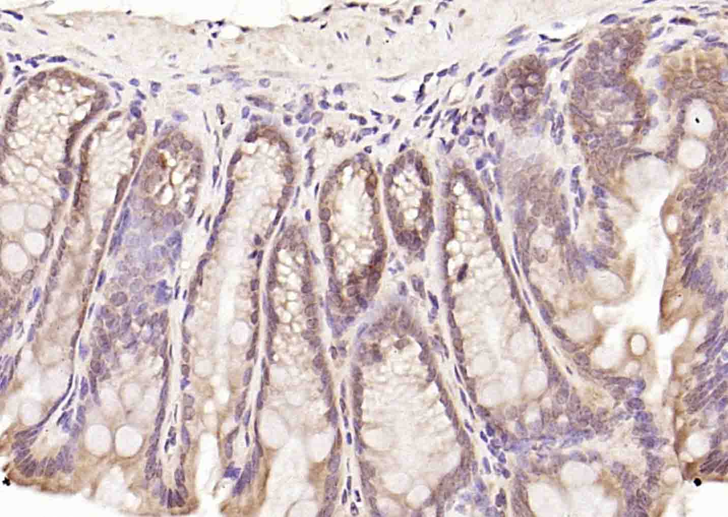
Paraformaldehyde-fixed, paraffin embedded (mouse colon); Antigen retrieval by boiling in sodium citrate buffer (pH6.0) for 15min; Block endogenous peroxidase by 3% hydrogen peroxide for 20 minutes; Blocking buffer (normal goat serum) at 37°C for 30min; Incubation with (Axin1) Polyclonal Antibody, Unconjugated (bs-2439R) at 1:200 overnight at 4°C, followed by operating according to SP Kit(Rabbit) (sp-0023) instructionsand DAB staining.
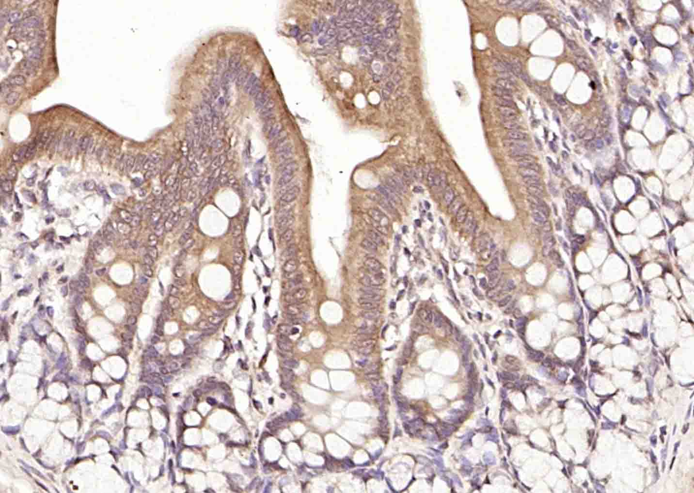
Paraformaldehyde-fixed, paraffin embedded (rat colon); Antigen retrieval by boiling in sodium citrate buffer (pH6.0) for 15min; Block endogenous peroxidase by 3% hydrogen peroxide for 20 minutes; Blocking buffer (normal goat serum) at 37°C for 30min; Incubation with (Axin1) Polyclonal Antibody, Unconjugated (bs-2439R) at 1:200 overnight at 4°C, followed by operating according to SP Kit(Rabbit) (sp-0023) instructionsand DAB staining.
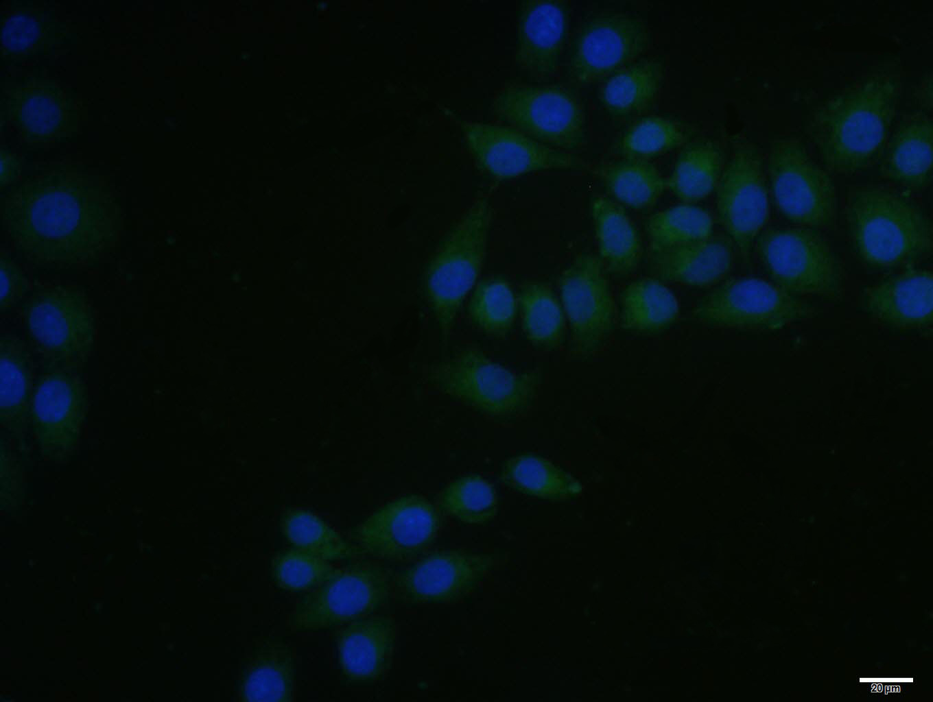
MCF7 cell; 4% Paraformaldehyde-fixed; Triton X-100 at room temperature for 20 min; Blocking buffer (normal goat serum, C-0005) at 37°C for 20 min; Antibody incubation with (Axin1) polyclonal Antibody, Unconjugated (bs-2439R) 1:100, 90 minutes at 37°C; followed by a conjugated Goat Anti-Rabbit IgG antibody at 37°C for 90 minutes, DAPI (blue, C02-04002) was used to stain the cell nuclei.
|
| 1、抗体溶解方法 | |
| 2、抗体修复方式 | |
| 3、常用试剂的配制 | |
| 4、免疫组化操作步骤 | |
| 5、免疫组化问题解答 | |
| 6、Western Blotting 操作步骤 | |
| 7、Western Blotting 问题解答 | |
| 8、关于肽链的设计 | |
| 9、多肽的溶解与保存 | |
| 10、酶标抗体效价测定程序 | |


