

| 产品编号 | bs-2157R |
| 英文名称 | Polycystin 1 Rabbit pAb |
| 中文名称 | 多囊肾蛋白1抗体 |
| 别 名 | PC1; PBP; Pc-1; TRPP1; eliosin; mFLJ00285; PKD1_HUMAN; PKD1; Autosomal dominant polycystic kidney disease 1 protein; PKD1_MOUSE; Autosomal dominant polycystic kidney disease 1 protein homolog; |
 |
Specific References (5) | bs-2157R has been referenced in 5 publications.
[IF=6.75] Ohata, Shinya, et al. "Mechanosensory Genes Pkd1 and Pkd2 Contribute to the Planar Polarization of Brain Ventricular Epithelium." The Journal of Neuroscience 35.31 (2015): 11153-11168. IHC-F ; Mouse.
[IF=6.721] Winokurow N et al. A role for polycystin-1 and polycystin-2 in neural progenitor cell differentiation.Cell Mol Life Sci. 2019 Mar 20. WB&IHC ; Mouse.
[IF=5.63] Chiou, Yi-Shiou, et al. "Peracetylated (−)-epigallocatechin-3-gallate (AcEGCG) potently prevents skin carcinogenesis by suppressing the PKD1-dependent signaling pathway in CD34+ skin stem cells and skin tumors." Carcinogenesis 34.6 (2013): 1315-1322. IP ; Mouse.
[IF=4.21] Kito, Yusuke, Chiemi Saigo, and Tamotsu Takeuchi. "Novel Transgenic Mouse Model of Polycystic Kidney Disease." The American Journal of Pathology (2017). WB ; Mouse.
[IF=2.79] Ren, Jian-gang, et al. "Down-regulation of polycystin in lymphatic malformations: Possible role in the proliferation of lymphatic endothelial cells." Human Pathology (2017). IHC-P ; Human.
|
| 研究领域 | 细胞生物 免疫学 发育生物学 |
| 抗体来源 | Rabbit |
| 克隆类型 | Polyclonal |
| 克 隆 号 | |
| 交叉反应 | Human (predicted: Mouse) |
| 产品应用 | Flow-Cyt=1μg/Test
not yet tested in other applications. optimal dilutions/concentrations should be determined by the end user. |
| 理论分子量 | 460 kDa |
| 细胞定位 | 细胞膜 |
| 性 状 | Liquid |
| 浓 度 | 1mg/ml |
| 免 疫 原 | KLH conjugated synthetic peptide derived from human Polycystin 1: 131-230/4303 <Extracellular> |
| 亚 型 | IgG |
| 纯化方法 | affinity purified by Protein A |
| 缓 冲 液 | 0.01M TBS (pH7.4) with 1% BSA, 0.02% Proclin300 and 50% Glycerol. |
| 保存条件 | Shipped at 4℃. Store at -20℃ for one year. Avoid repeated freeze/thaw cycles. |
| 注意事项 | This product as supplied is intended for research use only, not for use in human, therapeutic or diagnostic applications. |
| PubMed | PubMed |
| 产品介绍 |
This gene encodes a member of the polycystin protein family. The encoded glycoprotein contains a large N-terminal extracellular region, multiple transmembrane domains and a cytoplasmic C-tail. It is an integral membrane protein that functions as a regulator of calcium permeable cation channels and intracellular calcium homoeostasis. It is also involved in cell-cell/matrix interactions and may modulate G-protein-coupled signal-transduction pathways. It plays a role in renal tubular development, and mutations in this gene cause autosomal dominant polycystic kidney disease type 1 (ADPKD1). ADPKD1 is characterized by the growth of fluid-filled cysts that replace normal renal tissue and result in end-stage renal failure. Splice variants encoding different isoforms have been noted for this gene. Also, six pseudogenes, closely linked in a known duplicated region on chromosome 16p, have been described. [provided by RefSeq]. Function: Involved in renal tubulogenesis. Involved in fluid-flow mechanosensation by the primary cilium in renal epithelium. Acts as a regulator of cilium length, together with PKD2. The dynamic control of cilium length is essential in the regulation of mechanotransductive signaling. The cilium length response creates a negative feedback loop whereby fluid shear-mediated deflection of the primary cilium, which decreases intracellular cAMP, leads to cilium shortening and thus decreases flow-induced signaling. May be an ion-channel regulator. Involved in adhesive protein-protein and protein-carbohydrate interactions. Subunit: Interacts with PKD2. Interacts with PRKX; involved in differentiation and controlled morphogenesis of the kidney. Interacts with NPHP1 (via SH3 domain). Subcellular Location: Membrane; Multi-pass membrane protein. Cell projection, cilium. Note=PKD1 localization to the plasma and ciliary membranes requires PKD2, is independent of PKD2 channel activity, and involves stimulation of PKD1 autoproteolytic cleavage at the GPS domain. Post-translational modifications: After synthesis, undergoes cleavage between Leu-3048 and Thr-3049 in the GPS domain. Cleavage at the GPS domain occurs through a cis-autoproteolytic mechanism involving an ester-intermediate via N-O acyl rearrangement. This process takes place in the early secretory pathway, depends on initial N-glycosylation, and requires the REJ domain. There is evidence that cleavage at GPS domain is incomplete. Uncleaved and cleaved products may have different functions in vivo. DISEASE: Defects in PKD1 are the cause of polycystic kidney disease autosomal dominant type 1 (ADPKD1) [MIM:173900]. ADPKD is characterized by progressive formation and enlargement of cysts in both kidneys, typically leading to end-stage renal disease in adult life. Cysts also occurs in the liver and other organs. Its prevalence is estimated at about 1/1000. Similarity: Contains 1 C-type lectin domain. Contains 1 GPS domain. Contains 1 LDL-receptor class A domain. Contains 2 LRR (leucine-rich) repeats. Contains 1 LRRCT domain. Contains 1 LRRNT domain. Contains 17 PKD domains. Contains 1 PLAT domain. Contains 1 REJ domain. Contains 1 WSC domain. SWISS: P98161 Gene ID: 5310 Database links: Entrez Gene: 5310 Human Entrez Gene: 18763 Mouse Omim: 601313 Human SwissProt: P98161 Human SwissProt: O08852 Mouse Unigene: 75813 Human Unigene: 290442 Mouse Unigene: 30435 Mouse 多囊肾(polycystic kidney disease)为遗传性疾病,是肾脏一种先天性异常。双侧肾脏皮髓质均可累及,但在程度上可不同。在遗传方式上表现为常染色体显性和常染色体隐性遗传两种。 囊内上皮细胞异常增殖是ADPKD的显著特特之一,处于一种成熟不完全或重发育状态,高度提示为细胞的发育成熟调控出现障碍,使细胞处于一种未成熟状态,从而显示强增殖性。表现为细胞转运密切相关的Na+-K+-ATP ase的亚单位组合,分布及活性表达的改变;细胞信号传导异常以及离子转运通道的变化。细胞外基质异常增生是ADPKD第三种显著特征。目前许多研究已证明:这些异常均有与细胞生长有关的活性因子的参与。但关键的异常环节和途径尚未明了。因基因缺陷而致的细胞生长改变和间质形成异常,是本病的重要发病机制之一。 |
| 产品图片 |
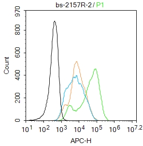
Blank control: SH-Sy5Y.
Primary Antibody (green line): Rabbit Anti-Polycystin 1 antibody (bs-2157R)
Dilution: 2μg /10^6 cells;
Isotype Control Antibody (orange line): Rabbit IgG .
Secondary Antibody : Goat anti-rabbit IgG-AF647
Dilution: 1μg /test.
Protocol
The cells were then incubated in 5%BSA to block non-specific protein-protein interactions for 30 min at at room temperature .Cells stained with Primary Antibody for 30 min at room temperature. The secondary antibody used for 40 min at room temperature. Acquisition of 20,000 events was performed.
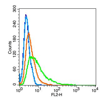
Blank control(blue): Hela(fixed with 2% paraformaldehyde(10 min)).
Primary Antibody:Rabbit Anti-Polycystin 1 antibody(bs-2157R), Dilution: 1μg in 100 μL 1X PBS containing 0.5% BSA;
Isotype Control Antibody: Rabbit IgG(orange) ,used under the same conditions ); Secondary Antibody: Goat anti-rabbit IgG-PE(white blue), Dilution: 1:200 in 1 X PBS containing 0.5% BSA.
|
| 1、抗体溶解方法 | |
| 2、抗体修复方式 | |
| 3、常用试剂的配制 | |
| 4、免疫组化操作步骤 | |
| 5、免疫组化问题解答 | |
| 6、Western Blotting 操作步骤 | |
| 7、Western Blotting 问题解答 | |
| 8、关于肽链的设计 | |
| 9、多肽的溶解与保存 | |
| 10、酶标抗体效价测定程序 | |


