

| 产品编号 | bs-2469R |
| 英文名称 | PERK Rabbit pAb |
| 中文名称 | 蛋白激酶样内质网激酶抗体 |
| 别 名 | PEK; PERK; WRS; E2AK3_HUMAN; EIF2AK3; PRKR-like endoplasmic reticulum kinase; Pancreatic eIF2-alpha kinase (HsPEK); Protein tyrosine kinase EIF2AK3; 2.7.11.1; E2AK3_MOUSE; Pancreatic eIF2-alpha kinase; E2AK3_RAT; |
 |
Specific References (29) | bs-2469R has been referenced in 29 publications.
|
| 研究领域 | 免疫学 染色质和核信号 信号转导 新陈代谢 表观遗传学 |
| 抗体来源 | Rabbit |
| 克隆类型 | Polyclonal |
| 克 隆 号 | |
| 交叉反应 | Human,Mouse,Rat |
| 产品应用 | WB=1:500-2000,IHC-P=1:100-500,IHC-F=1:100-500,IF=1:100-500,Flow-Cyt=2μg/Test
not yet tested in other applications. optimal dilutions/concentrations should be determined by the end user. |
| 理论分子量 | 122 kDa |
| 检测分子量 | 145 |
| 细胞定位 | 细胞浆 |
| 性 状 | Liquid |
| 浓 度 | 1mg/ml |
| 免 疫 原 | KLH conjugated synthetic peptide derived from human PERK: 1001-1116/1116 |
| 亚 型 | IgG |
| 纯化方法 | affinity purified by Protein A |
| 缓 冲 液 | 0.01M TBS (pH7.4) with 1% BSA, 0.02% Proclin300 and 50% Glycerol. |
| 保存条件 | Shipped at 4℃. Store at -20℃ for one year. Avoid repeated freeze/thaw cycles. |
| 注意事项 | This product as supplied is intended for research use only, not for use in human, therapeutic or diagnostic applications. |
| PubMed | PubMed |
| 产品介绍 |
The protein encoded by this gene phosphorylates the alpha subunit of eukaryotic translation-initiation factor 2 (EIF2), leading to its inactivation, and thus to a rapid reduction of translational initiation and repression of global protein synthesis. It is a type I membrane protein located in the endoplasmic reticulum (ER), where it is induced by ER stress caused by malfolded proteins. Mutations in this gene are associated with Wolcott-Rallison syndrome. [provided by RefSeq, Jan 2010] Function: Phosphorylates the alpha subunit of eukaryotic translation-initiation factor 2 (EIF2), leading to its inactivation and thus to a rapid reduction of translational initiation and repression of global protein synthesis. Serves as a critical effector of unfolded protein response (UPR)-induced G1 growth arrest due to the loss of cyclin-D1 (CCND1). Subunit: Forms dimers with HSPA5/BIP in resting cells. Oligomerizes in ER-stressed cells. Interacts with DNAJC3. Subcellular Location: Endoplasmic reticulum membrane; Single-pass type I membrane protein. Tissue Specificity: Ubiquitous. A high level expression is seen in secretory tissues. Post-translational modifications: Oligomerization of the N-terminal ER luminal domain by ER stress promotes PERK trans-autophosphorylation of the C-terminal cytoplasmic kinase domain at multiple residues including Thr-982 on the kinase activation loop. Autophosphorylated. Phosphorylated at Tyr-619 following endoplasmic reticulum stress, leading to activate its tyrosine-protein kinase activity. Dephosphorylated by PTPN1/TP1B, leading to inactivate its enzyme activity. N-glycosylated. ADP-ribosylated by PARP16 upon ER stress, which increases kinase activity. DISEASE: Wolcott-Rallison syndrome (WRS) [MIM:226980]: A rare autosomal recessive disorder, characterized by permanent neonatal or early infancy insulin-dependent diabetes and, at a later age, epiphyseal dysplasia, osteoporosis, growth retardation and other multisystem manifestations, such as hepatic and renal dysfunctions, mental retardation and cardiovascular abnormalities. Note=The disease is caused by mutations affecting the gene represented in this entry. Similarity: Belongs to the protein kinase superfamily. Ser/Thr protein kinase family. GCN2 subfamily. Contains 1 protein kinase domain. SWISS: Q9NZJ5 Gene ID: 9451 Database links: Entrez Gene: 9451 Human Entrez Gene: 13666 Mouse Omim: 604032 Human SwissProt: Q9NZJ5 Human SwissProt: Q9Z2B5 Mouse Unigene: 591589 Human Unigene: 247167 Mouse Unigene: 24897 Rat
|
| 产品图片 |
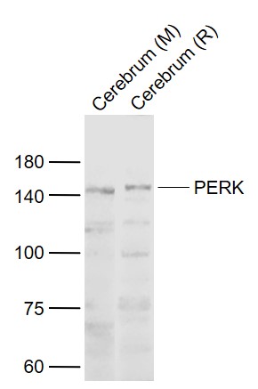
Sample:
Lane 1: Cerebrum (Mouse) Lysate at 40 ug
Lane 2: Cerebrum (Rat) Lysate at 40 ug
Primary: Anti-PERK (bs-2469R) at 1/1000 dilution
Secondary: IRDye800CW Goat Anti-Rabbit IgG at 1/20000 dilution
Predicted band size: 122 kD
Observed band size: 145 kD
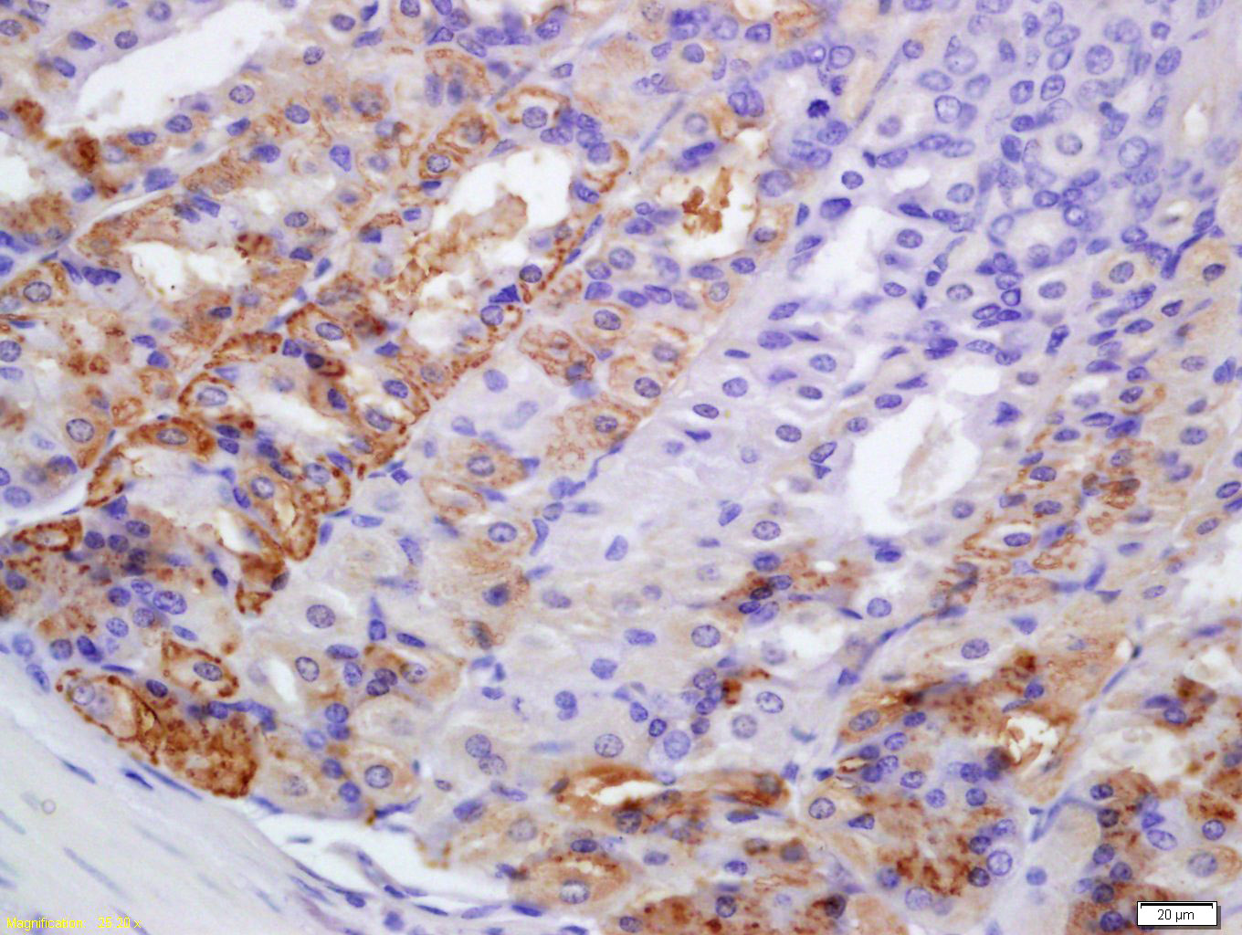
Tissue/cell: mouse stomach tissue; 4% Paraformaldehyde-fixed and paraffin-embedded;
Antigen retrieval: citrate buffer ( 0.01M, pH 6.0 ), Boiling bathing for 15min; Block endogenous peroxidase by 3% Hydrogen peroxide for 30min; Blocking buffer (normal goat serum,C-0005) at 37℃ for 20 min;
Incubation: Anti-PERK Polyclonal Antibody, Unconjugated(bs-2469R) 1:200, overnight at 4°C, followed by conjugation to the secondary antibody(SP-0023) and DAB(C-0010) staining
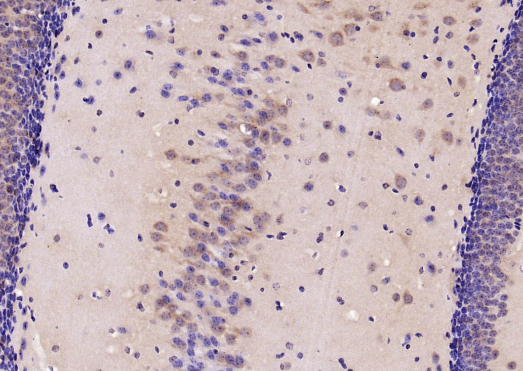
Paraformaldehyde-fixed, paraffin embedded (mouse brain); Antigen retrieval by boiling in sodium citrate buffer (pH6.0) for 15min; Block endogenous peroxidase by 3% hydrogen peroxide for 20 minutes; Blocking buffer (normal goat serum) at 37°C for 30min; Antibody incubation with (PERK) Polyclonal Antibody, Unconjugated (bs-2469R) at 1:200 overnight at 4°C, followed by operating according to SP Kit(Rabbit) (sp-0023) instructionsand DAB staining.
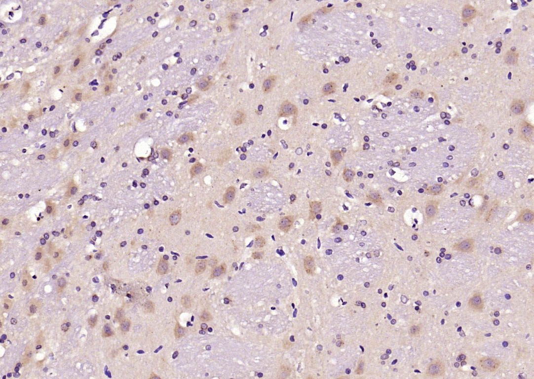
Paraformaldehyde-fixed, paraffin embedded (rat brain); Antigen retrieval by boiling in sodium citrate buffer (pH6.0) for 15min; Block endogenous peroxidase by 3% hydrogen peroxide for 20 minutes; Blocking buffer (normal goat serum) at 37°C for 30min; Antibody incubation with (PERK) Polyclonal Antibody, Unconjugated (bs-2469R) at 1:200 overnight at 4°C, followed by operating according to SP Kit(Rabbit) (sp-0023) instructionsand DAB staining.
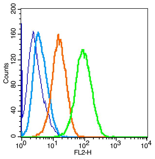
Blank control: U-87MG(blue). Primary Antibody:Rabbit Anti-PERK antibody(bs-2469R), Dilution: 1μg in 100 μL 1X PBS containing 0.5% BSA; Isotype Control Antibody: Rabbit IgG(orange) ,used under the same conditions ); Secondary Antibody: Goat anti-rabbit IgG-PE(white blue), Dilution: 1:200 in 1 X PBS containing 0.5% BSA.
Protocol
The cells were fixed with 2% paraformaldehyde (10 min) , then permeabilized with 90% ice-cold methanol for 30 min on ice. Primary antibody (bs-2469R,1μg /1x10^6 cells) were incubated for 30 min on the ice, followed by 1 X PBS containing 0.5% BSA + 1 0% goat serum (15 min) to block non-specific protein-protein interactions. Then the Goat Anti-rabbit IgG/PE antibody was added into the blocking buffer mentioned above to react with the primary antibody at 1/200 dilution for 30 min on ice. Acquisition of 20,000 events was performed.
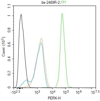
Blank control(black line):K562.
Primary Antibody (green line): Rabbit Anti-PERK antibody (bs-2469R)
Dilution:2ug/Test;
Secondary Antibody(white blue line): Goat anti-rabbit IgG-AF488
Dilution: 0.5ug/Test.
Isotype control(orange line): Normal Rabbit IgG
Protocol
The cells were fixed with 4% PFA (10min at room temperature)and then permeabilized with 90% ice-cold methanol for 20 min at -20℃, The cells were then incubated in 5%BSA to block non-specific protein-protein interactions for 30 min at room temperature .Cells stained with Primary Antibody for 30 min at room temperature. The secondary antibody used for 40 min at room temperature. Acquisition of 20,000 events was performed.
|
| 1、抗体溶解方法 | |
| 2、抗体修复方式 | |
| 3、常用试剂的配制 | |
| 4、免疫组化操作步骤 | |
| 5、免疫组化问题解答 | |
| 6、Western Blotting 操作步骤 | |
| 7、Western Blotting 问题解答 | |
| 8、关于肽链的设计 | |
| 9、多肽的溶解与保存 | |
| 10、酶标抗体效价测定程序 | |


