

| 产品编号 | bs-2489R |
| 英文名称 | CD9 Rabbit pAb |
| 中文名称 | CD9蛋白抗体 |
| 别 名 | BTCC-1; DRAP-27; MIC3; MRP-1; TSPAN-29; TSPAN29; CD9_HUMAN; CD9; 5H9 antigen; Cell growth-inhibiting gene 2 protein; Leukocyte antigen MIC3; Motility-related protein (MRP-1); Tetraspanin-29 (Tspan-29); p24; CD9_MOUSE; CD9_RAT; |
 |
Specific References (14) | bs-2489R has been referenced in 14 publications.
|
| 研究领域 | 肿瘤 细胞生物 免疫学 发育生物学 细胞表面分子 |
| 抗体来源 | Rabbit |
| 克隆类型 | Polyclonal |
| 克 隆 号 | |
| 交叉反应 | Human,Mouse,Rat (predicted: Rabbit,Pig,Sheep,Cow,Dog) |
| 产品应用 | WB=1:500-2000,Flow-Cyt=1μg/Test
not yet tested in other applications. optimal dilutions/concentrations should be determined by the end user. |
| 理论分子量 | 24 kDa |
| 检测分子量 | 24 |
| 细胞定位 | 细胞膜 |
| 性 状 | Liquid |
| 浓 度 | 1mg/ml |
| 免 疫 原 | KLH conjugated synthetic peptide derived from human CD9: 101-200/228 <Extracellular> |
| 亚 型 | IgG |
| 纯化方法 | affinity purified by Protein A |
| 缓 冲 液 | 0.01M TBS (pH7.4) with 1% BSA, 0.02% Proclin300 and 50% Glycerol. |
| 保存条件 | Shipped at 4℃. Store at -20℃ for one year. Avoid repeated freeze/thaw cycles. |
| 注意事项 | This product as supplied is intended for research use only, not for use in human, therapeutic or diagnostic applications. |
| PubMed | PubMed |
| 产品介绍 |
CD9 antigen is a glycoprotein expressed on the surface of developing B lymphocytes, platelets, monocytes, eosinophils, basophil, stimulated T lymphocytes and by neurons and glial cells in the peripheral nervous system. It belongs to a family of membrane proteins termed tetraspanins which transverse the membrane four times. In pre B cells and platelets, CD9 antigen regulates cell activation and aggregation possibly through an association with the integrin CD41 / CD61 (GPIIb / GPIIIa). It also regulates cell motility in a variety of cell lines, and appears to be an important regulator of Schwann cell behaviour in peripheral nerve. Function: Involved in platelet activation and aggregation. Regulates paranodal junction formation. Involved in cell adhesion, cell motility and tumor metastasis. Required for sperm-egg fusion. Subunit: Forms both disulfide-linked homodimers and higher homooligomers as well as heterooligomers with other members of the tetraspanin family. Associates with CR2/CD21 and with PTGFRN/CD9P1. Interacts directly with IGSF8. Subcellular Location: Membrane; Multi-pass membrane protein. Tissue Specificity: Expressed by a variety of hematopoietic and epithelial cells. Post-translational modifications: Protein exists in three forms with molecular masses between 22 and 27 kDa, and is known to carry covalently linked fatty acids. Similarity: Belongs to the tetraspanin (TM4SF) family. SWISS: P21926 Gene ID: 928 Database links: Entrez Gene: 928 Human Entrez Gene: 12527 Mouse SwissProt: P21926 Human SwissProt: P40240 Mouse CD9是一种普遍存在于细胞膜表面属于四跨膜超蛋白家族的细胞表面糖蛋白, 参与质膜的融合过程,在细胞膜生物学中发挥重要作用。 近年来经研究发现,CD9与多种肿瘤转移有关,故称肿瘤转移抑制基因亦称抗肿瘤转移基因。是一个新的肿瘤转移抑制基因膜运动相关蛋白-1(CD9/MRP-1),其分子功能还有待于进一步研究。CD9主要在早B细胞、活化T淋巴细胞、嗜酸性粒细胞、嗜碱性粒细胞和血小板表达。 |
| 产品图片 |
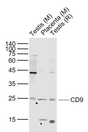
Sample:
Lane 1: Testis (Mouse) Lysate at 40 ug
Lane 2: Placenta (Mouse) Lysate at 40 ug
Lane 3: Testis (Rat) Lysate at 40 ug
Primary:
Anti-CD9 (bs-2489R) at 1/1000 dilution
Secondary: IRDye800CW Goat Anti-Rabbit IgG at 1/20000 dilution
Predicted band size: 24 kD
Observed band size: 24 kD
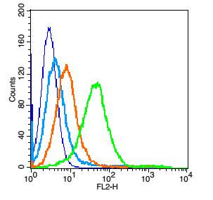
Blank control: Mouse spleen cells(blue). Primary Antibody:Rabbit Anti-CD9 antibody(bs-2489R), Dilution: 1μg in 100 μL 1X PBS containing 0.5% BSA; Isotype Control Antibody: Rabbit IgG(orange) ,used under the same conditions ); Secondary Antibody: Goat anti-rabbit IgG-PE(white blue), Dilution: 1:200 in 1 X PBS containing 0.5% BSA.
Protocol
Primary antibody were incubated for 30 min on the ice, followed by 1 X PBS containing 0.5% BSA + 1 0% goat serum (15 min) to block non-specific protein-protein interactions. Then the Goat Anti-rabbit IgG/PE antibody was added into the blocking buffer mentioned above to react with the primary antibody at 1/200 dilution for 30 min on ice. Acquisition of 20,000 events was performed.
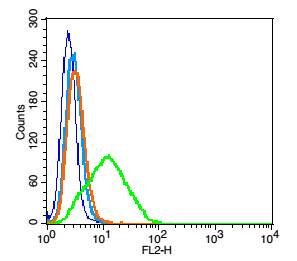
Blank control: RSC96 cells (blue). Primary Antibody:Rabbit Anti-CD9 antibody(bs-2489R), Dilution: 1μg in 100 μL 1X PBS containing 0.5% BSA; Isotype Control Antibody: Rabbit IgG(orange) ,used under the same conditions ); Secondary Antibody: Goat anti-rabbit IgG-PE(white blue), Dilution: 1:200 in 1 X PBS containing 0.5% BSA.
Protocol
Primary antibody were incubated for 30 min on the ice, followed by 1 X PBS containing 0.5% BSA + 10% goat serum (15 min) to block non-specific protein-protein interactions. Then the Goat Anti-rabbit IgG/PE antibody was added into the blocking buffer mentioned above to react with the primary antibody at 1/200 dilution for 30 min on ice. Acquisition of 20,000 events was performed.
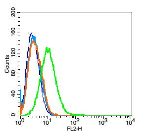
Blank control: Raji(blue).
Primary Antibody: Rabbit Anti-CD9 antibody(bs-2489R), Dilution: 5μg in 100 μL 1X PBS containing 0.5% BSA;
Isotype Control Antibody: Rabbit IgG (orange) ,used under the same conditions.
Secondary Antibody: Goat anti-rabbit IgG-PE(white blue), Dilution: 1:200 in 1 X PBS containing 0.5% BSA.
|
| 1、抗体溶解方法 | |
| 2、抗体修复方式 | |
| 3、常用试剂的配制 | |
| 4、免疫组化操作步骤 | |
| 5、免疫组化问题解答 | |
| 6、Western Blotting 操作步骤 | |
| 7、Western Blotting 问题解答 | |
| 8、关于肽链的设计 | |
| 9、多肽的溶解与保存 | |
| 10、酶标抗体效价测定程序 | |


