

| 产品编号 | bs-2592R |
| 英文名称 | JNK1+JNK2+JNK3 Rabbit pAb |
| 中文名称 | 氨基末端激酶1/2/3抗体 |
| 别 名 | JNK; JNK-46; JNK1; JNK1A2; JNK21B1/2; PRKM8; SAPK1; SAPK1c; MK08_HUMAN; MAPK8; MAP kinase 8; MAPK 8; Stress-activated protein kinase 1c (SAPK1c); Stress-activated protein kinase JNK1; c-Jun N-terminal kinase 1; 2.7.11.24; MK08_MOUSE; MK08_RAT; SAPK gamma; |
 |
Specific References (35) | bs-2592R has been referenced in 35 publications.
|
| 研究领域 | 细胞生物 细胞凋亡 糖尿病 |
| 抗体来源 | Rabbit |
| 克隆类型 | Polyclonal |
| 克 隆 号 | |
| 交叉反应 | Human,Mouse,Rat (predicted: Pig,Cow,Chicken,Dog) |
| 产品应用 | WB=1:500-2000,IHC-P=1:100-500,IHC-F=1:100-500,IF=1:100-500,Flow-Cyt=1ug/Test,ICC/IF=1:100-500
not yet tested in other applications. optimal dilutions/concentrations should be determined by the end user. |
| 理论分子量 | 42-47 kDa |
| 检测分子量 | 42-52 |
| 细胞定位 | 细胞核 细胞浆 |
| 性 状 | Liquid |
| 浓 度 | 1mg/ml |
| 免 疫 原 | KLH conjugated synthetic peptide derived from human JNK1/2/3: 151-250/384 |
| 亚 型 | IgG |
| 纯化方法 | affinity purified by Protein A |
| 缓 冲 液 | 0.01M TBS (pH7.4) with 1% BSA, 0.02% Proclin300 and 50% Glycerol. |
| 保存条件 | Shipped at 4℃. Store at -20℃ for one year. Avoid repeated freeze/thaw cycles. |
| 注意事项 | This product as supplied is intended for research use only, not for use in human, therapeutic or diagnostic applications. |
| PubMed | PubMed |
| 产品介绍 |
JNK1(MAPK8) is a member of the MAP kinase family. JNK1 is activated by threonine and tyrosine phosphorylation by either of two dual specificity kinases, MAP2K4 and MAP2K7. JNK2 (p54a, SAPK1a), along with JNK1 and JNK3, is thought to play an important role in nuclear signal transduction through its environmental stress activation and subsequent phosphorylation of the nuclear transcription factor p53. JNK3 is a neuron-specific form of c-Jun N-terminal kinases. Through its phosphorylation and nuclear localization, this kinase plays regulatory roles in the signaling pathways of neuronal apoptosis. The JNK pathway is critically involved in diabetes and levels are abnormally elevated in obesity. SWISS: P45983 Gene ID: 5599 Database links: Entrez Gene: 5599 Human Entrez Gene: 26419 Mouse Omim: 601158 Human JNK1 SwissProt: P45983 Human JNK1 Unigene: 138211 Human JNK1 Unigene: 21495 Mouse JNK1 Unigene: 4090 Rat JNK1 Entrez Gene: 5601 Human JNK2 |
| 产品图片 |
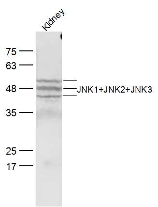
Sample:
Kidney (Mouse) Lysate at 40 ug
Primary: Anti-JNK1+JNK2+JNK3 (bs-2592R) at 1/300 dilution
Secondary: IRDye800CW Goat Anti-Rabbit IgG at 1/20000 dilution
Predicted band size: 42-47 kD
Observed band size:42-52 kD
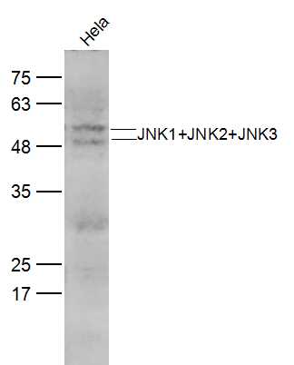
Sample:
Hela(Human) CellLysate at 30 ug
Primary: Anti-JNK1+JNK2+JNK3 (bs-2592R) at 1/300 dilution
Secondary: IRDye800CW Goat Anti-Rabbit IgG at 1/20000 dilution
Predicted band size: 42-47 kD
Observed band size:42-52 kD
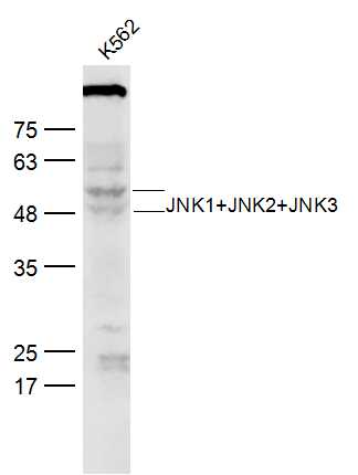
Sample:
K562 (Human) Lysate at 30 ug
Primary: Anti-JNK1+JNK2+JNK3 (bs-2592R) at 1/300 dilution
Secondary: IRDye800CW Goat Anti-Rabbit IgG at 1/20000 dilution
Predicted band size: 42-47 kD
Observed band size:42-52 kD
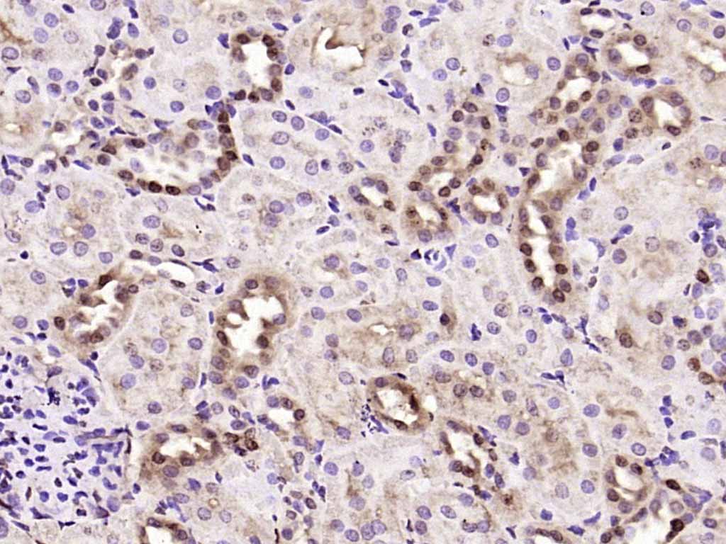
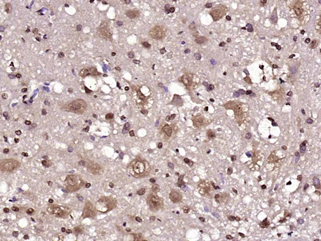
Paraformaldehyde-fixed, paraffin embedded (rat kidney); Antigen retrieval by boiling in sodium citrate buffer (pH6.0) for 15min; Block endogenous peroxidase by 3% hydrogen peroxide for 20 minutes; Blocking buffer (normal goat serum) at 37°C for 30min; Antibody incubation with (JNK1+JNK2+JNK3) Polyclonal Antibody, Unconjugated (bs-2592R) at 1:200 overnight at 4°C, followed by operating according to SP Kit(Rabbit) (sp-0023) instructionsand DAB staining.
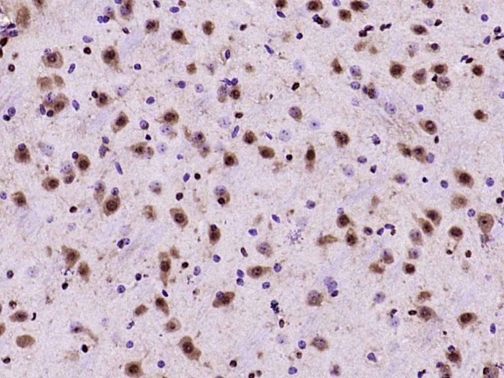
Paraformaldehyde-fixed, paraffin embedded (mouse brain); Antigen retrieval by boiling in sodium citrate buffer (pH6.0) for 15min; Block endogenous peroxidase by 3% hydrogen peroxide for 20 minutes; Blocking buffer (normal goat serum) at 37°C for 30min; Antibody incubation with (JNK1+JNK2+JNK3) Polyclonal Antibody, Unconjugated (bs-2592R) at 1:200 overnight at 4°C, followed by operating according to SP Kit(Rabbit) (sp-0023) instructionsand DAB staining.
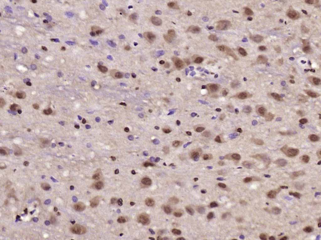
Paraformaldehyde-fixed, paraffin embedded (rat cerebellum); Antigen retrieval by boiling in sodium citrate buffer (pH6.0) for 15min; Block endogenous peroxidase by 3% hydrogen peroxide for 20 minutes; Blocking buffer (normal goat serum) at 37°C for 30min; Antibody incubation with (JNK1+JNK2+JNK3) Polyclonal Antibody, Unconjugated (bs-2592R) at 1:200 overnight at 4°C, followed by operating according to SP Kit(Rabbit) (sp-0023) instructionsand DAB staining.
Paraformaldehyde-fixed, paraffin embedded (rat brain); Antigen retrieval by boiling in sodium citrate buffer (pH6.0) for 15min; Block endogenous peroxidase by 3% hydrogen peroxide for 20 minutes; Blocking buffer (normal goat serum) at 37°C for 30min; Antibody incubation with (JNK1+JNK2+JNK3) Polyclonal Antibody, Unconjugated (bs-2592R) at 1:200 overnight at 4°C, followed by operating according to SP Kit(Rabbit) (sp-0023) instructionsand DAB staining.
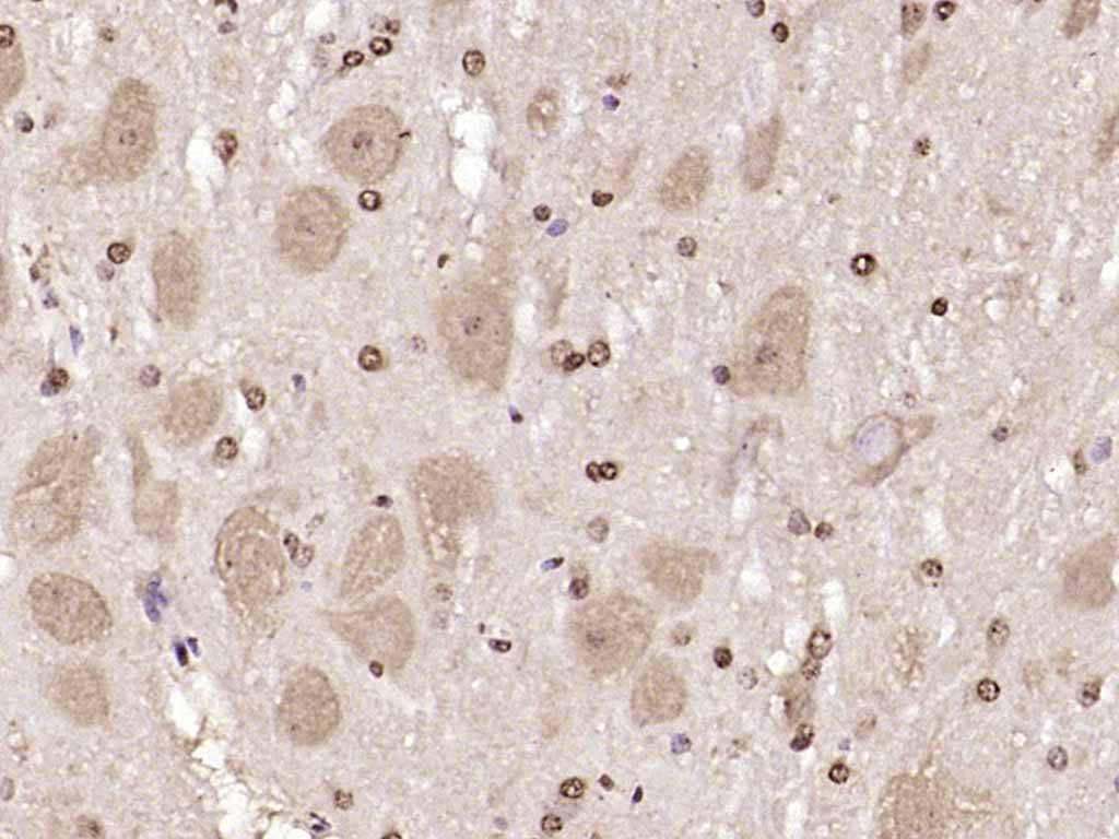
Paraformaldehyde-fixed, paraffin embedded (mouse cerebellum); Antigen retrieval by boiling in sodium citrate buffer (pH6.0) for 15min; Block endogenous peroxidase by 3% hydrogen peroxide for 20 minutes; Blocking buffer (normal goat serum) at 37°C for 30min; Antibody incubation with (JNK1+JNK2+JNK3) Polyclonal Antibody, Unconjugated (bs-2592R) at 1:200 overnight at 4°C, followed by operating according to SP Kit(Rabbit) (sp-0023) instructionsand DAB staining.
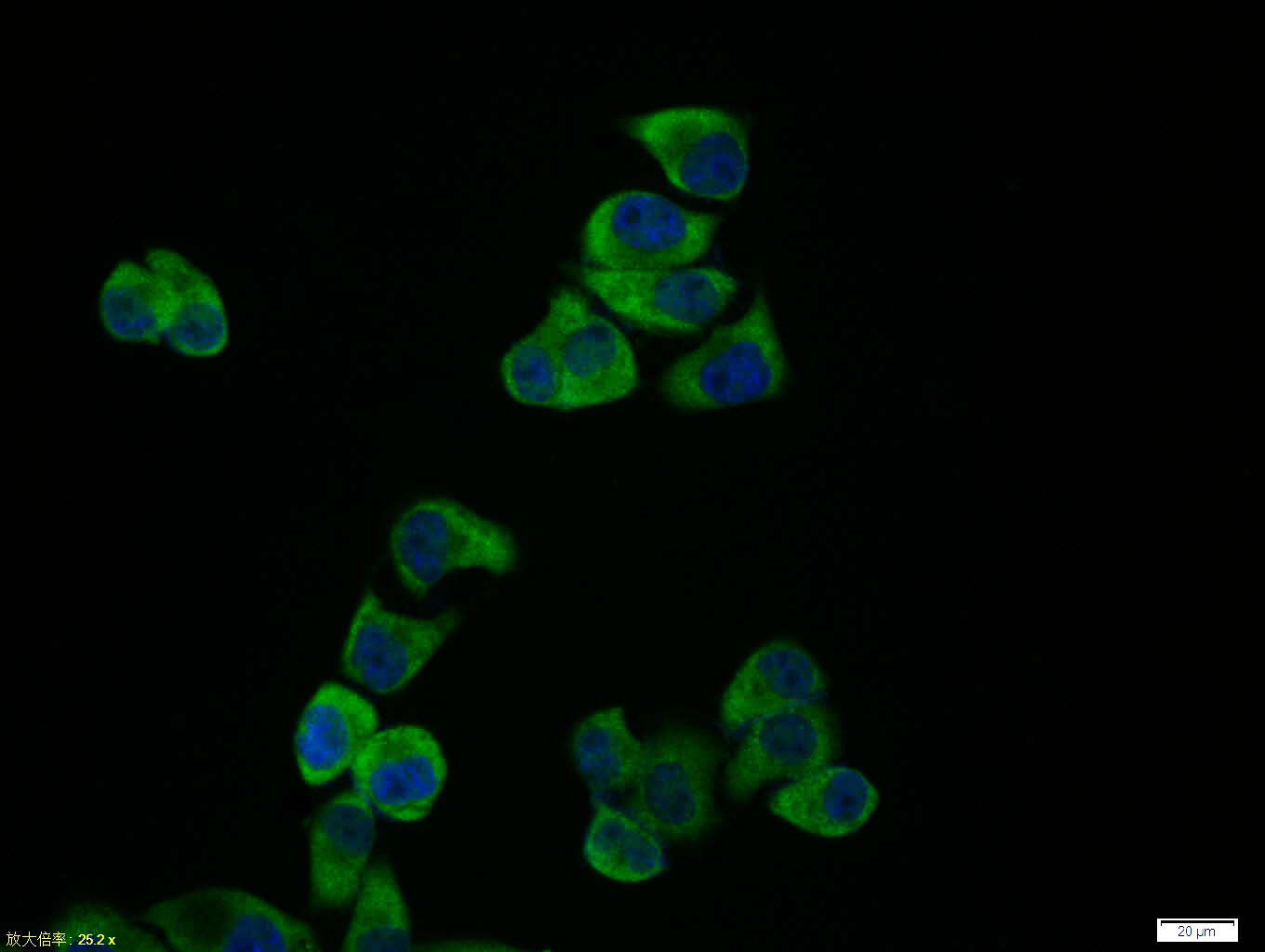
Hela cell; 4% Paraformaldehyde-fixed; Triton X-100 at room temperature for 20 min; Blocking buffer (normal goat serum, C-0005) at 37°C for 20 min; Antibody incubation with (JNK1+JNK2+JNK3) polyclonal Antibody, Unconjugated (bs-2592R) 1:100, 90 minutes at 37°C; followed by a conjugated Goat Anti-Rabbit IgG antibody at 37°C for 90 minutes, DAPI (blue, C02-04002) was used to stain the cell nuclei.
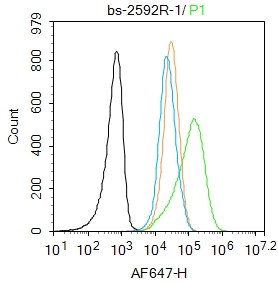
Blank control: Jurkat.
Primary Antibody (green line): Rabbit Anti-JNK1+JNK2+JNK3 antibody (bs-2592R)
Dilution: 1μg /10^6 cells;
Isotype Control Antibody (orange line): Rabbit IgG .
Secondary Antibody : Goat anti-rabbit IgG-AF647
Dilution: 1μg /test.
Protocol
The cells were fixed with 4% PFA (10min at room temperature)and then permeabilized with 90% ice-cold methanol for 20 min at-20℃. The cells were then incubated in 5%BSA to block non-specific protein-protein interactions for 30 min at room temperature .Cells stained with Primary Antibody for 30 min at room temperature. The secondary antibody used for 40 min at room temperature. Acquisition of 20,000 events was performed.
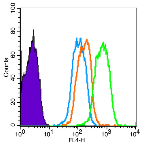
Blank control (Black line): HUVEC (Black).
Primary Antibody (green line): Rabbit Anti-JNK1+JNK2+JNK3 antibody (bs-2592R)
Dilution: 3μg /10^6 cells;
Isotype Control Antibody (orange line): Rabbit IgG .
Secondary Antibody (white blue line): Goat anti-rabbit IgG-AF647
Dilution: 1μg /test.
Protocol
The cells were fixed with 4% PFA (10min at room temperature)and then permeabilized with 90% ice-cold methanol for 20 min at room temperature. The cells were then incubated in 5%BSA to block non-specific protein-protein interactions for 30 min at room temperature .Cells stained with Primary Antibody for 30 min at room temperature. The secondary antibody used for 40 min at room temperature. Acquisition of 20,000 events was performed.
|
| 1、抗体溶解方法 | |
| 2、抗体修复方式 | |
| 3、常用试剂的配制 | |
| 4、免疫组化操作步骤 | |
| 5、免疫组化问题解答 | |
| 6、Western Blotting 操作步骤 | |
| 7、Western Blotting 问题解答 | |
| 8、关于肽链的设计 | |
| 9、多肽的溶解与保存 | |
| 10、酶标抗体效价测定程序 | |


