

| 产品编号 | bs-2700R |
| 英文名称 | CK10 Rabbit pAb |
| 中文名称 | 细胞角蛋白10抗体 |
| 别 名 | BCIE; BIE; CK10; EHK; EHK2; EHK2A; EHK2B; IHL; K10; KPP; D130054E02Rik; K1C1; Krt-1.10; Krt1-10; Ka10; K1C10_HUMAN; KRT10; Cytokeratin-10 (CK-10); Keratin-10 (K10); K1C10_MOUSE; 56 kDa cytokeratin; Keratin, type I cytoskeletal 59 kDa; K1C10_RAT; Type I keratin Ka10; keratin 10; keratosis palmaris et plantaris; keratin 10, type I; cytokeratin 10; epidermolytic hyperkeratosis |
| 研究领域 | 肿瘤 细胞周期蛋白 |
| 抗体来源 | Rabbit |
| 克隆类型 | Polyclonal |
| 交叉反应 | Human |
| 产品应用 | Flow-Cyt=1μg/Test,ICC/IF=1:100-500
not yet tested in other applications. optimal dilutions/concentrations should be determined by the end user. |
| 理论分子量 | 56kDa |
| 细胞定位 | 细胞核 细胞浆 细胞膜 |
| 性 状 | Liquid |
| 浓 度 | 1mg/ml |
| 免 疫 原 | KLH conjugated synthetic peptide derived from human CK10: 151-250/584 |
| 亚 型 | IgG |
| 纯化方法 | affinity purified by Protein A |
| 缓 冲 液 | 0.01M TBS (pH7.4) with 1% BSA, 0.02% Proclin300 and 50% Glycerol. |
| 保存条件 | Shipped at 4℃. Store at -20℃ for one year. Avoid repeated freeze/thaw cycles. |
| 注意事项 | This product as supplied is intended for research use only, not for use in human, therapeutic or diagnostic applications. |
| PubMed | PubMed |
| 产品介绍 |
Cytokeratin 10 is a heterotetramer of two type I and two type II keratins. Cytokeratin 10 is generally associated with keratin 1. It is seen in all suprabasal cell layers including stratum corneum. A number of alleles are known that mainly differ in the Gly-rich region (positions 490-560). Defects in cytokeratin 10 are a cause of epidermolytic hyperkeratosis (EHK), also known as bullous congenital ichthyosiform erythroderma (BCIE) or bullous erythroderma ichthyosiformis congenita of Brocq. EHK is an hereditary skin disorder characterized by blistering and a marked thickening of the stratum corneum. At birth, affected individuals usually present with redness, blisters and superficial erosions due to cytolysis. Within a few weeks, the erythroderma and blister formation diminish and hyperkeratoses develop. Transmission is autosomal dominant, but most cases are sporadic. Defects in cytokeratin 10 are also a cause of annular epidermolytic ichthyosis (AEI), also known as cyclic ichthyosis with epidermolytic hyperkeratosis. AEI resembles clinical and histologic features of both epidermolytic hyperkeratosis and ichthyosis bullosa of Siemens. Subunit: Heterotetramer of two type I and two type II keratins. keratin-10 is generally associated with keratin-1. Tissue Specificity: Seen in all suprabasal cell layers including stratum corneum. DISEASE: Epidermolytic hyperkeratosis (EHK) [MIM:113800]: An autosomal dominant skin disorder characterized by widespread blistering and an ichthyotic erythroderma at birth that persist into adulthood. Histologically there is a diffuse epidermolytic degeneration in the lower spinous layer of the epidermis. Within a few weeks from birth, erythroderma and blister formation diminish and hyperkeratoses develop. Note=The disease is caused by mutations affecting the gene represented in this entry. Ichthyosis annular epidermolytic (AEI) [MIM:607602]: A skin disorder resembling bullous congenital ichthyosiform erythroderma. Affected individuals present with bullous ichthyosis in early childhood and hyperkeratotic lichenified plaques in the flexural areas and extensor surfaces at later ages. The feature that distinguishes AEI from BCIE is dramatic episodes of flares of annular polycyclic plaques with scale, which coalesce to involve most of the body surface and can persist for several weeks or even months. Note=The disease is caused by mutations affecting the gene represented in this entry. Erythroderma, ichthyosiform, congenital reticular (CRIE) [MIM:609165]: A rare skin condition characterized by slowly enlarging islands of normal skin surrounded by erythematous ichthyotic patches in a reticulated pattern. The condition starts in infancy as a lamellar ichthyosis, with small islands of normal skin resembling confetti appearing in late childhood and at puberty. Histopathologic findings include band-like parakeratosis, psoriasiform acanthosis, and vacuolization of keratinocytes with binucleated cells in the upper epidermis, sometimes associated with amyloid deposition in the dermis. Ultrastructural abnormalities include perinuclear shells formed from a network of fine filaments in the upper epidermis. Note=The disease is caused by mutations affecting the gene represented in this entry. Similarity: Belongs to the intermediate filament family. SWISS: P13645 Gene ID: 3858 Database links: Entrez Gene: 3858 Human Entrez Gene: 16661 Mouse Omim: 148080 Human SwissProt: P13645 Human SwissProt: P02535 Mouse Unigene: 99936 Human Unigene: 22662 Mouse Unigene: 125065 Rat 细胞角蛋白是形成上皮细胞细胞骨架中间纤维的一类结构相关蛋白。CK10属于I型角蛋白,常常与CK1一起存在。缺失CK10与表皮松解性角化症(EHK)以及表皮松解性鳞癣病(AEI)密切相关。主要标记上皮的基底上层和颗粒细胞层细胞,同时CK10表达与细胞的分化程度呈正比,高分化者常阳性更强,故常用于鳞状细胞癌的诊断。 |
| 产品图片 |
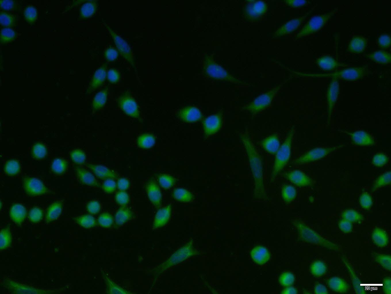
A431 cell; 4% Paraformaldehyde-fixed; Triton X-100 at room temperature for 20 min; Blocking buffer (normal goat serum, C-0005) at 37°C for 20 min; Antibody incubation with (CK10) polyclonal Antibody, Unconjugated (bs-2700R) 1:100, 90 minutes at 37°C; followed by a conjugated Goat Anti-Rabbit IgG antibody at 37°C for 90 minutes, DAPI (blue, C02-04002) was used to stain the cell nuclei.
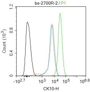
Blank control(black line):A431.
Primary Antibody (green line): Rabbit Anti-CK10 antibody (bs-2700R)
Dilution:2ug/Test;
Secondary Antibody(white blue line): Goat anti-rabbit IgG-AF488
Dilution: 0.5ug/Test.
Isotype control(orange line): Normal Rabbit IgG
Protocol
The cells were fixed with 4% PFA (10min at room temperature)and then permeabilized with 90% ice-cold methanol for 20 min at -20℃, The cells were then incubated in 5%BSA to block non-specific protein-protein interactions for 30 min at room temperature .Cells stained with Primary Antibody for 30 min at room temperature. The secondary antibody used for 40 min at room temperature. Acquisition of 20,000 events was performed.
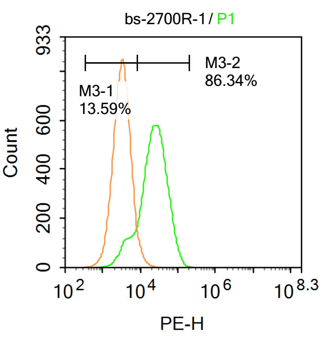
Blank control: Hela.
Primary Antibody (green line): Rabbit Anti-CK10 antibody (bs-2700R)
Dilution: 1μg /10^6 cells;
Isotype Control Antibody (orange line): Rabbit IgG .
Secondary Antibody : Goat anti-rabbit IgG-PE
Dilution: 1μg /test.
Protocol
The cells were fixed with 4% PFA (10min at room temperature)and then permeabilized with 90% ice-cold methanol for 20 min at-20℃. The cells were then incubated in 5%BSA to block non-specific protein-protein interactions for 30 min at at room temperature .Cells stained with Primary Antibody for 30 min at room temperature. The secondary antibody used for 40 min at room temperature. Acquisition of 20,000 events was performed.
|
| 1、抗体溶解方法 | |
| 2、抗体修复方式 | |
| 3、常用试剂的配制 | |
| 4、免疫组化操作步骤 | |
| 5、免疫组化问题解答 | |
| 6、Western Blotting 操作步骤 | |
| 7、Western Blotting 问题解答 | |
| 8、关于肽链的设计 | |
| 9、多肽的溶解与保存 | |
| 10、酶标抗体效价测定程序 | |


