

| 产品编号 | bs-3031R |
| 英文名称 | phospho-ASK1 (Thr845) Rabbit pAb |
| 中文名称 | 磷酸化细胞凋亡信号调节激酶1抗体 |
| 别 名 | MAP3K5 | ASK1 (phospho-T845); p-ASK1; phospho-ASK1; p-MAP3K5; MAP3K5 | ASK1 (phospho-Thr845); ASK1; MAPKKK5; MEKK5; 7420452D20Rik; ASK; M3K5_HUMAN; MAP3K5; Apoptosis signal-regulating kinase 1 (ASK-1); MAPK/ERK kinase kinase 5 (MEK kinase 5 | MEKK 5); 2.7 |
 |
Specific References (7) | bs-3031R has been referenced in 7 publications.
|
| 产品类型 | 磷酸化抗体 |
| 研究领域 | 肿瘤 细胞生物 免疫学 神经生物学 信号转导 细胞凋亡 激酶和磷酸酶 |
| 抗体来源 | Rabbit |
| 克隆类型 | Polyclonal |
| 克 隆 号 | |
| 交叉反应 | Human,Mouse,Rat |
| 产品应用 | WB=1:500-2000,Flow-Cyt=1μg /test
not yet tested in other applications. optimal dilutions/concentrations should be determined by the end user. |
| 理论分子量 | 155 kDa |
| 检测分子量 | 155 |
| 细胞定位 | 细胞浆 |
| 性 状 | Liquid |
| 浓 度 | 1mg/ml |
| 免 疫 原 | KLH conjugated Synthesised phosphopeptide derived from human ASK1 around the phosphorylation site of Thr845: TE(p-T)FT |
| 亚 型 | IgG |
| 纯化方法 | affinity purified by Protein A |
| 缓 冲 液 | 0.01M TBS (pH7.4) with 1% BSA, 0.02% Proclin300 and 50% Glycerol. |
| 保存条件 | Shipped at 4℃. Store at -20℃ for one year. Avoid repeated freeze/thaw cycles. |
| 注意事项 | This product as supplied is intended for research use only, not for use in human, therapeutic or diagnostic applications. |
| PubMed | PubMed |
| 产品介绍 |
Mitogen-activated protein kinase (MAPK) signaling cascades include MAPK or extracellular signal-regulated kinase (ERK), MAPK kinase (MKK or MEK), and MAPK kinase kinase (MAPKKK or MEKK). MAPKK kinase/MEKK phosphorylates and activates its downstream protein kinase, MAPK kinase/MEK, which in turn activates MAPK. The kinases of these signaling cascades are highly conserved, and homologs exist in yeast, Drosophila, and mammalian cells. MAPKKK5 contains 1,374 amino acids with all 11 kinase subdomains. Northern blot analysis shows that MAPKKK5 transcript is abundantly expressed in human heart and pancreas. The MAPKKK5 protein phosphorylates and activates MKK4 (aliases SERK1, MAPKK4) in vitro, and activates c-Jun N-terminal kinase (JNK)/stress-activated protein kinase (SAPK) during transient expression in COS and 293 cells; MAPKKK5 does not activate MAPK/ERK.
Function: Serine/threonine kinase which acts as an essential component of the MAP kinase signal transduction pathway. Plays an important role in the cascades of cellular responses evoked by changes in the environment. Mediates signaling for determination of cell fate such as differentiation and survival. Plays a crucial role in the apoptosis signal transduction pathway through mitochondria-dependent caspase activation. MAP3K5/ASK1 is required for the innate immune response, which is essential for host defense against a wide range of pathogens. Mediates signal transduction of various stressors like oxidative stress as well as by receptor-mediated inflammatory signals, such as the tumor necrosis factor (TNF) or lipopolysaccharide (LPS). Once activated, acts as an upstream activator of the MKK/JNK signal transduction cascade and the p38 MAPK signal transduction cascade through the phosphorylation and activation of several MAP kinase kinases like MAP2K4/SEK1, MAP2K3/MKK3, MAP2K6/MKK6 and MAP2K7/MKK7. These MAP2Ks in turn activate p38 MAPKs and c-jun N-terminal kinases (JNKs). Both p38 MAPK and JNKs control the transcription factors activator protein-1 (AP-1). Subunit: Homodimer when inactive. Binds both upstream activators and downstream substrates in multimolecular complexes. Associates with and inhibited by HIV-1 Nef. Part of a cytoplasmic complex made of HIPK1, DAB2IP and MAP3K5 in response to TNF. This complex formation promotes MAP3K5-JNK activation and subsequent apoptosis. Interacts with SOCS1 which recognizes phosphorylation of Tyr-718 and induces MAP3K5/ASK1 degradation in endothelial cells. Interacts with the 14-3-3 family proteins such as YWHAB, YWHAE, YWHAQ, YWHAH, YWHAZ and SFN. Interacts with ARRB2, BIRC2, DAB2IP, IGF1R, MAP3K6/ASK2, PGAM5, PIM1, PPP5C, SOCS1, STUB1, TRAF2, TRAF6 and TXN. Interacts with ERN1 in a TRAF2-dependent manner. Interacts with calcineurin subunit PPP3R1 and with PPM1L (By similarity). Interacts (via N-terminus) with RAF1 and this interaction inhibits the proapoptotic function of MAP3K5. Interacts with DAB2IP (via N-terminus C2 domain); the interaction occurs in a TNF-alpha-dependent manner. Subcellular Location: Cytoplasm. Endoplasmic reticulum. Note=Interaction with 14-3-3 proteins alters the distribution of MAP3K5/ASK1 and restricts it to the perinuclear endoplasmic reticulum region. Tissue Specificity: Abundantly expressed in heart and pancreas. Post-translational modifications: Phosphorylated at Thr-838 through autophosphorylation and by MAP3K6/ASK2 which leads to activation. Thr-838 is dephosphorylated by PPP5C. Ser-83 and Ser-1033 are inactivating phosphorylation sites, the former of which is phosphorylated by AKT1 and AKT2. Phosphorylated at Ser-966 which induces association of MAP3K5/ASK1 with the 14-3-3 family proteins and suppresses MAP3K5/ASK1 activity. Calcineurin (CN) dephosphorylates this site. Also dephosphorylated and activated by PGAM5. Ubiquitinated. Tumor necrosis factor (TNF) induces TNFR2-dependent ubiquitination leading to proteasomal degradation. Similarity: Belongs to the protein kinase superfamily. STE Ser/Thr protein kinase family. MAP kinase kinase kinase subfamily. Contains 1 protein kinase domain. SWISS: Q99683 Gene ID: 4217 Database links: Entrez Gene: 4217 Human Entrez Gene: 26408 Mouse Omim: 602448 Human SwissProt: Q99683 Human SwissProt: O35099 Mouse Unigene: 186486 Human Unigene: 6595 Mouse |
| 产品图片 |
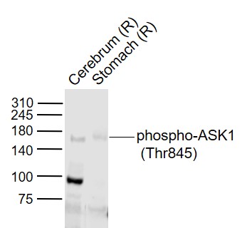
Sample:
Lane 8: Cerebrum (Rat) Lysate at 40 ug
Lane 9: Stomach (Rat) Lysate at 40 ug
Primary: Anti-phospho-ASK1 (Thr845) (bs-3031R) at 1/1000 dilution
Secondary: IRDye800CW Goat Anti-Rabbit IgG at 1/20000 dilution
Predicted band size: 155 kD
Observed band size: 155 kD
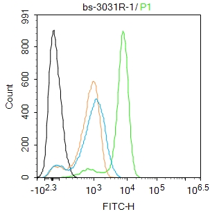
Blank control:Mouse spleen.
Primary Antibody (green line): Rabbit Anti-phospho-RelB (Ser551) antibody (bs-3031R)
Dilution: 2μg /10^6 cells;
Isotype Control Antibody (orange line): Rabbit IgG .
Secondary Antibody : Goat anti-rabbit IgG-AF488
Dilution: 1μg /test.
Protocol
The cells were fixed with 4% PFA (10min at room temperature)and then permeabilized with 90% ice-cold methanol for 20 min at-20℃. The cells were then incubated in 5%BSA to block non-specific protein-protein interactions for 30 min at room temperature .Cells stained with Primary Antibody for 30 min at room temperature. The secondary antibody used for 40 min at room temperature. Acquisition of 20,000 events was performed.
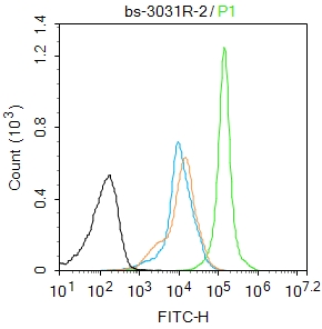
Blank control:U937.
Primary Antibody (green line): Rabbit Anti-phospho-ASK1 (Thr845) antibody (bs-3031R)
Dilution: 1ug/Test;
Secondary Antibody : Goat anti-rabbit IgG-FITC
Dilution: 0.5ug/Test.
Protocol
The cells were fixed with 4% PFA (10min at room temperature)and then permeabilized with 0.1% PBST for 20 min at room temperature.The cells were then incubated in 5%BSA to block non-specific protein-protein interactions for 30 min at room temperature .Cells stained with Primary Antibody for 30 min at room temperature. The secondary antibody used for 40 min at room temperature. Acquisition of 20,000 events was performed.
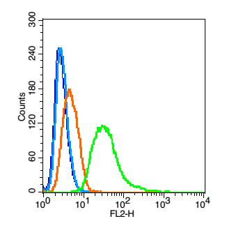
Blank control(blue): Hela(fixed with 2% paraformaldehyde (10 min), then permeabilized with 90% ice-cold methanol for 30 min on ice).
Primary Antibody:Rabbit Anti- phospho-ASK1 (Thr845) antibody(bs-3031R), Dilution: 0.2μg in 100 μL 1X PBS containing 0.5% BSA;
Isotype Control Antibody: Rabbit IgG(orange) ,used under the same conditions );
Secondary Antibody: Goat anti-rabbit IgG-PE(white blue), Dilution: 1:200 in 1 X PBS containing 0.5% BSA.
|
| 1、抗体溶解方法 | |
| 2、抗体修复方式 | |
| 3、常用试剂的配制 | |
| 4、免疫组化操作步骤 | |
| 5、免疫组化问题解答 | |
| 6、Western Blotting 操作步骤 | |
| 7、Western Blotting 问题解答 | |
| 8、关于肽链的设计 | |
| 9、多肽的溶解与保存 | |
| 10、酶标抗体效价测定程序 | |


