

| 产品编号 | bs-0719R |
| 英文名称 | CEA Rabbit pAb |
| 中文名称 | 癌胚抗原抗体 |
| 别 名 | CD66e; CEA; CEAM5_HUMAN; CEACAM5; Carcinoembryonic antigen (CEA); Carcinoembryonic antigen-related cell adhesion molecule 5 (CEA cell adhesion molecule 5); Meconium antigen 100; |
 |
Specific References (7) | bs-0719R has been referenced in 7 publications.
|
| 研究领域 | 肿瘤 信号转导 |
| 抗体来源 | Rabbit |
| 克隆类型 | Polyclonal |
| 克 隆 号 | |
| 交叉反应 | Human |
| 产品应用 | IHC-P=1:100-500,IHC-F=1:100-500,IF=1:100-500,Flow-Cyt=1μg/test
not yet tested in other applications. optimal dilutions/concentrations should be determined by the end user. |
| 理论分子量 | 150-200 kDa |
| 细胞定位 | 细胞膜 |
| 性 状 | Liquid |
| 浓 度 | 1mg/ml |
| 免 疫 原 | KLH conjugated synthetic peptide derived from human CEA/CD66e/CEACAM5: 301-400/702 |
| 亚 型 | IgG |
| 纯化方法 | affinity purified by Protein A |
| 缓 冲 液 | 0.01M TBS (pH7.4) with 1% BSA, 0.02% Proclin300 and 50% Glycerol. |
| 保存条件 | Shipped at 4℃. Store at -20℃ for one year. Avoid repeated freeze/thaw cycles. |
| 注意事项 | This product as supplied is intended for research use only, not for use in human, therapeutic or diagnostic applications. |
| PubMed | PubMed |
| 产品介绍 |
CEA-related cell adhesion molecules (CEACAM) belong to the carcinoembryonic antigen (CEA) family. It consists of seven CEACAM (CEACAM 1, CEACAM 3-CEACAM 8) and 11 pregnancy-specific glyco-protein (PSG 1-PSG 11) members. The CEA family proteins belong to the immunoglobulin (Ig) superfamily and are composed of one Ig variable-like (IgV) and a varying number (0-6) of Ig constant-like (IgC) domains. CEACAM molecules are membrane-bound either via a transmembrane domain or a glycosyl phosphatidyl inositol (GPI) anchor. CEACAM molecules are differentially expressed in epithelial cells or in leucocytes. Over-expression of CEA/ CEACAM 5 in tumors of epithelial origin is the basis of its wide-spread use as a tumor marker. The function of CEACAM family members varies widely: they function as cell adhesion molecules, tumor suppressors, regulators of lymphocyte and dendritic cell activation, receptors of Neisseria species and other bacteria. Function: Cell surface glycoprotein that plays a role in cell adhesion and in intracellular signaling. Receptor for E.coli Dr adhesins. Subunit: Homodimer. Binding of E.coli Dr adhesins leads to dissociation of the homodimer. Subcellular Location: Cell membrane; Lipid-anchor, GPI-anchor. Tissue Specificity: Found in adenocarcinomas of endodermally derived digestive system epithelium and fetal colon. Post-translational modifications: Complex immunoreactive glycoprotein with a MW of 180 kDa comprising 60% carbohydrate. Similarity: Belongs to the immunoglobulin superfamily. CEA family. Contains 7 Ig-like (immunoglobulin-like) domains. SWISS: P06731 Gene ID: 1048 Database links: Entrez Gene: 1048 Human Omim: 114890 Human SwissProt: P06731 Human Unigene: 709196 Human CEA是一种胚胎性抗原,主要存在于胎儿消化道上皮组织,胰腺和肝癌中。CEA广泛存在各种上皮性肿瘤,尤其是胃肠道恶性肿瘤,CEA分布阳性类型与肿瘤的恶性度有关。CEA与EMA一样可作为上皮性恶性肿瘤的重要标记。 |
| 产品图片 |
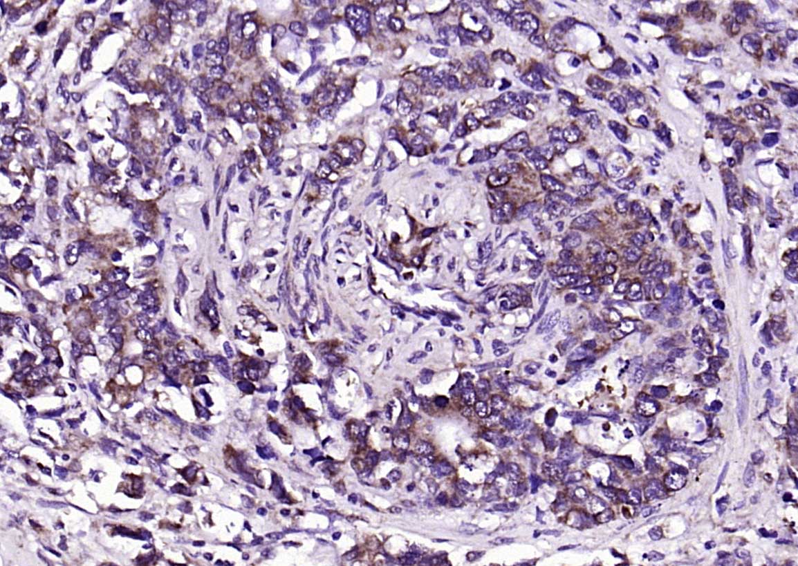
Paraformaldehyde-fixed, paraffin embedded (human rectal carcinoma); Antigen retrieval by boiling in sodium citrate buffer (pH6.0) for 15min; Block endogenous peroxidase by 3% hydrogen peroxide for 20 minutes; Blocking buffer (normal goat serum) at 37°C for 30min; Antibody incubation with (CEA) Polyclonal Antibody, Unconjugated (bs-0719R) at 1:200 overnight at 4°C, followed by operating according to SP Kit(Rabbit) (sp-0023) instructionsand DAB staining.

Paraformaldehyde-fixed, paraffin embedded (human rectal carcinoma); Antigen retrieval by boiling in sodium citrate buffer (pH6.0) for 15min; Blocking buffer (normal goat serum) at 37°C for 30min; Antibody incubation with (CEA) Polyclonal Antibody, Unconjugated (bs-0719R) at 1:200 overnight at 4°C, followed by a conjugated Goat Anti-Rabbit IgG antibody (bs-0295G-FITC) for 90 minutes, and DAPI for nuclei staining.
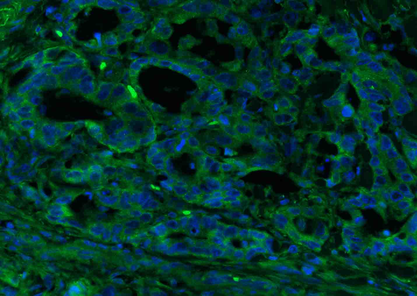
Paraformaldehyde-fixed, paraffin embedded (human colon carcinoma); Antigen retrieval by boiling in sodium citrate buffer (pH6.0) for 15min; Blocking buffer (normal goat serum) at 37°C for 30min; Antibody incubation with (CEA) Polyclonal Antibody, Unconjugated (bs-0719R) at 1:200 overnight at 4°C, followed by a conjugated Goat Anti-Rabbit IgG antibody (bs-0295G-FITC) for 90 minutes, and DAPI for nuclei staining.
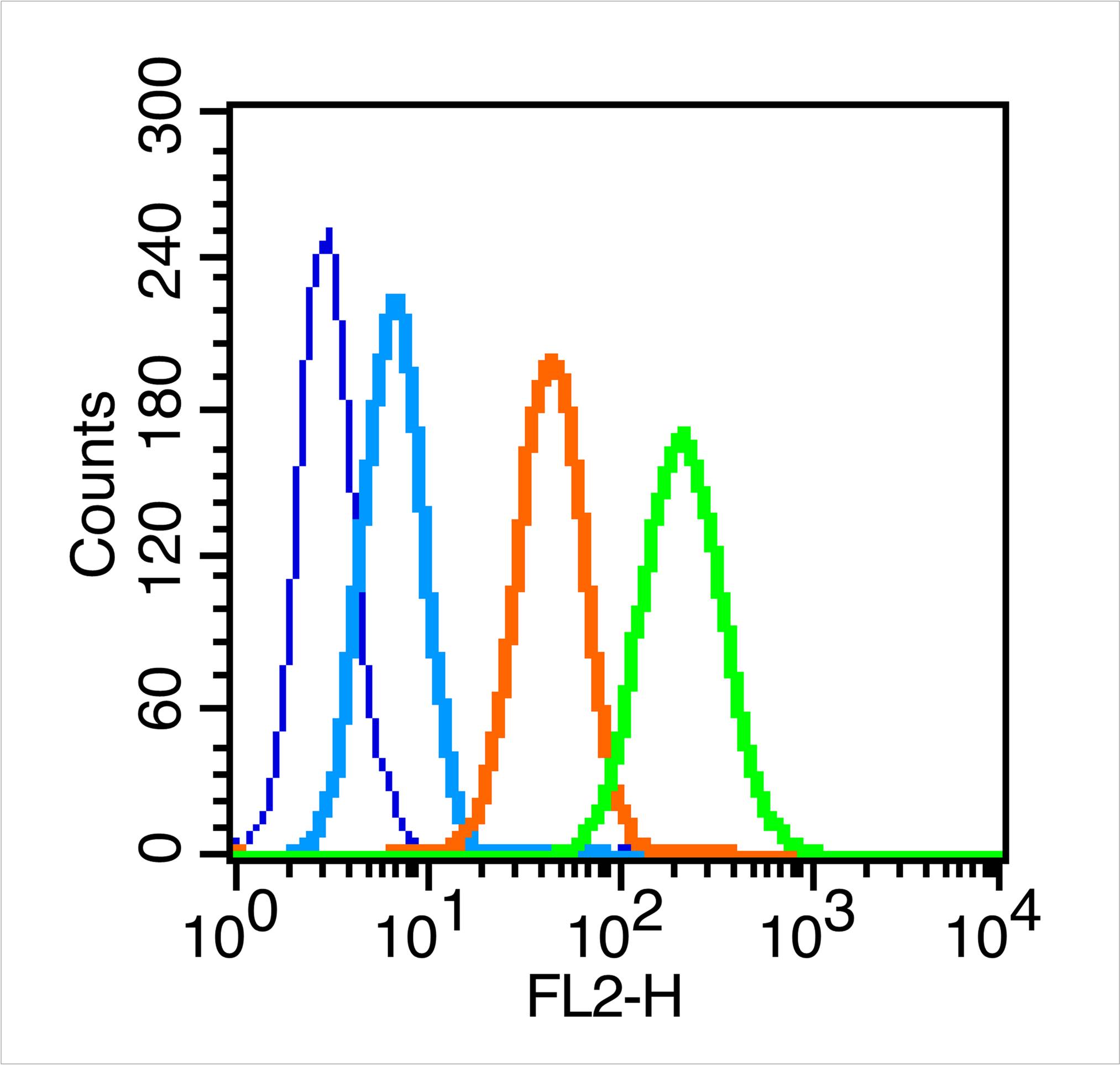
Blank control (blue line): MCF7 (fixed with 70% methanol overnight at 4℃).
Primary Antibody (green line): Rabbit Anti-CEA antibody (bs-0719R)
Dilution: 0.2μg /10^6 cells;
Isotype Control Antibody (orange line): Rabbit IgG .
Secondary Antibody (white blue line): Goat anti-rabbit IgG-PE
Dilution: 1μg /test.
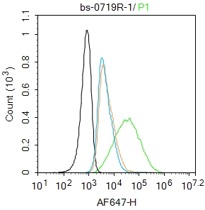
Blank control: MCF7.
Primary Antibody (green line): Rabbit Anti-CEA antibody (bs-0719R)
Dilution: 1μg /10^6 cells;
Isotype Control Antibody (orange line): Rabbit IgG .
Secondary Antibody : Goat anti-rabbit IgG-AF647
Dilution: 1μg /test.
Protocol
The cells were incubated in 5%BSA to block non-specific protein-protein interactions for 30 min at room temperature .Cells stained with Primary Antibody for 30 min at room temperature. The secondary antibody used for 40 min at room temperature. Acquisition of 20,000 events was performed.
|
| 1、抗体溶解方法 | |
| 2、抗体修复方式 | |
| 3、常用试剂的配制 | |
| 4、免疫组化操作步骤 | |
| 5、免疫组化问题解答 | |
| 6、Western Blotting 操作步骤 | |
| 7、Western Blotting 问题解答 | |
| 8、关于肽链的设计 | |
| 9、多肽的溶解与保存 | |
| 10、酶标抗体效价测定程序 | |


