

| 产品编号 | bs-3174R |
| 英文名称 | phospho-HDAC3 (Ser424) Rabbit pAb |
| 中文名称 | 磷酸化组蛋白去乙酰化酶3抗体 |
| 别 名 | HDAC3 (phospho-S424); p-HDAC3; phospho-HDAC3; HDAC3 (phospho-Ser424); HD3; KDAC3; RPD3; RPD3-2; HDAC3_HUMAN; HDAC3; Protein deacetylase HDAC3; Protein deacylase HDAC3; SMAP45; 3.5.1.98; HDAC3_MOUSE; HDAC3_RAT; |
| 产品类型 | 磷酸化抗体 |
| 研究领域 | 肿瘤 发育生物学 信号转导 |
| 抗体来源 | Rabbit |
| 克隆类型 | Polyclonal |
| 克 隆 号 | |
| 交叉反应 | Human,Mouse,Rat |
| 产品应用 | WB=1:500-2000,IHC-P=1:100-500,IHC-F=1:100-500,IF=1:100-500,Flow-Cyt=1ug/Test
not yet tested in other applications. optimal dilutions/concentrations should be determined by the end user. |
| 理论分子量 | 47 kDa |
| 检测分子量 | 50 |
| 细胞定位 | 细胞核 细胞浆 |
| 性 状 | Liquid |
| 浓 度 | 1mg/ml |
| 免 疫 原 | KLH conjugated Synthesised phosphopeptide derived from human HDAC3 around the phosphorylation site of Ser424: KE(p-S)DV |
| 亚 型 | IgG |
| 纯化方法 | affinity purified by Protein A |
| 缓 冲 液 | 0.01M TBS (pH7.4) with 1% BSA, 0.02% Proclin300 and 50% Glycerol. |
| 保存条件 | Shipped at 4℃. Store at -20℃ for one year. Avoid repeated freeze/thaw cycles. |
| 注意事项 | This product as supplied is intended for research use only, not for use in human, therapeutic or diagnostic applications. |
| PubMed | PubMed |
| 产品介绍 |
Histones play a critical role in transcriptional regulation, cell cycle progression, and developmental events. Histone acetylation/deacetylation alters chromosome structure and affects transcription factor access to DNA. The protein encoded by this gene belongs to the histone deacetylase/acuc/apha family. It has histone deacetylase activity and represses transcription when tethered to a promoter. It may participate in the regulation of transcription through its binding with the zinc-finger transcription factor YY1. This protein can also down-regulate p53 function and thus modulate cell growth and apoptosis. This gene is regarded as a potential tumor suppressor gene. [provided by RefSeq, Jul 2008] Function: Responsible for the deacetylation of lysine residues on the N-terminal part of the core histones (H2A, H2B, H3 and H4), and some other non-histone substrates. Histone deacetylation gives a tag for epigenetic repression and plays an important role in transcriptional regulation, cell cycle progression and developmental events. Histone deacetylases act via the formation of large multiprotein complexes. Probably participates in the regulation of transcription through its binding to the zinc-finger transcription factor YY1; increases YY1 repression activity. Required to repress transcription of the POU1F1 transcription factor. Acts as a molecular chaperone for shuttling phosphorylated NR2C1 to PML bodies for sumoylation. Subunit: Interacts with HDAC7 and HDAC9. Forms a heterologous complex at least with YY1. Interacts with DAXX, HDAC10 and DACH1. Found in a complex with NCOR1 and NCOR2. Component of the N-Cor repressor complex, at least composed of NCOR1, NCOR2, HDAC3, TBL1X, TBL1R, CORO2A and GPS2. Interacts with BCOR, MJD2A/JHDM3A, NRIP1, PRDM6 and SRY. Interacts with BTBD14B. Interacts with GLIS2. Interacts (via the DNA-binding domain) with NR2C1; the interaction recruits phosphorylated NR2C1 to PML bodies for sumoylation. Component of the Notch corepressor complex. Interacts with CBFA2T3 and NKAP. Interacts with APEX1; the interaction is not dependent on the acetylated status of APEX1. Interacts with and deacetylates MAPK14. Interacts with ZMYND15. Subcellular Location: Nucleus. Tissue Specificity: Widely expressed. Post-translational modifications: Sumoylated in vitro. Similarity: Belongs to the histone deacetylase family. HD type 1 subfamily. SWISS: O15379 Gene ID: 8841 Database links: Entrez Gene: 8841 Human Entrez Gene: 15183 Mouse Omim: 605166 Human SwissProt: O15379 Human SwissProt: O88895 Mouse Unigene: 519632 Human Unigene: 20521 Mouse Unigene: 17284 Rat |
| 产品图片 |
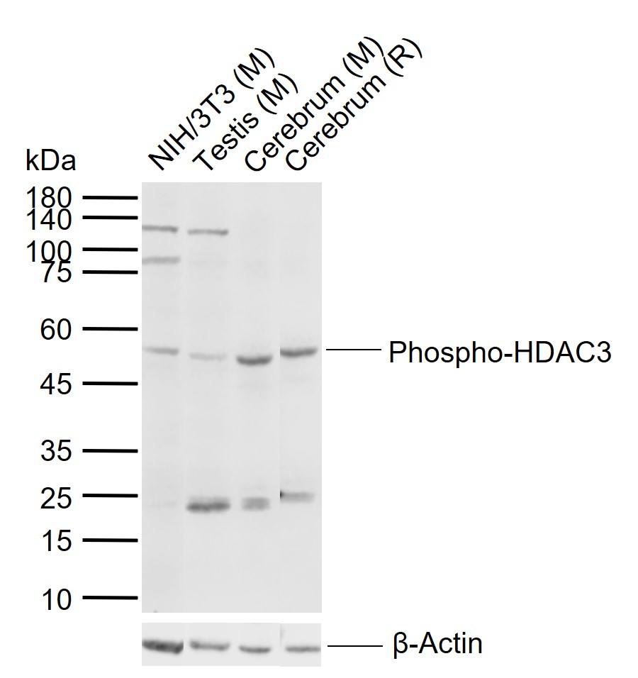
Sample:
Lane 1: Mouse NIH/3T3 cell lysates
Lane 2: Mouse Testis tissue lysates
Lane 3: Mouse Cerebrum tissue lysates
Lane 4: Rat Cerebrum tissue lysates
Primary: Anti-Phospho-HDAC3 (Ser424) (bs-3174R) at 1/1000 dilution
Secondary: IRDye800CW Goat Anti-Rabbit IgG at 1/20000 dilution
Predicted band size: 47 kDa
Observed band size: 50 kDa
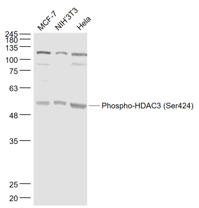
Sample:
MCF-7(Human) Cell Lysate at 30 ug
NIH/3T3(Mouse) Cell Lysate at 30 ug
Hela(Human) Cell Lysate at 30 ug
Primary: Anti- Phospho-HDAC3 (Ser424) (bs-3174R) at 1/1000 dilution
Secondary: IRDye800CW Goat Anti-Rabbit IgG at 1/20000 dilution
Predicted band size: 47 kD
Observed band size: 50 kD
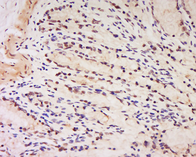
Tissue/cell: Rat rectum tissue; 4% Paraformaldehyde-fixed and paraffin-embedded;
Antigen retrieval: citrate buffer ( 0.01M, pH 6.0 ), Boiling bathing for 15min; Block endogenous peroxidase by 3% Hydrogen peroxide for 30min; Blocking buffer (normal goat serum,C-0005) at 37℃ for 20 min;
Incubation: Anti-Phospho-HDAC3, Unconjugated(bs-3174R) 1:200, overnight at 4°C, followed by conjugation to the secondary antibody(SP-0023) and DAB(C-0010) staining
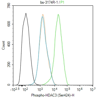
Blank control(black line):NIH/3T3.
Primary Antibody (green line): Rabbit Anti-Phospho-HDAC3 (Ser424) antibody (bs-3174R)
Dilution:1ug/Test;
Secondary Antibody(white blue line): Goat anti-rabbit IgG-AF488
Dilution: 0.5ug/Test.
Isotype control(orange line): Normal Rabbit IgG
Protocol
The cells were fixed with 4% PFA (10min at room temperature)and then permeabilized with 90% ice-cold methanol for 20 min at -20℃, The cells were then incubated in 5%BSA to block non-specific protein-protein interactions for 30 min at room temperature .Cells stained with Primary Antibody for 30 min at room temperature. The secondary antibody used for 40 min at room temperature. Acquisition of 20,000 events was performed.

Blank control(black line):Molt4.
Primary Antibody (green line): Rabbit Anti-Phospho-HDAC3 (Ser424) antibody (bs-3174R)
Dilution:1ug/Test;
Secondary Antibody(white blue line): Goat anti-rabbit IgG-AF488
Dilution: 0.5ug/Test.
Isotype control(orange line): Normal Rabbit IgG
Protocol
The cells were fixed with 4% PFA (10min at room temperature)and then permeabilized with 90% ice-cold methanol for 20 min at -20℃, The cells were then incubated in 5%BSA to block non-specific protein-protein interactions for 30 min at room temperature .Cells stained with Primary Antibody for 30 min at room temperature. The secondary antibody used for 40 min at room temperature. Acquisition of 20,000 events was performed.
|
| 1、抗体溶解方法 | |
| 2、抗体修复方式 | |
| 3、常用试剂的配制 | |
| 4、免疫组化操作步骤 | |
| 5、免疫组化问题解答 | |
| 6、Western Blotting 操作步骤 | |
| 7、Western Blotting 问题解答 | |
| 8、关于肽链的设计 | |
| 9、多肽的溶解与保存 | |
| 10、酶标抗体效价测定程序 | |


