

| 产品编号 | bs-3154R |
| 英文名称 | phospho-c-Fos (Thr325) Rabbit pAb |
| 中文名称 | 磷酸化c-fos抗体 |
| 别 名 | FOS | c-Fos (phospho-T325); p-c-Fos; phospho-c-Fos; p-FOS; FOS | c-Fos (phospho-Thr325); AP-1; C-FOS; p55; D12Rfj1; cFos; FOS_HUMAN; FOS; Cellular oncogene fos; Fos proto-oncogene, AP-1 transcription factor subunit; G0/G1 switch regulatory protein 7; Prot |
 |
Specific References (3) | bs-3154R has been referenced in 3 publications.
[IF=6.551] Mei Ha. et al. PKCα mediated by the PI3K/Akt-FOXA1 cascade facilitates cypermethrin-induced hyperthyroidism. Sci Total Environ. 2021 Feb;757:143727 WB ; Rat.
[IF=5.22] Tavares, Raquel, and Sushil Kumar Pathak. "Helicobacter pylori Secreted Protein HP1286 Triggers Apoptosis in Macrophages via TNF-Independent and ERK MAPK-Dependent Pathways." Frontiers in Cellular and Infection Microbiology 7 (2017): 58. WB ; Human.
[IF=3.362] Zheng N et al. Chlamydia pneumoniae infection promotes vascular smooth muscle cell migration via c-Fos/interleukin-17C signaling. International Journal of Medical Microbiology,2019, 151340. WB ; Rat.
|
| 产品类型 | 磷酸化抗体 |
| 研究领域 | 肿瘤 细胞生物 免疫学 神经生物学 信号转导 转录调节因子 肿瘤细胞生物标志物 表观遗传学 |
| 抗体来源 | Rabbit |
| 克隆类型 | Polyclonal |
| 克 隆 号 | |
| 交叉反应 | Human,Mouse,Rat (predicted: Pig,Sheep,Chicken,Dog) |
| 产品应用 | WB=1:500-2000,IHC-P=1:100-500,IHC-F=1:100-500,IF=1:100-500,Flow-Cyt=1μg /Test
not yet tested in other applications. optimal dilutions/concentrations should be determined by the end user. |
| 理论分子量 | 41 kDa |
| 检测分子量 | 41 |
| 细胞定位 | 细胞核 细胞浆 |
| 性 状 | Liquid |
| 浓 度 | 1mg/ml |
| 免 疫 原 | KLH conjugated Synthesised phosphopeptide derived from human c-Fos around the phosphorylation site of Thr325: LC(p-T)PV |
| 亚 型 | IgG |
| 纯化方法 | affinity purified by Protein A |
| 缓 冲 液 | 0.01M TBS (pH7.4) with 1% BSA, 0.02% Proclin300 and 50% Glycerol. |
| 保存条件 | Shipped at 4℃. Store at -20℃ for one year. Avoid repeated freeze/thaw cycles. |
| 注意事项 | This product as supplied is intended for research use only, not for use in human, therapeutic or diagnostic applications. |
| PubMed | PubMed |
| 产品介绍 |
The Fos gene family consists of 4 members: FOS, FOSB, FOSL1, and FOSL2. These genes encode leucine zipper proteins that can dimerize with proteins of the JUN family, thereby forming the transcription factor complex AP-1. As such, the FOS proteins have been implicated as regulators of cell proliferation, differentiation, and transformation. In some cases, expression of the FOS gene has also been associated with apoptotic cell death. [provided by RefSeq, Jul 2008]. Function: Nuclear phosphoprotein which forms a tight butnon-covalently linked complex with the JUN/AP-1 transcriptionfactor. In the heterodimer, FOS and JUN/AP-1 basic regions eachseems to interact with symmetrical DNA half sites. On TGF-betaactivation, forms a multimeric SMAD3/SMAD4/JUN/FOS complex at theAP1/SMAD-binding site to regulate TGF-beta-mediated signaling. Hasa critical function in regulating the Has a critical function inregulating the development of cells destined to form and maintainthe skeleton. It is thought to have an important role in signaltransduction, cell proliferation and differentiation. Subunit: Heterodimer; with JUN (By similarity). Interacts withMAFB. Component of the SMAD3/SMAD4/JUN/FOS complexrequired for syngernistic TGF-beta-mediated transcription at theAP1 promoter site. Interacts with SMAD3; the interaction is weakeven on TGF-beta activation. Interacts with MAFB. Interacts withDSIPI; this interaction inhibits the binding of active AP1 to itstarget DNA. Subcellular Location: Nucleus. Post-translational modifications: Phosphorylated in the C-terminal upon stimulation by nerve growth factor (NGF) and epidermal growth factor (EGF). Phosphorylated, in vitro, by MAPK and RSK1. Phosphorylation on both Ser-362 and Ser-374 by MAPK1/2 and RSK1/2 leads to protein stabilization with phosphorylation on Ser-374 being the major site for protein stabilization on NGF stimulation. Phosphorylation on Ser-362 and Ser-374 primes further phosphorylations on Thr-325 and Thr-331 through promoting docking of MAPK to the DEF domain. Phosphorylation on Thr-232, induced by HA-RAS, activates the transcriptional activity and antagonizes sumoylation. Phosphorylation on Ser-362 by RSK2 in osteoblasts contributes to osteoblast transformation. Constitutively sumoylated by SUMO1, SUMO2 and SUMO3. Desumoylated by SENP2. Sumoylation requires heterodimerization with JUN and is enhanced by mitogen stimulation. Sumoylation inhibits the AP-1 transcriptional activity and is, itself, inhibited by Ras-activated phosphorylation on Thr-232. Similarity: Belongs to the bZIP family. Fos subfamily. Contains 1 bZIP domain SWISS: P01100 Gene ID: 2353 Database links: Entrez Gene: 2353 Human Entrez Gene: 14281 Mouse Omim: 164810 Human SwissProt: P01100 Human SwissProt: P01101 Mouse Unigene: 246513 Mouse Unigene: 103750 Rat |
| 产品图片 |
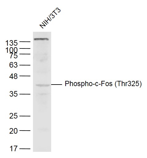
Sample:
NIH/3T3(Mouse) Cell Lysate at 40 ug
Primary: Anti-Phospho-c-Fos (Thr325) (bs-3154R) at 1/300 dilution
Secondary: IRDye800CW Goat Anti-Rabbit IgG at 1/20000 dilution
Predicted band size: 41 kD
Observed band size: 41 kD
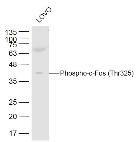
Sample:
LOVO(Human) Cell Lysate at 40 ug
Primary: Anti-Phospho-c-Fos (Thr325) (bs-3154R) at 1/300 dilution
Secondary: IRDye800CW Goat Anti-Rabbit IgG at 1/20000 dilution
Predicted band size: 41 kD
Observed band size: 41 kD
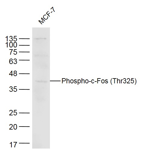
Sample:
MCF-7(Human)Cell Lysate at 40 ug
Primary: Anti-Phospho-c-Fos (Thr325) (bs-3154R) at 1/300 dilution
Secondary: IRDye800CW Goat Anti-Rabbit IgG at 1/20000 dilution
Predicted band size: 41 kD
Observed band size: 41 kD
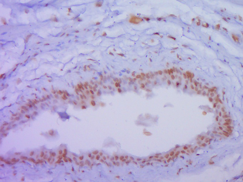
Paraformaldehyde-fixed, paraffin embedded (Rat urinary bladder); Antigen retrieval by boiling in sodium citrate buffer (pH6.0) for 15min; Block endogenous peroxidase by 3% hydrogen peroxide for 20 minutes; Blocking buffer (normal goat serum) at 37°C for 30min; Antibody incubation with (P-c-Fos (Thr325)) Polyclonal Antibody, Unconjugated (bs-3154R) at 1:400 overnight at 4°C, followed by operating according to SP Kit(Rabbit) (sp-0023) instructionsand DAB staining.

Tissue/cell: mouse intestine tissue; 4% Paraformaldehyde-fixed and paraffin-embedded;
Antigen retrieval: citrate buffer ( 0.01M, pH 6.0 ), Boiling bathing for 15min; Block endogenous peroxidase by 3% Hydrogen peroxide for 30min; Blocking buffer (normal goat serum,C-0005) at 37℃ for 20 min;
Incubation: Anti-Phospho-c-Fos (Thr325) Polyclonal Antibody, Unconjugated(bs-3154R) 1:200, overnight at 4°C, followed by conjugation to the secondary antibody(SP-0023) and DAB(C-0010) staining
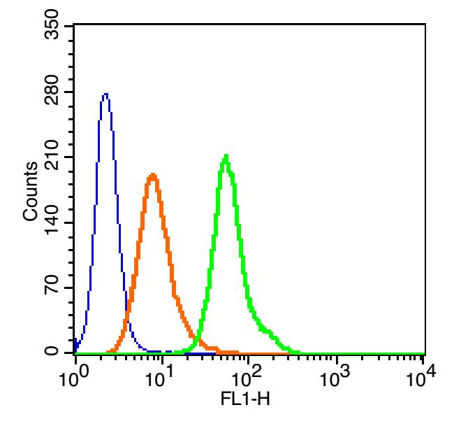
Blank control(blue): 293T(fixed with 2% paraformaldehyde (10 min) and then permeabilized with ice-cold 90% methanol for 30 min on ice). Primary Antibody: Rabbit Anti- Phospho-c-Fos (Thr325)/AF488 Conjugated antibody (bs-3154R /AF488), Dilution: 1μg in 100 μL 1X PBS containing 0.5% BSA; Isotype Control Antibody: Rabbit IgG/FITC(orange) ,used under the same conditions.
|
| 1、抗体溶解方法 | |
| 2、抗体修复方式 | |
| 3、常用试剂的配制 | |
| 4、免疫组化操作步骤 | |
| 5、免疫组化问题解答 | |
| 6、Western Blotting 操作步骤 | |
| 7、Western Blotting 问题解答 | |
| 8、关于肽链的设计 | |
| 9、多肽的溶解与保存 | |
| 10、酶标抗体效价测定程序 | |


