

| 产品编号 | bs-3164R |
| 英文名称 | phospho-FAK (Tyr407) Rabbit pAb |
| 中文名称 | 磷酸化粘着斑激酶抗体 |
| 别 名 | PTK2 | FAK (phospho-Y407); p-FAK; phospho-FAK; p-PTK2; PTK2 | FAK (phospho-Tyr407); FADK; FADK 1; FAK; FAK1; FRNK; PPP1R71; p125FAK; pp125FAK; FAK1_HUMAN; PTK2; Focal adhesion kinase-related nonkinase (FRNK); Protein phosphatase 1 regulatory subunit 71 (P |
| 产品类型 | 磷酸化抗体 |
| 研究领域 | 肿瘤 细胞生物 神经生物学 信号转导 激酶和磷酸酶 |
| 抗体来源 | Rabbit |
| 克隆类型 | Polyclonal |
| 克 隆 号 | |
| 交叉反应 | Human,Mouse (predicted: Rat,Rabbit,Sheep,Cow,Dog,Horse) |
| 产品应用 | WB=1:500-2000,IHC-P=1:100-500,IHC-F=1:100-500,IF=1:100-500,Flow-Cyt=1μg /Test
not yet tested in other applications. optimal dilutions/concentrations should be determined by the end user. |
| 理论分子量 | 116 kDa |
| 检测分子量 | 116 |
| 细胞定位 | 细胞核 细胞浆 细胞膜 |
| 性 状 | Liquid |
| 浓 度 | 1mg/ml |
| 免 疫 原 | KLH conjugated Synthesised phosphopeptide derived from human FAK around the phosphorylation site of Tyr407: DT(p-Y)TM |
| 亚 型 | IgG |
| 纯化方法 | affinity purified by Protein A |
| 缓 冲 液 | 0.01M TBS (pH7.4) with 1% BSA, 0.02% Proclin300 and 50% Glycerol. |
| 保存条件 | Shipped at 4℃. Store at -20℃ for one year. Avoid repeated freeze/thaw cycles. |
| 注意事项 | This product as supplied is intended for research use only, not for use in human, therapeutic or diagnostic applications. |
| PubMed | PubMed |
| 产品介绍 |
Non-receptor protein-tyrosine kinase implicated in signaling pathways involved in cell motility, proliferation and apoptosis. Activated by tyrosine-phosphorylation in response to either integrin clustering induced by cell adhesion or antibody cross-linking, or via G-protein coupled receptor (GPCR) occupancy by ligands such as bombesin or lysophosphatidic acid, or via LDL receptor occupancy. Plays a potential role in oncogenic transformations resulting in increased kinase activity. [SUBCELLULAR LOCATION] Cell junction, focal adhesion. Cell membrane; Peripheral membrane protein; Cytoplasmic side. Note=Constituent of focal adhesions. Function: Non-receptor protein-tyrosine kinase that plays an essential role in regulating cell migration, adhesion, spreading, reorganization of the actin cytoskeleton, formation and disassembly of focal adhesions and cell protrusions, cell cycle progression, cell proliferation and apoptosis. Required for early embryonic development and placenta development. Required for embryonic angiogenesis, normal cardiomyocyte migration and proliferation, and normal heart development. Regulates axon growth and neuronal cell migration, axon branching and synapse formation; required for normal development of the nervous system. Plays a role in osteogenesis and differentiation of osteoblasts. Functions in integrin signal transduction, but also in signaling downstream of numerous growth factor receptors, G-protein coupled receptors (GPCR), EPHA2, netrin receptors and LDL receptors. Forms multisubunit signaling complexes with SRC and SRC family members upon activation; this leads to the phosphorylation of additional tyrosine residues, creating binding sites for scaffold proteins, effectors and substrates. Regulates numerous signaling pathways. Promotes activation of phosphatidylinositol 3-kinase and the AKT1 signaling cascade. Promotes activation of MAPK1/ERK2, MAPK3/ERK1 and the MAP kinase signaling cascade. Promotes localized and transient activation of guanine nucleotide exchange factors (GEFs) and GTPase-activating proteins (GAPs), and thereby modulates the activity of Rho family GTPases. Signaling via CAS family members mediates activation of RAC1. Recruits the ubiquitin ligase MDM2 to P53/TP53 in the nucleus, and thereby regulates P53/TP53 activity, P53/TP53 ubiquitination and proteasomal degradation. Phosphorylates SRC; this increases SRC kinase activity. Phosphorylates ACTN1, ARHGEF7, GRB7, RET and WASL. Promotes phosphorylation of PXN and STAT1; most likely PXN and STAT1 are phosphorylated by a SRC family kinase that is recruited to autophosphorylated PTK2/FAK1, rather than by PTK2/FAK1 itself. Promotes phosphorylation of BCAR1; GIT2 and SHC1; this requires both SRC and PTK2/FAK1. Promotes phosphorylation of BMX and PIK3R1. Isoform 6 (FRNK) does not contain a kinase domain and inhibits PTK2/FAK1 phosphorylation and signaling. Its enhanced expression can attenuate the nuclear accumulation of LPXN and limit its ability to enhance serum response factor (SRF)-dependent gene transcription. Subunit: Interacts (via first Pro-rich region) with CAS family members (via SH3 domain), including BCAR1, BCAR3, CASS4 and NEDD9. Interacts with GIT1. Interacts with SORBS1. Interacts with RGNEF. Interacts with SHB. Interacts with PXN and TLN1. Interacts with STAT1. Interacts with DCC. Interacts with WASL. Interacts with ARHGEF7. Interacts with GRB2 and GRB7 (By similarity). Component of a complex that contains at least FER, CTTN and PTK2/FAK1. Interacts with BMX. Interacts with TGFB1I1. Interacts with STEAP4. Interacts with ZFYVE21. Interacts with ESR1. Interacts with PIK3R1 or PIK3R2. Interacts with SRC, FGR, FLT4 and RET. Interacts with EPHA2 in resting cells; activation of EPHA2 recruits PTPN11, leading to dephosphorylation of PTK2/FAK1 and dissociation of the complex. Interacts with EPHA1 (kinase activity-dependent). Interacts with CD4; this interaction requires the presence of HIV-1 gp120. Interacts with PIAS1. Interacts with ARHGAP26 and SHC1. Interacts with RB1CC1; this inhibits PTK2/FAK1 activity and activation of downstream signaling pathways. Interacts with P53/TP53 and MDM2. Interacts with LPXN (via LD motif 3). Subcellular Location: Cell junction, focal adhesion. Cell membrane; Peripheral membrane protein; Cytoplasmic side. Cytoplasm, cell cortex. Cytoplasm, cytoskeleton. Cytoplasm, cytoskeleton, centrosome. Nucleus. Note=Constituent of focal adhesions. Detected at microtubules. Tissue Specificity: Detected in B and T-lymphocytes. Isoform 1 and isoform 6 are detected in lung fibroblasts (at protein level). Ubiquitous. Post-translational modifications: Phosphorylated on tyrosine residues upon activation, e.g. upon integrin signaling. Tyr-397 is the major autophosphorylation site, but other kinases can also phosphorylate this residue. Phosphorylation at Tyr-397 promotes interaction with SRC and SRC family members, leading to phosphorylation at Tyr-576, Tyr-577 and at additional tyrosine residues. FGR promotes phosphorylation at Tyr-397 and Tyr-576. FER promotes phosphorylation at Tyr-577, Tyr-861 and Tyr-925, even when cells are not adherent. Tyr-397, Tyr-576 and Ser-722 are phosphorylated only when cells are adherent. Phosphorylation at Tyr-397 is important for interaction with BMX, PIK3R1 and SHC1. Phosphorylation at Tyr-925 is important for interaction with GRB2. Dephosphorylated by PTPN11; PTPN11 is recruited to PTK2 via EPHA2 (tyrosine phosphorylated). Microtubule-induced dephosphorylation at Tyr-397 is crucial for the induction of focal adhesion disassembly; this dephosphorylation could be catalyzed by PTPN11 and regulated by ZFYVE21. Sumoylated; this enhances autophosphorylation. DISEASE: Note=Aberrant PTK2/FAK1 expression may play a role in cancer cell proliferation, migration and invasion, in tumor formation and metastasis. PTK2/FAK1 overexpression is seen in many types of cancer. Similarity: Belongs to the protein kinase superfamily. Tyr protein kinase family. FAK subfamily. Contains 1 FERM domain. SWISS: Q05397 Gene ID: 5747 Database links: Entrez Gene: 5747 Human Entrez Gene: 14083 Mouse Omim: 600758 Human SwissProt: Q05397 Human SwissProt: P34152 Mouse Unigene: 395482 Human Unigene: 254494 Mouse Unigene: 2809 Rat FAK是整合蛋白介导的信号转导中的重要成员,有酪氨酸蛋白激酶活性,并可自身磷酸化,FAK本身是胱冬肽酶(caspase)的底物。作为信号分子的FAK参与抑制细胞凋亡并直接参与细胞多种功能的调节。 1.FAK 局部粘着斑激酶,是一种酪氨酸激酶;肿瘤细胞的侵袭性生长是一个多步骤的复杂过程,有多种生物化学因子参与其中,局部粘着斑激酶(focal adhesion kinase, FAK)介导的信号转导系统就是其中最为重要的细胞信号转导途径之一。肿瘤细胞必须黏附于细胞外基质,通过促进依赖于PTK激酶活性的细胞外基质信号转导,进而影响细胞的黏附、运动与迁移。 2.粘着斑激酶(focal adhesion kinase,FAK)是整合蛋白介导的信号转导中的重要成员,有酪氨酸蛋白激酶活性,并可自身磷酸化;为信号分子的FAK,还与细胞内其他信号转导通路存在串话(crosstalk),直接参与了细胞多种功能的调节。 3.尽管FAK的确切功能尚不清楚,但若干实验均提示FAK可能有两个作用,一是在细胞铺展和移动时,FAK参与粘着斑形成和调节;二是FAK参与信号转导过程,以告知细胞核其细胞已锚定了。近年有关FAK在细胞凋亡中的作用也业已肯定。 |
| 产品图片 |
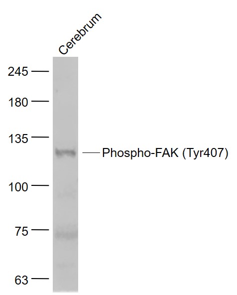
Sample:
Cerebrum (Mouse) Lysate at 40 ug
Primary: Anti- Phospho-FAK (Tyr407) (bs-3164R) at 1/1000 dilution
Secondary: IRDye800CW Goat Anti-Rabbit IgG at 1/20000 dilution
Predicted band size: 116 kD
Observed band size: 116 kD
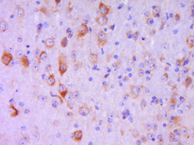
Paraformaldehyde-fixed, paraffin embedded (mouse brain tissue); Antigen retrieval by boiling in sodium citrate buffer (pH6.0) for 15min; Block endogenous peroxidase by 3% hydrogen peroxide for 20 minutes; Blocking buffer (normal goat serum) at 37°C for 30min; Antibody incubation with (P-FAK (Tyr407)) Polyclonal Antibody, Unconjugated (bs-3164R) at 1:400 overnight at 4°C, followed by a conjugated secondary (sp-0023) for 20 minutes and DAB staining.
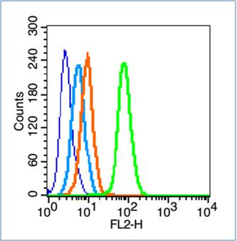
Blank control (blue line): Hep G2 (fixed with 70% ethanol (Overmight at 4℃) and then permeabilized with 90% ice-cold methanol for 30 min at -20℃).
Primary Antibody (green line): Rabbit Anti-Phospho-FAK (Tyr407) antibody (bs-3164R),Dilution: 1μg /10^6 cells;
Isotype Control Antibody (orange line): Rabbit IgG .
Secondary Antibody (white blue line): Goat anti-rabbit IgG-PE,Dilution: 1μg /test.
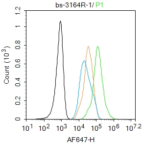
Blank control:A431.
Primary Antibody (green line): Rabbit Anti-Phospho-FAK (Tyr407) antibody (bs-3164R)
Dilution: 1μg /10^6 cells;
Isotype Control Antibody (orange line): Rabbit IgG .
Secondary Antibody : Goat anti-rabbit IgG-AF647
Dilution: 1μg /test.
Protocol
The cells were fixed with 4% PFA (10min at room temperature)and then permeabilized with 90% ice-cold methanol for 20 min at -20℃.The cells were then incubated in 5%BSA to block non-specific protein-protein interactions for 30 min at room temperature.Cells stained with Primary Antibody for 30 min at room temperature. The secondary antibody used for 40 min at room temperature. Acquisition of 20,000 events was performed.
|
| 1、抗体溶解方法 | |
| 2、抗体修复方式 | |
| 3、常用试剂的配制 | |
| 4、免疫组化操作步骤 | |
| 5、免疫组化问题解答 | |
| 6、Western Blotting 操作步骤 | |
| 7、Western Blotting 问题解答 | |
| 8、关于肽链的设计 | |
| 9、多肽的溶解与保存 | |
| 10、酶标抗体效价测定程序 | |


