

| 产品编号 | bs-2775R |
| 英文名称 | LIMK1 Rabbit pAb |
| 中文名称 | 单丝氨酸蛋白激酶1抗体 |
| 别 名 | LIMK; LIMK-1; KIZ-1; LIMK1_HUMAN; LIMK1; 2.7.11.1; LIMK1_MOUSE; LIMK1_RAT; |
 |
Specific References (2) | bs-2775R has been referenced in 2 publications.
[IF=3.29] Zhou et al. MiR-20a inhibits cutaneous squamous cell carcinoma metastasis and proliferation by directly targeting LIMK1. (2014) Cancer.Biol.Ther. 15:1340-9 WB ; Human.
[IF=2.894] Wufuer R et al. Downregulation of Rac1/PAK1/LIMK1/cofilin signaling pathway in colon cancer SW620 cells treated with Chlorin e6 photodynamic therapyPhotodiagnosis Photodyn Ther.2020 Dec 8;102143. WB ; Human.
|
| 研究领域 | 肿瘤 细胞生物 神经生物学 信号转导 激酶和磷酸酶 表观遗传学 |
| 抗体来源 | Rabbit |
| 克隆类型 | Polyclonal |
| 克 隆 号 | |
| 交叉反应 | Human,Mouse,Rat (predicted: Rabbit,Cow) |
| 产品应用 | WB=1:500-2000,IHC-P=1:20-100,IHC-F=1:20-100,IF=1:20-100,Flow-Cyt=1ug/Test
not yet tested in other applications. optimal dilutions/concentrations should be determined by the end user. |
| 理论分子量 | 71 kDa |
| 检测分子量 | 75 |
| 细胞定位 | 细胞核 细胞浆 |
| 性 状 | Liquid |
| 浓 度 | 1mg/ml |
| 免 疫 原 | KLH conjugated synthetic peptide derived from human LIMK1: 451-550/647 |
| 亚 型 | IgG |
| 纯化方法 | affinity purified by Protein A |
| 缓 冲 液 | 0.01M TBS (pH7.4) with 1% BSA, 0.02% Proclin300 and 50% Glycerol. |
| 保存条件 | Shipped at 4℃. Store at -20℃ for one year. Avoid repeated freeze/thaw cycles. |
| 注意事项 | This product as supplied is intended for research use only, not for use in human, therapeutic or diagnostic applications. |
| PubMed | PubMed |
| 产品介绍 |
LIMK1 is a protein kinase which regulates actin filament dynamics. Phosphorylates and inactivates the actin binding/depolymerizing factor cofilin, thereby stabilizing the actin cytoskeleton. LIMK1 may be involved in brain development; it is highly expressed in both adult and fetal nervous system. Detected ubiquitously throughout the different regions of adult brain, with highest levels in the cerebral cortex. Expressed to a lesser extent in heart and skeletal muscle. Function: Serine/threonine-protein kinase that plays an essential role in the regulation of actin filament dynamics. Acts downstream of several Rho family GTPase signal transduction pathways. Activated by upstream kinases including ROCK1, PAK1 and PAK4, which phosphorylate LIMK1 on a threonine residue located in its activation loop. LIMK1 subsequently phosphorylates and inactivates the actin binding/depolymerizing factors cofilin-1/CFL1, cofilin-2/CFL2 and destrin/DSTN, thereby preventing the cleavage of filamentous actin (F-actin), and stabilizing the actin cytoskeleton. In this way LIMK1 regulates several actin-dependent biological processes including cell motility, cell cycle progression, and differentiation. Phosphorylates TPPP on serine residues, thereby promoting microtubule disassembly. Stimulates axonal outgrowth and may be involved in brain development. Isoform 3 has a dominant negative effect on actin cytoskeletal changes. Subunit: Interacts (via LIM domain) with the cytoplasmic domain of NRG1. Interacts with NISCH. Interacts with RLIM and RNF6. Self-associates to form homodimers. Interacts with HSP90AA1; this interaction promotes LIMK1 dimerization and subsequent transphosphorylation. Interacts with CDN1C. Interacts with SSH1. Interacts with ROCK1. Subcellular Location: Cytoplasm. Nucleus. Note=Predominantly found in the cytoplasm. Tissue Specificity: Highest expression in both adult and fetal nervous system. Detected ubiquitously throughout the different regions of adult brain, with highest levels in the cerebral cortex. Expressed to a lesser extent in heart and skeletal muscle. Post-translational modifications: Autophosphorylated. Phosphorylated on Thr-508 by ROCK1 and PAK1, resulting in activation. Phosphorylated by PAK4 which increases the ability of LIMK1 to phosphorylate cofilin. Phosphorylated at Ser-323 by MAPKAPK2 during activation of VEGFA-induced signaling, which results in activation of LIMK1 and promotion of actin reorganization, cell migration, and tubule formation of endothelial cells. Dephosphorylated and inactivated by SSH1. Phosphorylated by CDC42BP. Ubiquitinated. 'Lys-48'-linked polyubiquitination by RNF6 leads to proteasomal degradation through the 26S proteasome, modulating LIMK1 levels in the growth cone and its effect on axonal outgrowth. Also polyubiquitinated by RLIM. DISEASE: Note=LIMK1 is located in the Williams-Beuren syndrome (WBS) critical region. WBS results from a hemizygous deletion of several genes on chromosome 7q11.23, thought to arise as a consequence of unequal crossing over between highly homologous low-copy repeat sequences flanking the deleted region. Similarity: Belongs to the protein kinase superfamily. TKL Ser/Thr protein kinase family. Contains 2 LIM zinc-binding domains. Contains 1 PDZ (DHR) domain. Contains 1 protein kinase domain. SWISS: P53667 Gene ID: 3984 Database links: Entrez Gene: 3984 Human Entrez Gene: 16885 Mouse Omim: 601329 Human SwissProt: P53667 Human SwissProt: P53668 Mouse Unigene: 647035 Human Unigene: 15409 Mouse Unigene: 11250 Rat LIM激酶1(LIMkinase1,LIMK-1)是单丝氨酸蛋白激酶,LIMK-1的活化受多种机制调控,成为联系细胞外刺激与细胞骨架稳定性的枢纽,在多种基本的生物学过程中起重要作用。LIMK-1包含LIM和PDZ蛋白和蛋白相互作用区、P/S结构域、激酶结构域,在神经元中呈高表达。它的主要作用是使肌动蛋白解聚因子cofilin磷酸化和失活,从而导致肌动蛋白细胞骨架的改变。 |
| 产品图片 |
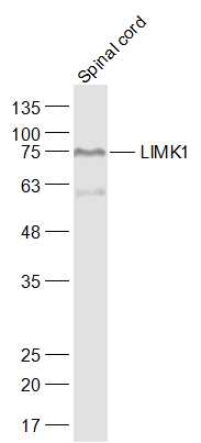
Sample:
Spinal cord (Mouse) Lysate at 40 ug
Primary: Anti-LIMK1 (bs-2775R) at 1/1000 dilution
Secondary: IRDye800CW Goat Anti-Rabbit IgG at 1/20000 dilution
Predicted band size: 71 kD
Observed band size: 75 kD
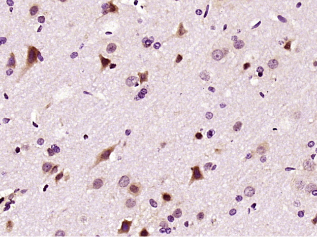
Paraformaldehyde-fixed, paraffin embedded (Rat brain); Antigen retrieval by boiling in sodium citrate buffer (pH6.0) for 15min; Block endogenous peroxidase by 3% hydrogen peroxide for 20 minutes; Blocking buffer (normal goat serum) at 37°C for 30min; Antibody incubation with (LIMK1) Polyclonal Antibody, Unconjugated (bs-2775R) at 1:400 overnight at 4°C, followed by a conjugated secondary antibody (sp-0023) for 20 minutes and DAB staining.
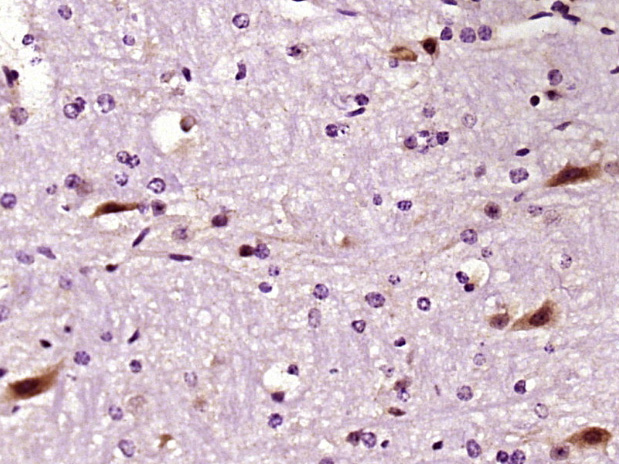
Paraformaldehyde-fixed, paraffin embedded (Mouse brain); Antigen retrieval by boiling in sodium citrate buffer (pH6.0) for 15min; Block endogenous peroxidase by 3% hydrogen peroxide for 20 minutes; Blocking buffer (normal goat serum) at 37°C for 30min; Antibody incubation with (LIMK1) Polyclonal Antibody, Unconjugated (bs-2775R) at 1:400 overnight at 4°C, followed by a conjugated secondary antibody (sp-0023) for 20 minutes and DAB staining.
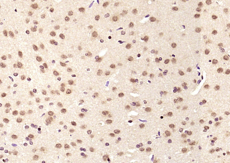
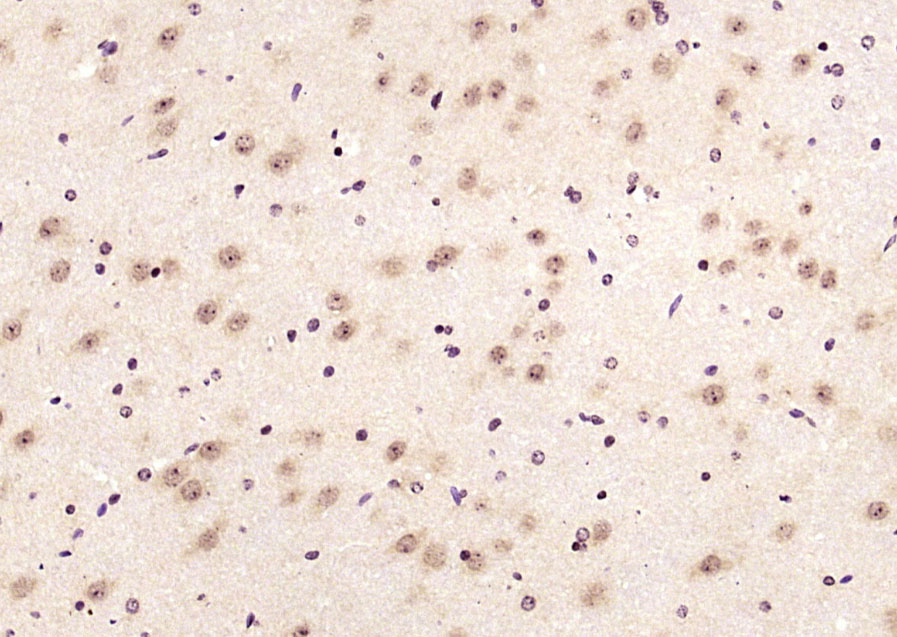
Paraformaldehyde-fixed, paraffin embedded (rat brain); Antigen retrieval by boiling in sodium citrate buffer (pH6.0) for 15min; Block endogenous peroxidase by 3% hydrogen peroxide for 20 minutes; Blocking buffer (normal goat serum) at 37°C for 30min; Antibody incubation with (LIMK1) Polyclonal Antibody, Unconjugated (bs-2775R) at 1:200 overnight at 4°C, followed by operating according to SP Kit(Rabbit) (sp-0023) instructionsand DAB staining.
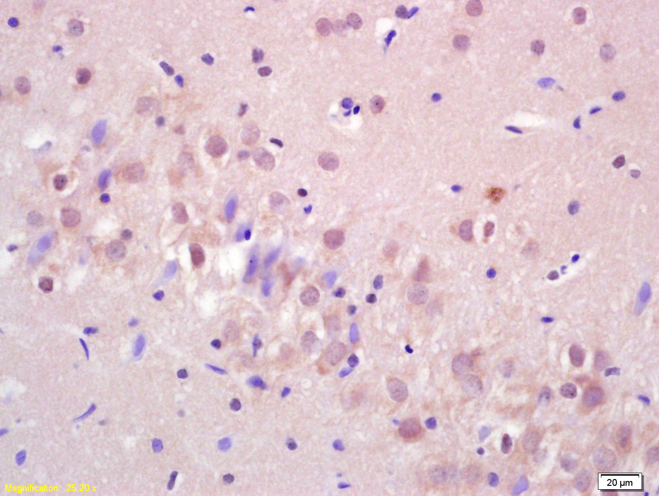
Paraformaldehyde-fixed, paraffin embedded (mouse brain); Antigen retrieval by boiling in sodium citrate buffer (pH6.0) for 15min; Block endogenous peroxidase by 3% hydrogen peroxide for 20 minutes; Blocking buffer (normal goat serum) at 37°C for 30min; Antibody incubation with (LIMK1) Polyclonal Antibody, Unconjugated (bs-2775R) at 1:200 overnight at 4°C, followed by operating according to SP Kit(Rabbit) (sp-0023) instructionsand DAB staining.
Tissue/cell: rat brain tissue; 4% Paraformaldehyde-fixed and paraffin-embedded;
Antigen retrieval: citrate buffer ( 0.01M, pH 6.0 ), Boiling bathing for 15min; Block endogenous peroxidase by 3% Hydrogen peroxide for 30min; Blocking buffer (normal goat serum,C-0005) at 37℃ for 20 min;
Incubation: Anti-LIMK1 Polyclonal Antibody, Unconjugated(bs-2775R) 1:200, overnight at 4°C, followed by conjugation to the secondary antibody(SP-0023) and DAB(C-0010) staining
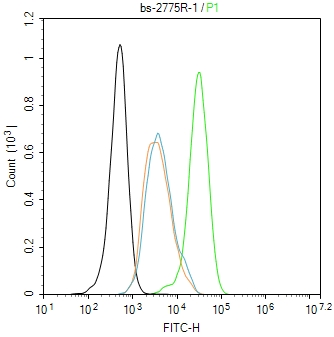
Blank control(black line):Hela.
Primary Antibody (green line): Rabbit Anti-LIMK1 antibody (bs-2775R)
Dilution:1ug/Test;
Secondary Antibody(white blue line): Goat anti-rabbit IgG-FITC
Dilution: 0.5ug/Test.
Isotype control(orange line): Normal Rabbit IgG
Protocol
The cells were fixed with 4% PFA (10min at room temperature)and then permeabilized with 90% ice-cold methanol for 20 min at -20℃, The cells were then incubated in 5%BSA to block non-specific protein-protein interactions for 30 min at room temperature .Cells stained with Primary Antibody for 30 min at room temperature. The secondary antibody used for 40 min at room temperature. Acquisition of 20,000 events was performed.
|
| 1、抗体溶解方法 | |
| 2、抗体修复方式 | |
| 3、常用试剂的配制 | |
| 4、免疫组化操作步骤 | |
| 5、免疫组化问题解答 | |
| 6、Western Blotting 操作步骤 | |
| 7、Western Blotting 问题解答 | |
| 8、关于肽链的设计 | |
| 9、多肽的溶解与保存 | |
| 10、酶标抗体效价测定程序 | |


