

| 产品编号 | bs-2781R |
| 英文名称 | CACNA1G Rabbit pAb |
| 中文名称 | 电压依赖性钙通道Cav3.1抗体 |
| 别 名 | calcium channel voltage dependent alpha 1G subunit; calcium channel voltage dependent T type alpha 1G subunit; calcium channel voltage dependent T type alpha1G subunit; CaV T1; Cav3 1; cav3 1c; NBR13; voltage dependent calcium channel alpha 1G subunit isoform 11; voltage dependent T type calcium channel subunit alpha 1G; CAC1G_HUMAN; Cav3.1 |
 |
Specific References (1) | bs-2781R has been referenced in 1 publications.
[IF=2.36] Lu, Yujie, et al. "Mibefradil reduces blood glucose concentration in db/db mice." Clinics 69.1 (2014): 61. WB ; Mouse.
|
| 研究领域 | 通道蛋白 |
| 抗体来源 | Rabbit |
| 克隆类型 | Polyclonal |
| 克 隆 号 | |
| 交叉反应 | Human,Mouse,Rat (predicted: Cow,Dog) |
| 产品应用 | IHC-P=1:100-500,IHC-F=1:100-500,IF=1:100-500
not yet tested in other applications. optimal dilutions/concentrations should be determined by the end user. |
| 理论分子量 | 262 kDa |
| 检测分子量 | |
| 细胞定位 | 细胞膜 |
| 性 状 | Liquid |
| 浓 度 | 1mg/ml |
| 免 疫 原 | KLH conjugated synthetic peptide derived from human CACNA1G/Cav31: 901-1000/2377 <Extracellular> |
| 亚 型 | IgG |
| 纯化方法 | affinity purified by Protein A |
| 缓 冲 液 | 0.01M TBS (pH7.4) with 1% BSA, 0.02% Proclin300 and 50% Glycerol. |
| 保存条件 | Shipped at 4℃. Store at -20℃ for one year. Avoid repeated freeze/thaw cycles. |
| 注意事项 | This product as supplied is intended for research use only, not for use in human, therapeutic or diagnostic applications. |
| PubMed | PubMed |
| 产品介绍 |
Voltage-sensitive calcium channels mediate the entry of calcium ions into excitable cells, and are also involved in a variety of calcium-dependent processes, including muscle contraction, hormone or neurotransmitter release, gene expression, cell motility, cell division, and cell death. This gene encodes a T-type, low-voltage activated calcium channel. The T-type channels generate currents that are both transient, owing to fast inactivation, and tiny, owing to small conductance. T-type channels are thought to be involved in pacemaker activity, low-threshold calcium spikes, neuronal oscillations and resonance, and rebound burst firing. Many alternatively spliced transcript variants encoding different isoforms have been described for this gene. [provided by RefSeq, Sep 2011] Function: Voltage-sensitive calcium channels (VSCC) mediate the entry of calcium ions into excitable cells and are also involved in a variety of calcium-dependent processes, including muscle contraction, hormone or neurotransmitter release, gene expression, cell motility, cell division and cell death. The isoform alpha-1G gives rise to T-type calcium currents. T-type calcium channels belong to the 'low-voltage activated (LVA)' group and are strongly blocked by mibefradil. A particularity of this type of channels is an opening at quite negative potentials and a voltage-dependent inactivation. T-type channels serve pacemaking functions in both central neurons and cardiac nodal cells and support calcium signaling in secretory cells and vascular smooth muscle. They may also be involved in the modulation of firing patterns of neurons which is important for information processing as well as in cell growth processes. Subcellular Location: Membrane; Multi-pass membrane protein. Tissue Specificity: Highly expressed in brain, in particular in the amygdala, subthalamic nuclei, cerebellum and thalamus. Moderate expression in heart; low expression in placenta, kidney and lung. Also expressed in colon and bone marrow and in tumoral cells to a lesser extent. Highly expressed in fetal brain, but also in peripheral fetal tissues as heart, kidney and lung, suggesting a developmentally regulated expression. Similarity: Belongs to the calcium channel alpha-1 subunit (TC 1.A.1.11) family. CACNA1G subfamily. SWISS: O43497 Gene ID: 8913 Database links: Entrez Gene: 8913 Human Entrez Gene: 12291 Mouse Omim: 604065 Human SwissProt: O43497 Human Unigene: 29585 Mouse Unigene: 86960 Rat |
| 产品图片 |
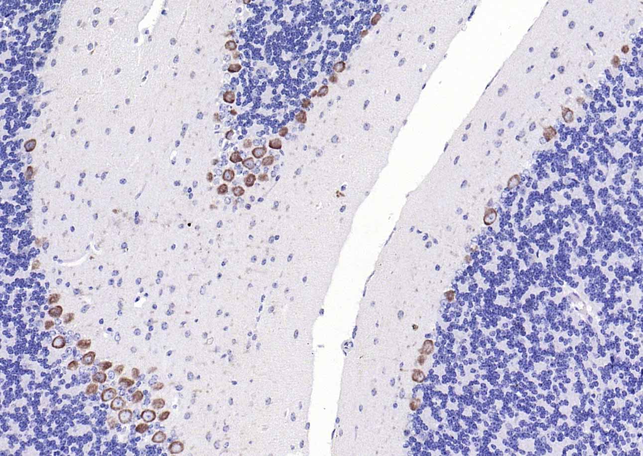
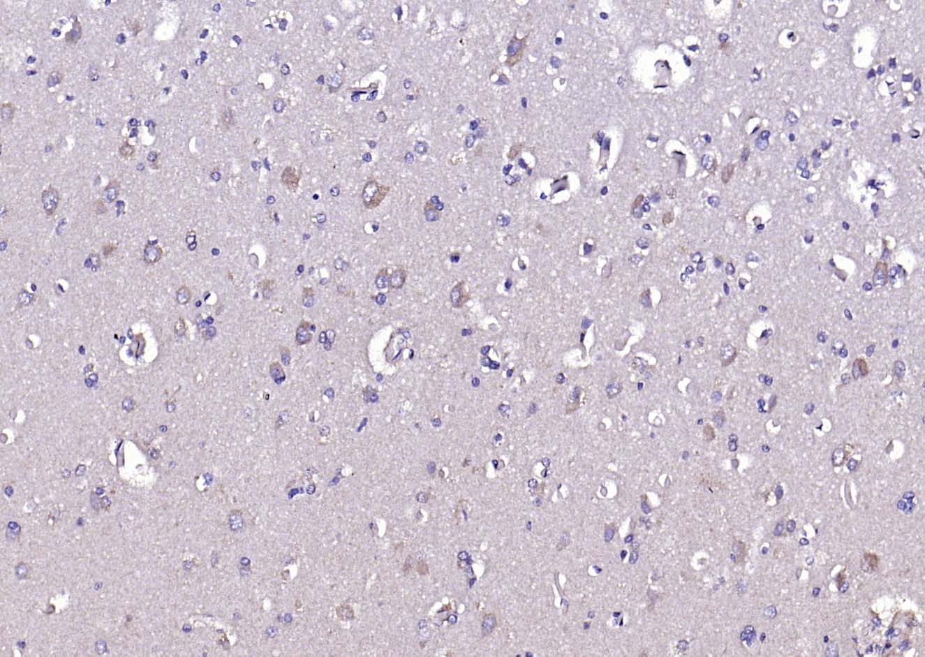
Paraformaldehyde-fixed, paraffin embedded Mouse Cerebellum; Antigen retrieval by boiling in sodium citrate buffer (pH6.0) for 15 min; Antibody incubation with CACNA1G Polyclonal Antibody, Unconjugated (bs-2781R) at 1:200 overnight at 4°C, followed by conjugation to the bs-0295G-HRP and DAB (C-0010) staining.
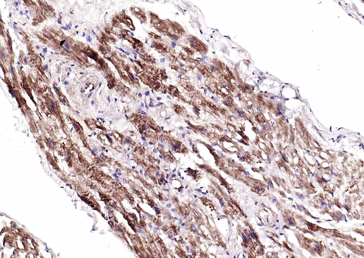
Paraformaldehyde-fixed, paraffin embedded Human Heart; Antigen retrieval by boiling in sodium citrate buffer (pH6.0) for 15 min; Antibody incubation with CACNA1G Polyclonal Antibody, Unconjugated (bs-2781R) at 1:200 overnight at 4°C, followed by conjugation to the bs-0295G-HRP and DAB (C-0010) staining.
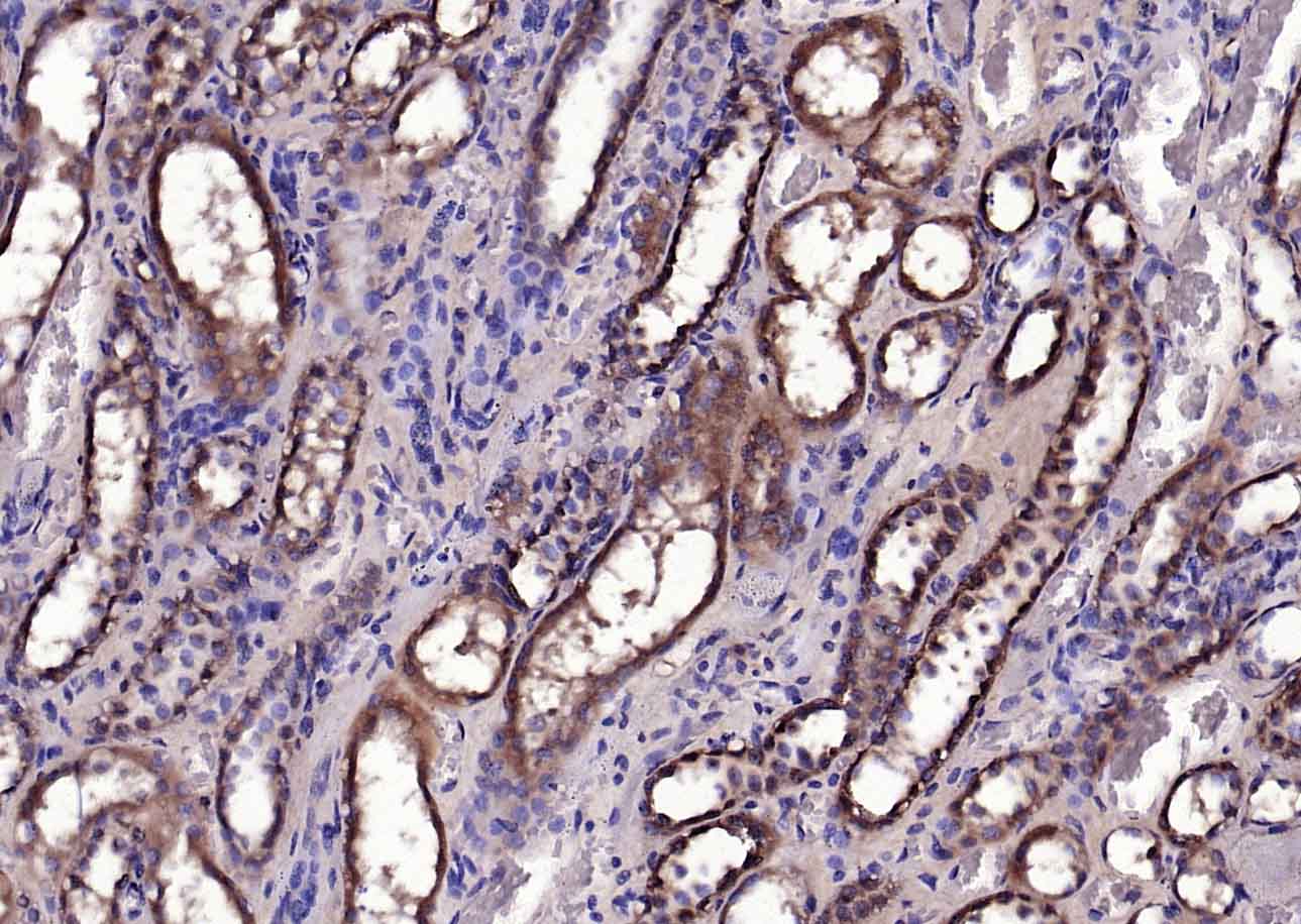
Paraformaldehyde-fixed, paraffin embedded Human Kidney; Antigen retrieval by boiling in sodium citrate buffer (pH6.0) for 15 min; Antibody incubation with CACNA1G Polyclonal Antibody, Unconjugated (bs-2781R) at 1:200 overnight at 4°C, followed by conjugation to the bs-0295G-HRP and DAB (C-0010) staining.
Paraformaldehyde-fixed, paraffin embedded Rat Cerebellum; Antigen retrieval by boiling in sodium citrate buffer (pH6.0) for 15 min; Antibody incubation with CACNA1G Polyclonal Antibody, Unconjugated (bs-2781R) at 1:200 overnight at 4°C, followed by conjugation to the bs-0295G-HRP and DAB (C-0010) staining.
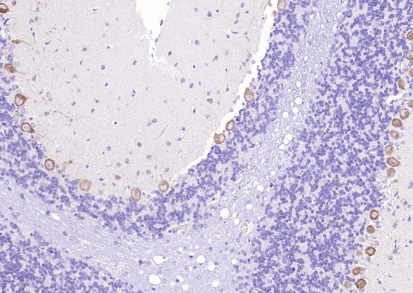
Paraformaldehyde-fixed, paraffin embedded Human Cerebrum; Antigen retrieval by boiling in sodium citrate buffer (pH6.0) for 15 min; Antibody incubation with CACNA1G Polyclonal Antibody, Unconjugated (bs-2781R) at 1:200 overnight at 4°C, followed by conjugation to the bs-0295G-HRP and DAB (C-0010) staining.
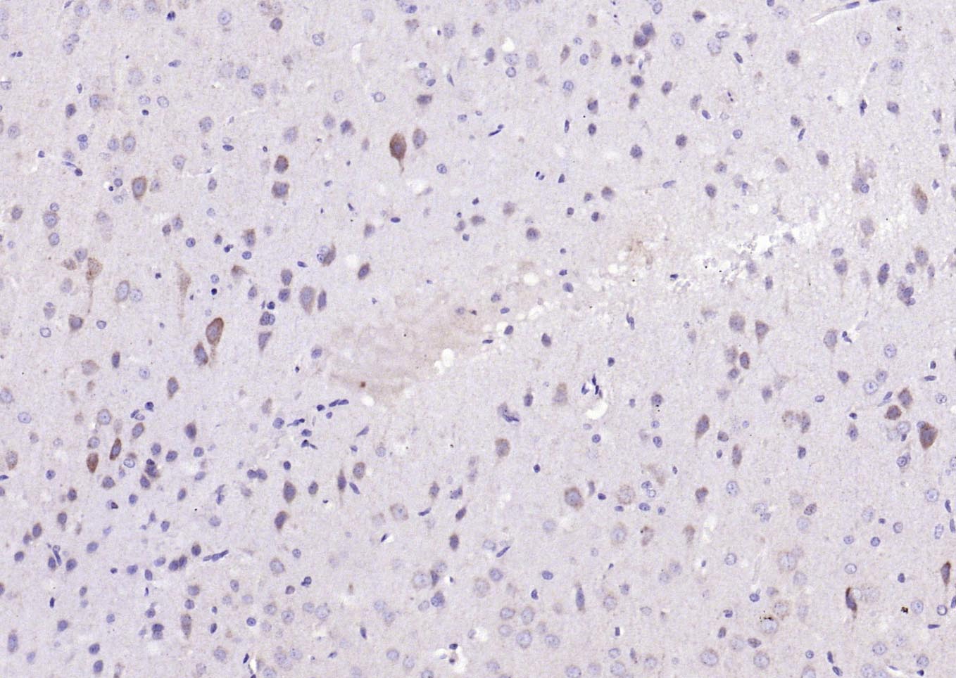
Paraformaldehyde-fixed, paraffin embedded Rat Cerebrum; Antigen retrieval by boiling in sodium citrate buffer (pH6.0) for 15 min; Antibody incubation with CACNA1G Polyclonal Antibody, Unconjugated (bs-2781R) at 1:200 overnight at 4°C, followed by conjugation to the bs-0295G-HRP and DAB (C-0010) staining.
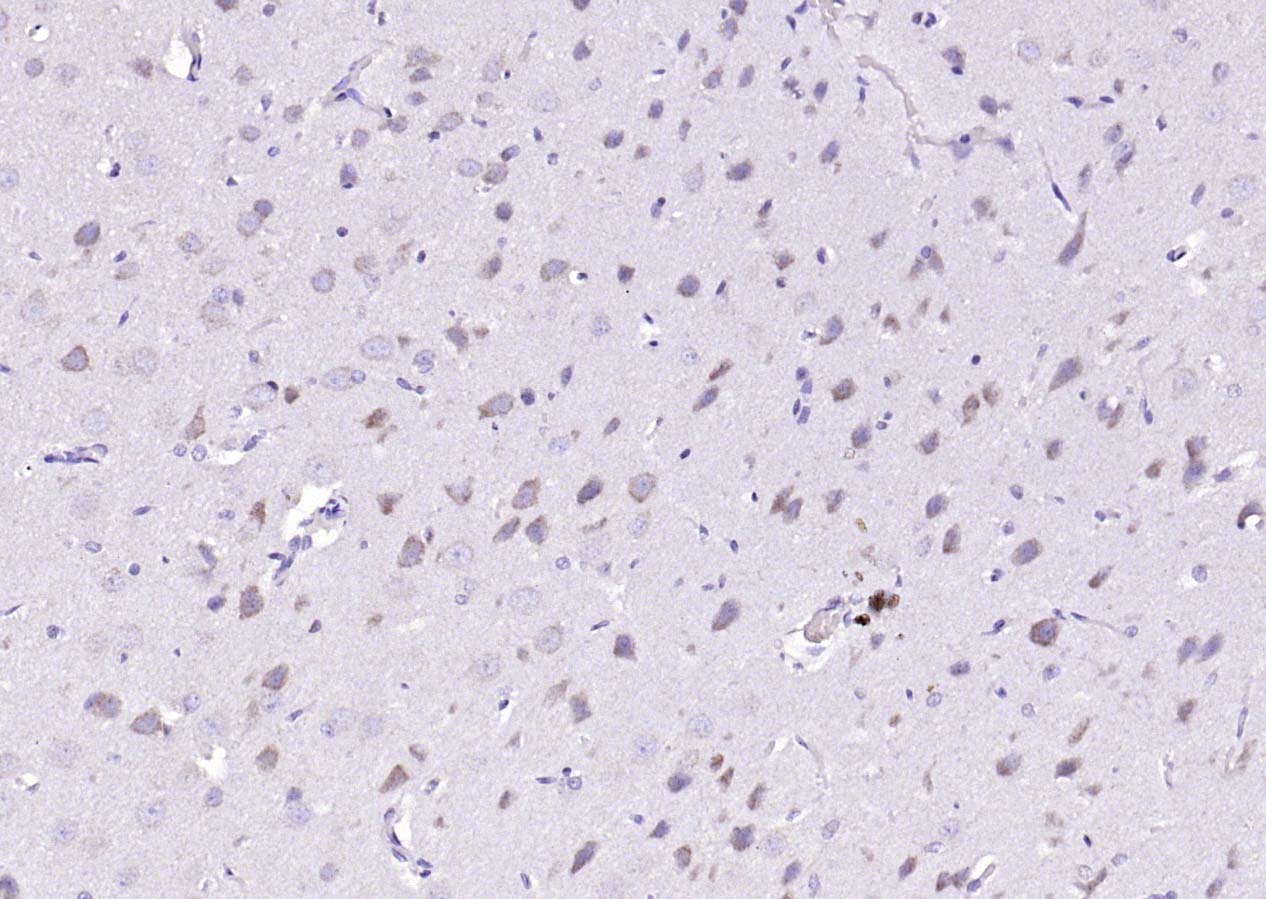
Paraformaldehyde-fixed, paraffin embedded Mouse Cerebrum; Antigen retrieval by boiling in sodium citrate buffer (pH6.0) for 15 min; Antibody incubation with CACNA1G Polyclonal Antibody, Unconjugated (bs-2781R) at 1:200 overnight at 4°C, followed by conjugation to the bs-0295G-HRP and DAB (C-0010) staining.
|
| 1、抗体溶解方法 | |
| 2、抗体修复方式 | |
| 3、常用试剂的配制 | |
| 4、免疫组化操作步骤 | |
| 5、免疫组化问题解答 | |
| 6、Western Blotting 操作步骤 | |
| 7、Western Blotting 问题解答 | |
| 8、关于肽链的设计 | |
| 9、多肽的溶解与保存 | |
| 10、酶标抗体效价测定程序 | |


