

| 产品编号 | bs-2808R |
| 英文名称 | Jak3 Rabbit pAb |
| 中文名称 | 蛋白酪氨酸激酶JAK-3抗体 |
| 别 名 | JAK-3; JAK3_HUMAN; JAKL; L-JAK; LJAK; fae; wil; JAK3; Janus kinase 3 (JAK-3); Leukocyte janus kinase (L-JAK); 2.7.10.2; JAK3_MOUSE; Janus kinase 3; tyrosine-protein kinase JAK3; leukocyte Janus kinase |
 |
Specific References (4) | bs-2808R has been referenced in 4 publications.
[IF=6.291] Changjiang Liu. et al. Cypermethrin triggers YY1-mediated testosterone biosynthesis suppression. Ecotox Environ Safe. 2021 Dec;225:112792 WB ; Rat.
[IF=5.195] Yong Wang. et al. An integrated network pharmacology approach reveals that Darutigenol reduces inflammation and cartilage degradation in a mouse collagen-induced arthritis model by inhibiting the JAK-STAT3 pathway. J ETHNOPHARMACOL. 2023 May;:116574 WB ; Mouse.
[IF=4.46] Xing Chang. et al. 1-(4-((5-chloro-4-((2-(isopropylsulfonyl)phenyl)amino)pyrimidin-2-yl)amino)-3-methoxyphenyl)-3-(2-(dimethylamino)ethyl)imidazolidin-2-one (ZX-42) inhibits cell proliferation and induces apoptosis via inhibiting ALK and its downstream pathways in Karpas299 cells. TOXICOL APPL PHARM. 2022 Sep;450:116156 WB ; Human.
[IF=1.55] Zhang, Zhongti, et al. "Jak3 is involved in CCR7-dependent migration and invasion in metastatic squamous cell carcinoma of the head and neck." Oncology Letters 13.5 (2017): 3191-3197. WB ; Human.
|
| 研究领域 | 肿瘤 免疫学 信号转导 转录调节因子 |
| 抗体来源 | Rabbit |
| 克隆类型 | Polyclonal |
| 交叉反应 | Human,Mouse,Rat (predicted: Pig,Cow,Dog,Horse) |
| 产品应用 | WB=1:500-2000,IHC-P=1:100-500,IHC-F=1:100-500,IF=1:100-500,Flow-Cyt=1ug/test
not yet tested in other applications. optimal dilutions/concentrations should be determined by the end user. |
| 理论分子量 | 125kDa |
| 检测分子量 | 125 |
| 细胞定位 | 细胞浆 |
| 性 状 | Liquid |
| 浓 度 | 1mg/ml |
| 免 疫 原 | KLH conjugated synthetic peptide derived from human Jak3: 601-700/1124 |
| 亚 型 | IgG |
| 纯化方法 | affinity purified by Protein A |
| 缓 冲 液 | 0.01M TBS (pH7.4) with 1% BSA, 0.02% Proclin300 and 50% Glycerol. |
| 保存条件 | Shipped at 4℃. Store at -20℃ for one year. Avoid repeated freeze/thaw cycles. |
| 注意事项 | This product as supplied is intended for research use only, not for use in human, therapeutic or diagnostic applications. |
| PubMed | PubMed |
| 产品介绍 |
The protein encoded by this gene is a member of the Janus kinase (JAK) family of tyrosine kinases involved in cytokine receptor-mediated intracellular signal transduction. It is predominantly expressed in immune cells and transduces a signal in response to its activation via tyrosine phosphorylation by interleukin receptors. Mutations in this gene are associated with autosomal SCID (severe combined immunodeficiency disease). [provided by RefSeq, Jul 2008] Function: Non-receptor tyrosine kinase involved in various processes such as cell growth, development, or differentiation. Mediates essential signaling events in both innate and adaptive immunity and plays a crucial role in hematopoiesis during T-cells development. In the cytoplasm, plays a pivotal role in signal transduction via its association with type I receptors sharing the common subunit gamma such as IL2R, IL4R, IL7R, IL9R, IL15R and IL21R. Following ligand binding to cell surface receptors, phosphorylates specific tyrosine residues on the cytoplasmic tails of the receptor, creating docking sites for STATs proteins. Subsequently, phosphorylates the STATs proteins once they are recruited to the receptor. Phosphorylated STATs then form homodimer or heterodimers and translocate to the nucleus to activate gene transcription. For example, upon IL2R activation by IL2, JAK1 and JAK3 molecules bind to IL2R beta (IL2RB) and gamma chain (IL2RG) subunits inducing the tyrosine phosphorylation of both receptor subunits on their cytoplasmic domain. Then, STAT5A AND STAT5B are recruited, phosphorylated and activated by JAK1 and JAK3. Once activated, dimerized STAT5 translocates to the nucleus and promotes the transcription of specific target genes in a cytokine-specific fashion. Subunit: Interacts with STAM2 and MYO18A. Interacts with SHB. Subcellular Location: Endomembrane system; Peripheral membrane protein. Cytoplasm. Tissue Specificity: In NK cells and an NK-like cell line but not in resting T-cells or in other tissues. The S-form is more commonly seen in hematopoietic lines, whereas the B-form is detected in cells both of hematopoietic and epithelial origins. Post-translational modifications: Tyrosine phosphorylated in response to IL-2 and IL-4. DISEASE: Defects in JAK3 are a cause of severe combined immunodeficiency autosomal recessive T-cell-negative/B-cell-positive/NK-cell-negative (T(-)B(+)NK(-) SCID) [MIM:600802]. A form of severe combined immunodeficiency (SCID), a genetically and clinically heterogeneous group of rare congenital disorders characterized by impairment of both humoral and cell-mediated immunity, leukopenia, and low or absent antibody levels. Patients present in infancy recurrent, persistent infections by opportunistic organisms. The common characteristic of all types of SCID is absence of T-cell-mediated cellular immunity due to a defect in T-cell development. Similarity: Belongs to the protein kinase superfamily. Tyr protein kinase family. JAK subfamily. Contains 1 FERM domain. Contains 1 protein kinase domain. Contains 1 SH2 domain. SWISS: P52333 Gene ID: 3718 Database links: Entrez Gene: 3718 Human Entrez Gene: 16453 Mouse Omim: 600173 Human SwissProt: P52333 Human SwissProt: Q62137 Mouse Unigene: 515247 Human Unigene: 249645 Mouse Unigene: 476857 Mouse |
| 产品图片 |
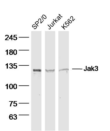
Sample:
SP2/0 Cell (Mouse) Lysate at 40 ug
Jurkat Cell (Human) Lysate at 40 ug
K562 Cell (Human) Lysate at 40 ug
Primary: Anti- Jak3 (bs-2808R) at 1/300 dilution
Secondary: IRDye800CW Goat Anti-Rabbit IgG at 1/20000 dilution
Predicted band size: 125 kD
Observed band size: 125 kD
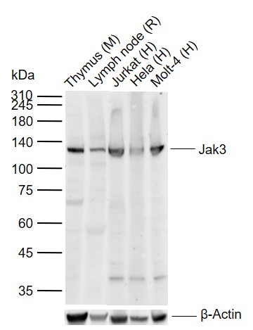
Sample:
Lane 1: Mouse Thymus tissue lysates
Lane 2: Rat Lymph node tissue lysates
Lane 3: Human Jurkat cell lysates
Lane 4: Human Hela cell lysates
Lane 5: Human Molt-4 cell lysates
Primary: Anti-Jak3 (bs-2808R) at 1/1000 dilution
Secondary: IRDye800CW Goat Anti-Rabbit IgG at 1/20000 dilution
Predicted band size: 125 kDa
Observed band size: 125 kDa
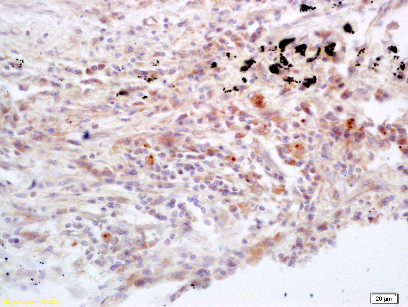
Tissue/cell: human lung carcinoma; 4% Paraformaldehyde-fixed and paraffin-embedded;
Antigen retrieval: citrate buffer ( 0.01M, pH 6.0 ), Boiling bathing for 15min; Block endogenous peroxidase by 3% Hydrogen peroxide for 30min; Blocking buffer (normal goat serum,C-0005) at 37℃ for 20 min;
Incubation: Anti-Jak3 Polyclonal Antibody, Unconjugated(bs-2808R) 1:200, overnight at 4°C, followed by conjugation to the secondary antibody(SP-0023) and DAB(C-0010) staining
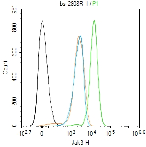
Blank control(black line):Molt4.
Primary Antibody (green line): Rabbit Anti-Jak3 antibody (bs-2808R)
Dilution:1ug/Test;
Secondary Antibody(white blue line): Goat anti-rabbit IgG-AF488
Dilution: 0.5ug/Test.
Isotype control(orange line): Normal Rabbit IgG
Protocol
The cells were fixed with 4% PFA (10min at room temperature)and then permeabilized with 90% ice-cold methanol for 20 min at -20℃, The cells were then incubated in 5%BSA to block non-specific protein-protein interactions for 30 min at room temperature .Cells stained with Primary Antibody for 30 min at room temperature. The secondary antibody used for 40 min at room temperature. Acquisition of 20,000 events was performed.
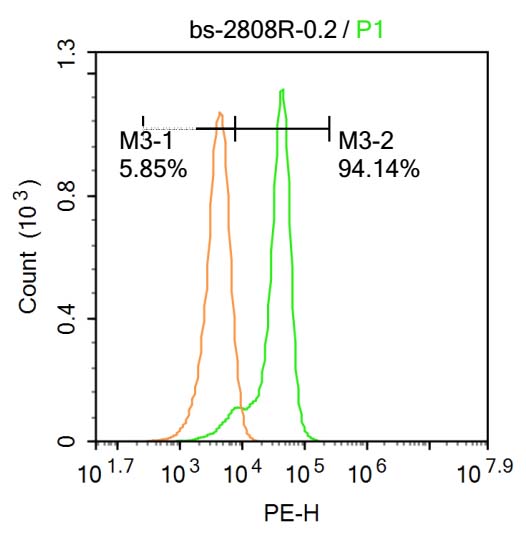
Blank control:Molt-4.
Primary Antibody (green line): Rabbit Anti-Jak3 antibody (bs-2808R)
Dilution: 0.2μg /10^6 cells;
Isotype Control Antibody (orange line): Rabbit IgG .
Secondary Antibody : Goat anti-rabbit IgG-PE
Dilution: 0.2μg /test.
Protocol
The cells were fixed with 4% PFA (10min at room temperature)and then permeabilized with 20% PBST for 20 min at-20℃. The cells were then incubated in 5%BSA to block non-specific protein-protein interactions for 30 min at at room temperature .Cells stained with Primary Antibody for 30 min at room temperature. The secondary antibody used for 40 min at room temperature. Acquisition of 20,000 events was performed.
|
| 1、抗体溶解方法 | |
| 2、抗体修复方式 | |
| 3、常用试剂的配制 | |
| 4、免疫组化操作步骤 | |
| 5、免疫组化问题解答 | |
| 6、Western Blotting 操作步骤 | |
| 7、Western Blotting 问题解答 | |
| 8、关于肽链的设计 | |
| 9、多肽的溶解与保存 | |
| 10、酶标抗体效价测定程序 | |


