

| 产品编号 | bs-3551R |
| 英文名称 | RIPK3 Rabbit pAb |
| 中文名称 | 受体结合丝氨酸苏氨酸激酶3抗体 |
| 别 名 | RIP3; RIPK3_HUMAN; RIPK3; RIP-like protein kinase 3; Receptor-interacting protein 3 (RIP-3); 2.7.11.1; RIPK3_MOUSE; Receptor-interacting protein 3 (RIP-3 | mRIP3); |
 |
Specific References (8) | bs-3551R has been referenced in 8 publications.
|
| 研究领域 | 肿瘤 细胞生物 免疫学 信号转导 细胞凋亡 转录调节因子 |
| 抗体来源 | Rabbit |
| 克隆类型 | Polyclonal |
| 克 隆 号 | |
| 交叉反应 | Human,Mouse,Rat (predicted: Rabbit,Pig,Cow,Dog,GuineaPig) |
| 产品应用 | WB=1:500-2000,IHC-P=1:100-500,IHC-F=1:100-500,IF=1:100-500
not yet tested in other applications. optimal dilutions/concentrations should be determined by the end user. |
| 理论分子量 | 57 kDa |
| 检测分子量 | 60 |
| 细胞定位 | 细胞浆 细胞膜 |
| 性 状 | Liquid |
| 浓 度 | 1mg/ml |
| 免 疫 原 | KLH conjugated synthetic peptide derived from human RIPK3: 101-230/518 |
| 亚 型 | IgG |
| 纯化方法 | affinity purified by Protein A |
| 缓 冲 液 | 0.01M TBS (pH7.4) with 1% BSA, 0.02% Proclin300 and 50% Glycerol. |
| 保存条件 | Shipped at 4℃. Store at -20℃ for one year. Avoid repeated freeze/thaw cycles. |
| 注意事项 | This product as supplied is intended for research use only, not for use in human, therapeutic or diagnostic applications. |
| PubMed | PubMed |
| 产品介绍 |
The product of this gene is a member of the receptor-interacting protein (RIP) family of serine/threonine protein kinases, and contains a C-terminal domain unique from other RIP family members. The encoded protein is predominantly localized to the cytoplasm, and can undergo nucleocytoplasmic shuttling dependent on novel nuclear localization and export signals. It is a component of the tumor necrosis factor (TNF) receptor-I signaling complex, and can induce apoptosis and weakly activate the NF-kappaB transcription factor. [provided by RefSeq, Jul 2008] Function: Essential for programmed necrosis in response to death-inducing TNF-alpha family members. Upon induction of necrosis, RIPK3 interacts with, and phosphorylates RIPK1 to form a necrosis-inducing complex. RIPK3 binds to and enhances the activity of three metabolic enzymes: GLUL, GLUD1, and PYGL. These metabolic enzymes may eventually stimulate the tricarboxylic acid cycle and oxidative phosphorylation, which could result in enhanced ROS production. Subunit: Interacts (via RIP homotypic interaction motif) with RIPK1 (via RIP homotypic interaction motif); this interaction induces RIPK1 phosphorylation and formation of a RIPK1-RIPK3 necrosis-inducing complex. Upon TNF-induced necrosis, the RIPK1-RIPK3 dimer further interacts with PGAM5 and MLKL; the formation of this complex leads to PGAM5 phosphorylation and increase in PGAM5 phosphatase activity. Binds TRAF2 and is recruited to the TNFR-1 signaling complex. Interacts with PYGL, GLUL and GLUD1; these interactions result in activation of these metabolic enzymes. Interacts with BIRC2/c-IAP1, BIRC3/c-IAP2 and XIAP/BIRC4. Subcellular Location: Cytoplasm. Cell membrane. Mitochondrion (Potential). Tissue Specificity: Highly expressed in the pancreas. Detected at lower levels in heart, placenta, lung and kidney. Isoform 3 is significantly increased in colon and lung cancers. Post-translational modifications: RIPK1 and RIPK3 undergo reciprocal auto- and trans-phosphorylation. Phosphorylation of Ser-199 plays a role in the necroptotic function of RIPK3. Polyubiquitinated with 'Lys-48' and 'Lys-63'-linked chains by BIRC2/c-IAP1 and BIRC3/c-IAP2, leading to activation of NF-kappa-B. Similarity: Belongs to the protein kinase superfamily. TKL Ser/Thr protein kinase family. Contains 1 protein kinase domain. SWISS: Q9Y572 Gene ID: 11035 Database links: Entrez Gene: 11035 Human Entrez Gene: 56532 Mouse Omim: 605817 Human SwissProt: Q9Y572 Human SwissProt: Q9QZL0 Mouse Unigene: 268551 Human Unigene: 46612 Mouse |
| 产品图片 |
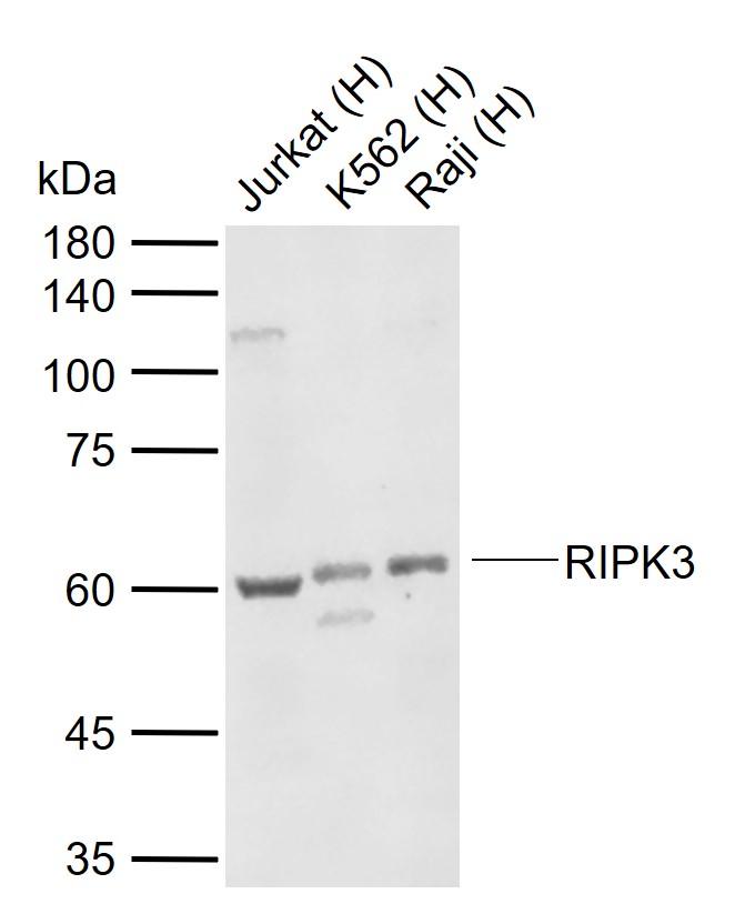
Sample:
Lane 1: Human Jurkat cell lysates
Lane 2: Human K562 cell lysates
Lane 3: Human Raji cell lysates
Primary: Anti-RIPK3 (bs-3551R) at 1/1000 dilution
Secondary: IRDye800CW Goat Anti-Rabbit IgG at 1/20000 dilution
Predicted band size: 57 kDa
Observed band size: 60 kDa
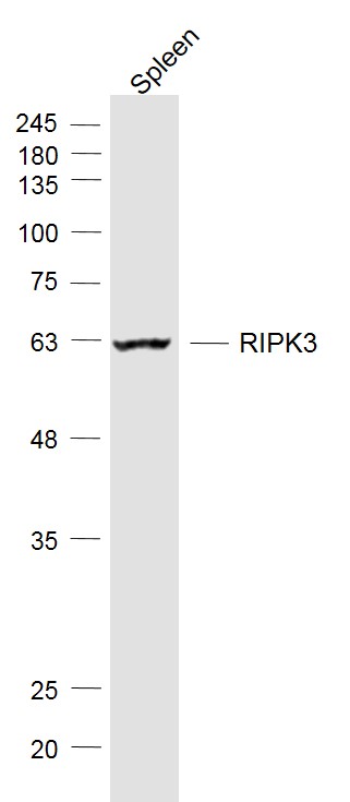
Sample:
Spleen (Mouse) Lysate at 40 ug
Primary: Anti-RIPK3 (bs-3551R) at 1/1000 dilution
Secondary: IRDye800CW Goat Anti-Rabbit IgG at 1/20000 dilution
Predicted band size: 57 kD
Observed band size: 60 kD
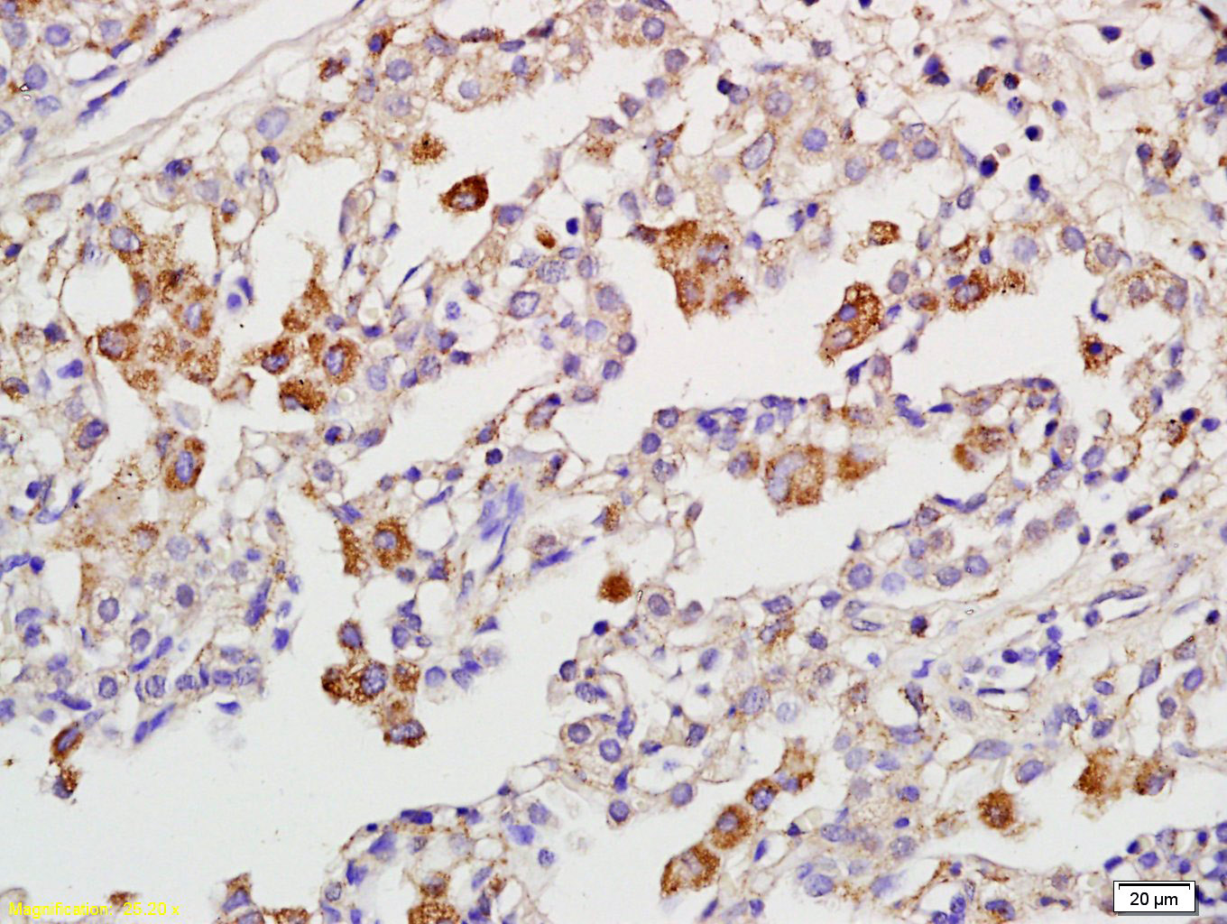
Tissue/cell: human pneumonia tissue; 4% Paraformaldehyde-fixed and paraffin-embedded;
Antigen retrieval: citrate buffer ( 0.01M, pH 6.0 ), Boiling bathing for 15min; Block endogenous peroxidase by 3% Hydrogen peroxide for 30min; Blocking buffer (normal goat serum,C-0005) at 37℃ for 20 min;
Incubation: Anti-RIPK3 Polyclonal Antibody, Unconjugated(bs-3551R) 1:200, overnight at 4°C, followed by conjugation to the secondary antibody(SP-0023) and DAB(C-0010) staining

Paraformaldehyde-fixed, paraffin embedded (Rat brain); Antigen retrieval by boiling in sodium citrate buffer (pH6.0) for 15min; Block endogenous peroxidase by 3% hydrogen peroxide for 20 minutes; Blocking buffer (normal goat serum) at 37°C for 30min; Antibody incubation with (RIPK3) Polyclonal Antibody, Unconjugated (bs-3551R) at 1:400 overnight at 4°C, followed by a conjugated secondary antibody (sp-0023) for 20 minutes and DAB staining.
|
| 1、抗体溶解方法 | |
| 2、抗体修复方式 | |
| 3、常用试剂的配制 | |
| 4、免疫组化操作步骤 | |
| 5、免疫组化问题解答 | |
| 6、Western Blotting 操作步骤 | |
| 7、Western Blotting 问题解答 | |
| 8、关于肽链的设计 | |
| 9、多肽的溶解与保存 | |
| 10、酶标抗体效价测定程序 | |


