

| 产品编号 | bs-3393R |
| 英文名称 | phospho-SIRT1 (Ser47) Rabbit pAb |
| 中文名称 | 磷酸化沉默调节蛋白1抗体 |
| 别 名 | SIRT1 (phospho-S47); p-SIRT1; phospho-SIRT1; SIRT1 (phospho-Ser47); SIR2; SIR2L1; SIR2alpha; Sir2a; SIR1_HUMAN; SIRT1; hSIRT1; NAD-dependent protein deacylase sirtuin-1; Regulatory protein SIR2 homolog 1; SIR2-like protein 1 (hSIR2); 2.3.1.286; SIR1_MOUSE |
 |
Specific References (7) | bs-3393R has been referenced in 7 publications.
|
| 产品类型 | 磷酸化抗体 |
| 研究领域 | 细胞生物 免疫学 染色质和核信号 微生物学 新陈代谢 表观遗传学 |
| 抗体来源 | Rabbit |
| 克隆类型 | Polyclonal |
| 克 隆 号 | |
| 交叉反应 | Human,Rat (predicted: Mouse,Rabbit,Pig,Dog) |
| 产品应用 | IHC-P=1:100-500,IHC-F=1:100-500,IF=1:100-500,Flow-Cyt=1ug/Test
not yet tested in other applications. optimal dilutions/concentrations should be determined by the end user. |
| 理论分子量 | 58/81 kDa |
| 细胞定位 | 细胞核 细胞浆 |
| 性 状 | Liquid |
| 浓 度 | 1mg/ml |
| 免 疫 原 | KLH conjugated Synthesised phosphopeptide derived from human SirT1 around the phosphorylation site of Ser47: R(p-S)PG |
| 亚 型 | IgG |
| 纯化方法 | affinity purified by Protein A |
| 缓 冲 液 | 0.01M TBS (pH7.4) with 1% BSA, 0.02% Proclin300 and 50% Glycerol. |
| 保存条件 | Shipped at 4℃. Store at -20℃ for one year. Avoid repeated freeze/thaw cycles. |
| 注意事项 | This product as supplied is intended for research use only, not for use in human, therapeutic or diagnostic applications. |
| PubMed | PubMed |
| 产品介绍 |
This gene encodes a member of the sirtuin family of proteins, homologs to the yeast Sir2 protein. Members of the sirtuin family are characterized by a sirtuin core domain and grouped into four classes. The functions of human sirtuins have not yet been determined; however, yeast sirtuin proteins are known to regulate epigenetic gene silencing and suppress recombination of rDNA. Studies suggest that the human sirtuins may function as intracellular regulatory proteins with mono-ADP-ribosyltransferase activity. The protein encoded by this gene is included in class I of the sirtuin family. Alternative splicing results in multiple transcript variants. Function: SirtT1 75 kDa fragment: catalytically inactive 75SirT1 may be involved in regulation of apoptosis. May be involved in protecting chondrocytes from apoptotic death by associating with cytochrome C and interfering with apoptosome assembly. Subunit: Found in a complex with PCAF and MYOD1 (By similarity). Component of the eNoSC complex, composed of SIRT1, SUV39H1 and RRP8. Interacts with HES1, HEY2 and PML. Interacts with RPS19BP1/AROS. Interacts with KIAA1967/DBC1 (via N-terminus); the interaction disrupts the interaction between SIRT1 and p53/TP53. Interacts with SETD7; the interaction induces the dissociation of SIRT1 from p53/TP53 and increases p53/TP53 activity. Interacts with MYCN, NR1I2, CREBZF, TSC2, TLE1, FOS, JUN, NR0B2, PPARG, NCOR, IRS1, IRS2 and NMNAT1. Interacts with HNF1A; the interaction occurs under nutrient restriction. Interacts with SUZ12; the interaction mediates the association with the PRC4 histone methylation complex which is specific as an association with PCR2 and PCR3 complex variants is not found. Interacts with HIV-1 tat. Subcellular Location: Nucleus, PML body. Cytoplasm. Note=Recruited to the nuclear bodies via its interaction with PML. Colocalized with APEX1 in the nucleus. May be found in nucleolus, nuclear euchromatin, heterochromatin and inner membrane. Shuttles between nucleus and cytoplasm. SirtT1 75 kDa fragment: Cytoplasm. Mitochondrion. Tissue Specificity: Widely expressed. Post-translational modifications: Methylated on multiple lysine residues; methylation is enhanced after DNA damage and is dispensable for deacetylase activity toward p53/TP53. Phosphorylated. Phosphorylated by STK4/MST1, resulting in inhibition of SIRT1-mediated p53/TP53 deacetylation. Phosphorylation by MAPK8/JNK1 at Ser-27, Ser-47, and Thr-530 leads to increased nuclear localization and enzymatic activity. Phosphorylation at Thr-530 by DYRK1A and DYRK3 acivates deacetylase activity and promotes cell survival. Phosphorylation by mammalian target of rapamycin complex 1 (mTORC1) at Ser-47 inhibits deacetylation activity. Phosphorylated by CaMK2, leading to increased p53/TP53 and NF-kappa-B p65/RELA deacetylation activity (By similarity). Phosphorylation at Ser-27 implicating MAPK9 is linked to protein stability. There is some ambiguity for some phosphosites: Ser-159/Ser-162 and Thr-544/Ser-545. Proteolytically cleaved by cathepsin B upon TNF-alpha treatment to yield catalytic inactive but stable SirtT1 75 kDa fragment (75SirT1). S-nitrosylated by GAPDH, leading to inhibit the NAD-dependent protein deacetylase activity (By similarity). Similarity: Belongs to the sirtuin family. Contains 1 deacetylase sirtuin-type domain. SWISS: Q96EB6 Gene ID: 23411 Database links: Entrez Gene: 23411 Human Entrez Gene: 93759 Mouse Omim: 604479 Human SwissProt: Q96EB6 Human SwissProt: Q923E4 Mouse Unigene: 369779 Human Unigene: 351459 Mouse 在Sirtuin蛋白家族中,sirtuin 1参与多种新陈代谢活动,包括DNA的自我保护和修复,抑制脂质过氧化积累,抑制其他细胞凋亡相关基因的表达以及和细胞寿命相关的活动。限制摄入的热量可以加强SIRT1的表达,从而延长了寿命。 |
| 产品图片 |
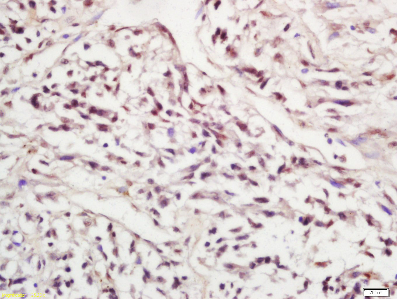
Paraformaldehyde-fixed, paraffin embedded (rat adrenal gland); Antigen retrieval by boiling in sodium citrate buffer (pH6.0) for 15min; Block endogenous peroxidase by 3% hydrogen peroxide for 20 minutes; Blocking buffer (normal goat serum) at 37°C for 30min; Antibody incubation with (Phospho-SIRT1 (Ser47)) Polyclonal Antibody, Unconjugated (bs-3393R) at 1:200 overnight at 4°C, followed by operating according to SP Kit(Rabbit) (sp-0023) instructionsand DAB staining.
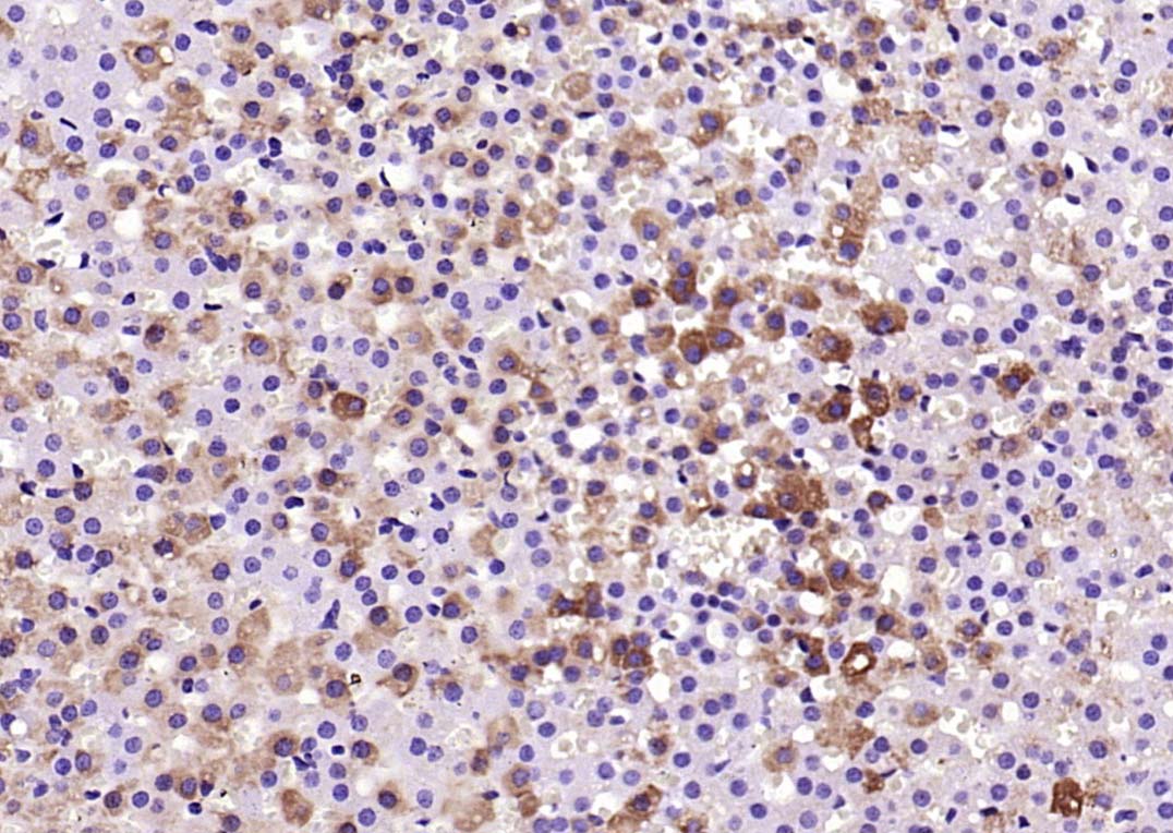
Tissue/cell: human lung cancer; 4% Paraformaldehyde-fixed and paraffin-embedded;
Antigen retrieval: citrate buffer ( 0.01M, pH 6.0 ), Boiling bathing for 15min; Block endogenous peroxidase by 3% Hydrogen peroxide for 30min; Blocking buffer (normal goat serum,C-0005) at 37℃ for 20 min;
Incubation: Anti-Phospho-SIRT1 (Ser47) Polyclonal Antibody, Unconjugated(bs-3393R) 1:200, overnight at 4°C, followed by conjugation to the secondary antibody(SP-0023) and DAB(C-0010) staining
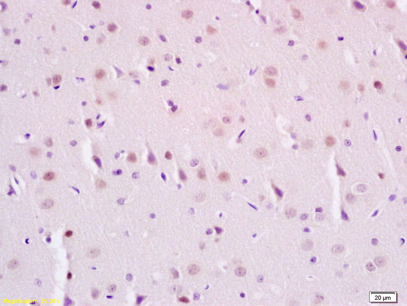
Tissue/cell: rat brain tissue; 4% Paraformaldehyde-fixed and paraffin-embedded;
Antigen retrieval: citrate buffer ( 0.01M, pH 6.0 ), Boiling bathing for 15min; Block endogenous peroxidase by 3% Hydrogen peroxide for 30min; Blocking buffer (normal goat serum,C-0005) at 37℃ for 20 min;
Incubation: Anti-Phospho-SIRT1(Ser47) Polyclonal Antibody, Unconjugated(bs-3393R) 1:200, overnight at 4°C, followed by conjugation to the secondary antibody(SP-0023) and DAB(C-0010) staining
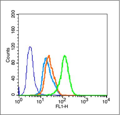
Blank control (blue line): MCF 7 (fixed with 70% methanol (Overnight at 4℃) and then permeabilized with 90% ice-cold methanol for 30 min on ice).
Primary Antibody (green line): Rabbit Anti-Phospho-SIRT1(Ser47) antibody (bs-3393R),Dilution: 3μg /10^5 cells.
Isotype Control Antibody (orange line): Rabbit IgG .
Secondary Antibody (white blue line): Goat anti-rabbit IgG-FITC,Dilution: 1μg /test.
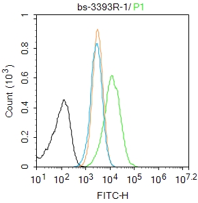
Blank control:HL-60.
Primary Antibody (green line): Rabbit Anti-Phospho-SIRT1 (Ser47) antibody (bs-3393R)
Dilution: 1μg /10^6 cells;
Isotype Control Antibody (orange line): Rabbit IgG .
Secondary Antibody : Goat anti-rabbit IgG-AF488
Dilution: 1μg /test.
Protocol
The cells were fixed with 4% PFA (10min at room temperature)and then permeabilized with 0.1% PBST for 20 min at room temperature. The cells were then incubated in 5%BSA to block non-specific protein-protein interactions for 30 min at room temperature .Cells stained with Primary Antibody for 30 min at room temperature. The secondary antibody used for 40 min at room temperature. Acquisition of 20,000 events was performed.
|
| 1、抗体溶解方法 | |
| 2、抗体修复方式 | |
| 3、常用试剂的配制 | |
| 4、免疫组化操作步骤 | |
| 5、免疫组化问题解答 | |
| 6、Western Blotting 操作步骤 | |
| 7、Western Blotting 问题解答 | |
| 8、关于肽链的设计 | |
| 9、多肽的溶解与保存 | |
| 10、酶标抗体效价测定程序 | |


