

| 产品编号 | bs-3438R |
| 英文名称 | phospho-TAK1 (Thr187) Rabbit pAb |
| 中文名称 | 磷酸化转化生长因子β活化激酶1 |
| 别 名 | MAP3K7 | TAK1 (phospho-T187); p-TAK1; phospho-TAK1; p-MAP3K7; MAP3K7 | TAK1 (phospho-Thr187); CSCF; FMD2; MEKK7; TAK1; TGF1a; B430101B05; M3K7_HUMAN; MAP3K7; Transforming growth factor-beta-activated kinase 1 (TGF-beta-activated kinase 1); 2.7.11.25; M3K7 |
 |
Specific References (3) | bs-3438R has been referenced in 3 publications.
[IF=3.53] Dvashi Z, Goldberg M, Adir O, Shapira M, Pollack A (2015) TGF-β1 Induced Transdifferentiation of RPE Cells is Mediated by TAK1. PLoS ONE 10(4): e0122229. Human.
[IF=3.067] Zhou G et al. TGF-β1 alleviates HgCl2 induced apoptosis via P38 MAPK signaling pathway in human trophoblast cells. Toxicol In Vitro. 2019 Aug 13;61:104626. ICF ; Human.
[IF=0.76] Dvashi, Z., et al. "Aberrant Activity of TAK1 is Associated with Retinal Pathology." Journal of Cytology and Histology 5 (2016): 2. IHC-P ; Human.
|
| 产品类型 | 磷酸化抗体 |
| 研究领域 | 肿瘤 免疫学 信号转导 细胞凋亡 转录调节因子 激酶和磷酸酶 |
| 抗体来源 | Rabbit |
| 克隆类型 | Polyclonal |
| 克 隆 号 | |
| 交叉反应 | Human,Mouse,Rat (predicted: Rabbit,Pig,Cow,Chicken,Horse) |
| 产品应用 | WB=1:500-2000,IHC-P=1:100-500,IHC-F=1:100-500,IF=1:100-500,Flow-Cyt=1μg/Test
not yet tested in other applications. optimal dilutions/concentrations should be determined by the end user. |
| 理论分子量 | 67 kDa |
| 检测分子量 | 72-75 |
| 细胞定位 | 细胞浆 细胞膜 |
| 性 状 | Liquid |
| 浓 度 | 1mg/ml |
| 免 疫 原 | KLH conjugated Synthesised phosphopeptide derived from human TAK1 around the phosphorylation site of Thr187: HM(p-T)NN |
| 亚 型 | IgG |
| 纯化方法 | affinity purified by Protein A |
| 缓 冲 液 | 0.01M TBS (pH7.4) with 1% BSA, 0.02% Proclin300 and 50% Glycerol. |
| 保存条件 | Shipped at 4℃. Store at -20℃ for one year. Avoid repeated freeze/thaw cycles. |
| 注意事项 | This product as supplied is intended for research use only, not for use in human, therapeutic or diagnostic applications. |
| PubMed | PubMed |
| 产品介绍 |
The protein encoded by this gene is a member of the serine/threonine protein kinase family. This kinase mediates the signaling transduction induced by TGF beta and morphogenetic protein (BMP), and controls a variety of cell functions including transcription regulation and apoptosis. In response to IL-1, this protein forms a kinase complex including TRAF6, MAP3K7P1/TAB1 and MAP3K7P2/TAB2; this complex is required for the activation of nuclear factor kappa B. This kinase can also activate MAPK8/JNK, MAP2K4/MKK4, and thus plays a role in the cell response to environmental stresses. Four alternatively spliced transcript variants encoding distinct isoforms have been reported. [provided by RefSeq, Jul 2008] Function: Component of a protein kinase signal transduction cascade. Mediator of TRAF6 and TGF-beta signal transduction. Activates IKBKB and MAPK8 in response to TRAF6 signaling. Stimulates NF-kappa-B activation and the p38 MAPK pathway. In osmotic stress signaling, plays a major role in the activation of MAPK8/JNK, but not that of NF-kappa-B. Subunit: Binds both upstream activators and downstream substrates in multimolecular complexes. Interacts with TAB1/MAP3K7IP1 and TAB2/MAP3K7IP2. Identified in the TRIKA2 complex composed of MAP3K7, TAB1/MAP3K7IP1 and TAB2/MAP3K7IP2. Interacts with PPM1L. Interaction with PP2A and PPP6C leads to its repressed activity. Interacts with TRAF6 and TAB1/MAP3K7IP1; during IL-1 signaling. Interacts with TAOK1 and TAOK2; interaction with TAOK2 interferEs with MAP3K7 interaction with IKKA, thus preventing NF-kappa-B activation. Interacts with WDR34 (via WD domains). Interacts with RBCK1. Interacts with CYLD. Subcellular Location: Cytoplasm. Cell membrane; Peripheral membrane protein; Cytoplasmic side. Note=Although the majority of MAP3K7/TAK1 is found in the cytosol, when complexed with TAB1/MAP3K7IP1 and TAB2/MAP3K7IP2, it is also localized at the cell membrane. Tissue Specificity: Isoform 1A is the most abundant in ovary, skeletal muscle, spleen and blood mononuclear cells. Isoform 1B is highly expressed in brain, kidney and small intestine. Isoform 1C is the major form in prostate. Isoform 1D is the less abundant form. Post-translational modifications: Association with TAB1/MAP3K7IP1 promotes autophosphorylation and subsequent activation. Association with TAB2/MAP3K7IP2, itself associated with free unanchored Lys-63 polyubiquitin chain, promotes autophosphorylation and subsequent activation of MAP3K7. Dephosphorylation at Thr-187 by PP2A and PPP6C leads to inactivation. Ubiquitinated, leading to proteasomal degradation. Requires 'Lys-63'-linked polyubiquitination for autophosphorylation and subsequent activation. 'Lys-63'-linked ubiquitination does not lead to proteasomal degradation. Deubiquitinated by CYLD, a protease that selectively cleaves 'Lys-63'-linked ubiquitin chains. Similarity: Belongs to the protein kinase superfamily. STE Ser/Thr protein kinase family. MAP kinase kinase kinase subfamily. Contains 1 protein kinase domain. SWISS: O43318 Gene ID: 6885 Database links: Entrez Gene: 6885 Human Entrez Gene: 26409 Mouse Omim: 602614 Human SwissProt: O43318 Human SwissProt: Q62073 Mouse Unigene: 722892 Human Unigene: 258589 Mouse Unigene: 24019 Rat |
| 产品图片 |
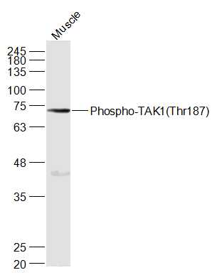
Sample:
Muscle (Mouse) Lysate at 40 ug
Primary: Anti-Phospho-TAK1(Thr187) (bs-3438R) at 1/1000 dilution
Secondary: IRDye800CW Goat Anti-Rabbit IgG at 1/20000 dilution
Predicted band size: 67 kD
Observed band size: 72 kD
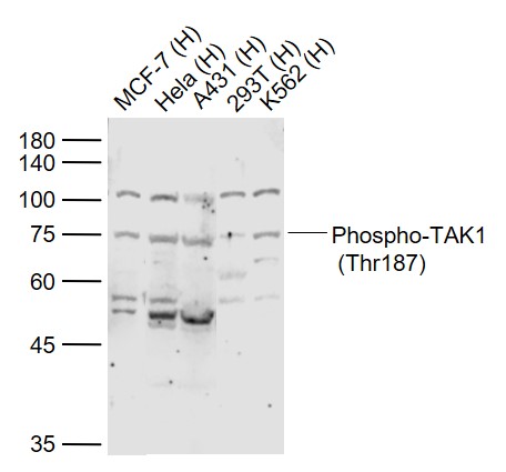
Sample:
Lane 1: MCF-7 (Human) Cell Lysate at 30 ug
Lane 2: Hela (Human) Cell Lysate at 30 ug
Lane 3: A431 (Human) Cell Lysate at 30 ug
Lane 4: 293T (Human) Cell Lysate at 30 ug
Lane 5: K562 (Human) Cell Lysate at 30 ug
Primary:
Anti-Phospho-TAK1 (Thr187) (bs-3438R) at 1/1000 dilution
Secondary: IRDye800CW Goat Anti-Rabbit IgG at 1/20000 dilution
Predicted band size: 67 kD
Observed band size: 75 kD
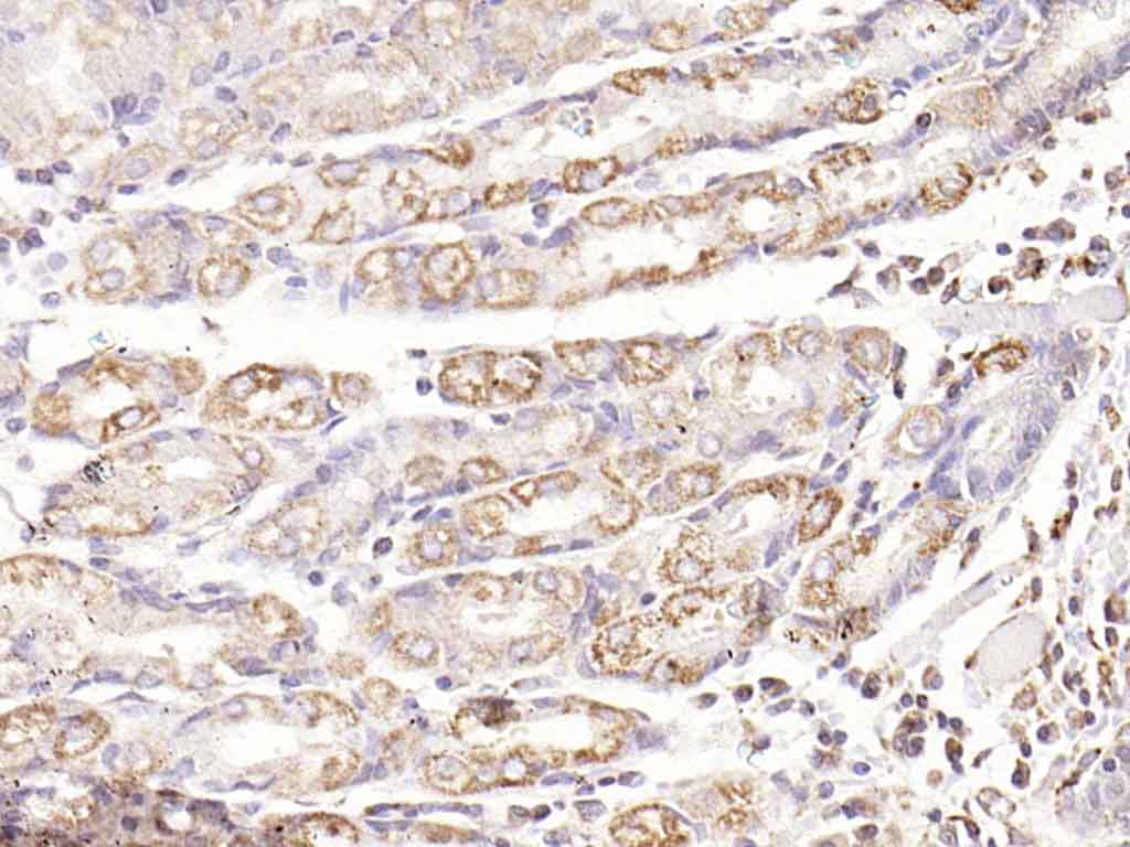
Paraformaldehyde-fixed, paraffin embedded (human gastric carcinoma); Antigen retrieval by boiling in sodium citrate buffer (pH6.0) for 15min; Block endogenous peroxidase by 3% hydrogen peroxide for 20 minutes; Blocking buffer (normal goat serum) at 37°C for 30min; Antibody incubation with (Phospho-TAK1 (Thr187)) Polyclonal Antibody, Unconjugated (bs-3438R) at 1:200 overnight at 4°C, followed by operating according to SP Kit(Rabbit) (sp-0023) instructionsand DAB staining.
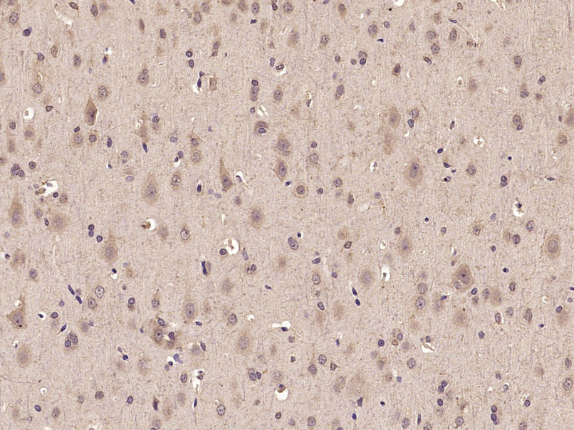
Paraformaldehyde-fixed, paraffin embedded (Rat brain); Antigen retrieval by microwave in sodium citrate buffer (pH6.0) ; Block endogenous peroxidase by 3% hydrogen peroxide for 30 minutes; Blocking buffer (3% BSA) at RT for 30min; Antibody incubation with (Phospho-TAK1(Thr187)) Polyclonal Antibody, Unconjugated (bs-3438R) at 1:400 overnight at 4℃, followed by conjugation to the secondary antibody (labeled with HRP)and DAB staining.
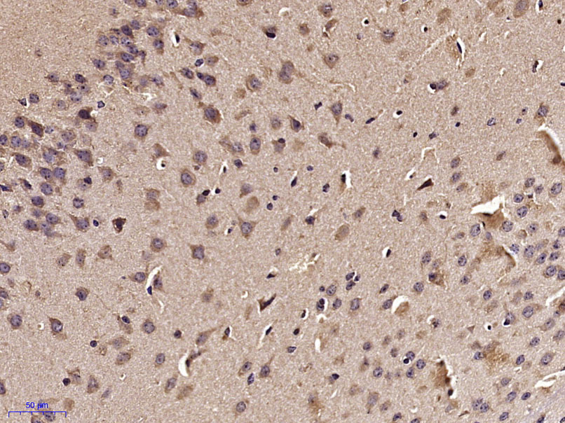
Paraformaldehyde-fixed, paraffin embedded (Mouse brain); Antigen retrieval by microwave in sodium citrate buffer (pH6.0) ; Block endogenous peroxidase by 3% hydrogen peroxide for 30 minutes; Blocking buffer (3% BSA) at RT for 30min; Antibody incubation with (Phospho-TAK1(Thr187)) Polyclonal Antibody, Unconjugated (bs-3438R) at 1:400 overnight at 4℃, followed by conjugation to the secondary antibody (labeled with HRP)and DAB staining.
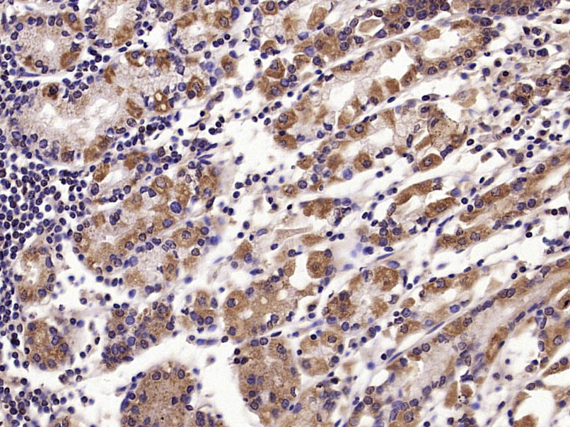
Paraformaldehyde-fixed, paraffin embedded (Human stomach); Antigen retrieval by microwave in sodium citrate buffer (pH6.0) ; Block endogenous peroxidase by 3% hydrogen peroxide for 30 minutes; Blocking buffer (3% BSA) at RT for 30min; Antibody incubation with (Phospho-TAK1(Thr187)) Polyclonal Antibody, Unconjugated (bs-3438R) at 1:400 overnight at 4℃, followed by conjugation to the secondary antibody (labeled with HRP)and DAB staining.
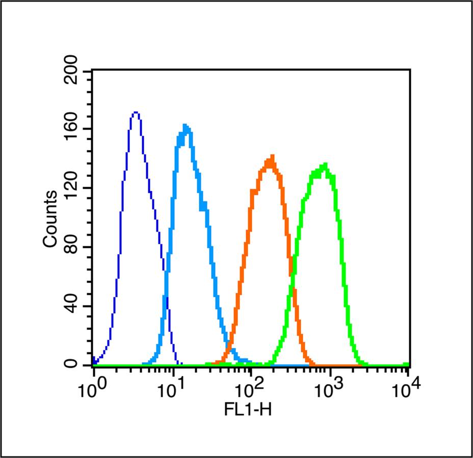
Blank control (Black line): Raji (Black).
Primary Antibody (green line): Rabbit Anti-Phospho-TAK1(Thr187) antibody (bs-3438R)
Dilution: 1μg /10^6 cells;
Isotype Control Antibody (orange line): Rabbit IgG .
Secondary Antibody (white blue line): Goat anti-rabbit IgG-PE
Dilution: 1μg /test.
Protocol
The cells were fixed with 70% ice-cold methanol overnight at 4℃ and then permeabilized with 0.1% PBS-Tween for 20 min at room temperature (The cells were fixed with 2% paraformaldehyde (10 min) , then permeabilized with 90% ice-cold methanol for 20 min on ice.). Cells stained with Primary Antibody for 30 min at room temperature. The cells were then incubated in 1 X PBS/2%BSA/10% goat serum to block non-specific protein-protein interactions followed by the antibody for 15 min at room temperature. The secondary antibody used for 40 min at room temperature. Acquisition of 20,000 events was performed.
|
| 1、抗体溶解方法 | |
| 2、抗体修复方式 | |
| 3、常用试剂的配制 | |
| 4、免疫组化操作步骤 | |
| 5、免疫组化问题解答 | |
| 6、Western Blotting 操作步骤 | |
| 7、Western Blotting 问题解答 | |
| 8、关于肽链的设计 | |
| 9、多肽的溶解与保存 | |
| 10、酶标抗体效价测定程序 | |


