

| 产品编号 | bs-3464R |
| 英文名称 | phospho-ATG1 (Ser556) Rabbit pAb |
| 中文名称 | 磷酸化自噬相关蛋白1抗体 |
| 别 名 | ULK1 | ATG1 (phospho-S556); p-ATG1; phospho-ATG1; p-ULK1; ULK1 | ATG1 (phospho-Ser556); ATG1; ATG1A; UNC51; Unc51.1; hATG1; mKIAA0722; ULK1_HUMAN; ULK1; Autophagy-related protein 1 homolog (ATG1 | hATG1); Unc-51-like kinase 1; 2.7.11.1; KIAA0722; ULK1_MOU |
 |
Specific References (3) | bs-3464R has been referenced in 3 publications.
[IF=7.675] Ramona D’Amico. et al. Complex Interplay between Autophagy and Oxidative Stress in the Development of Endometriosis. ANTIOXIDANTS-BASEL. 2022 Dec;11(12):2484 WB ; Rat.
[IF=4.571] Peng Lin. et al. Polystyrene nanoplastics exacerbate lipopolysaccharide-induced myocardial fibrosis and autophagy in mice via ROS/TGF-β1/Smad. TOXICOLOGY. 2022 Oct;480:153338 WB ; Mouse.
[IF=3.322] Chunli Yang. et al. BMAL1 involved in autophagy and injury of thoracic aortic endothelial cells of rats induced by intermittent heat stress through the AMPK/mTOR/ULK1 pathway. BIOCHEM BIOPH RES CO. 2023 Jun;661:34 WB ; Rat.
|
| 产品类型 | 磷酸化抗体 |
| 研究领域 | 细胞生物 免疫学 信号转导 细胞凋亡 转录调节因子 激酶和磷酸酶 |
| 抗体来源 | Rabbit |
| 克隆类型 | Polyclonal |
| 克 隆 号 | |
| 交叉反应 | Human,Rat,Rabbit (predicted: Mouse,Pig,Horse) |
| 产品应用 | IHC-P=1:100-500,IHC-F=1:100-500,IF=1:100-500,Flow-Cyt=3ug/test,ICC/IF=1:100-500,ELISA=1:5000-10000
not yet tested in other applications. optimal dilutions/concentrations should be determined by the end user. |
| 理论分子量 | 116 kDa |
| 细胞定位 | 细胞浆 |
| 性 状 | Liquid |
| 浓 度 | 1mg/ml |
| 免 疫 原 | KLH conjugated Synthesised phosphopeptide derived from human ATG1/ULK1 around the phosphorylation site of Ser556: LH(p-S)AP |
| 亚 型 | IgG |
| 纯化方法 | affinity purified by Protein A |
| 缓 冲 液 | 0.01M TBS (pH7.4) with 1% BSA, 0.02% Proclin300 and 50% Glycerol. |
| 保存条件 | Shipped at 4℃. Store at -20℃ for one year. Avoid repeated freeze/thaw cycles. |
| 注意事项 | This product as supplied is intended for research use only, not for use in human, therapeutic or diagnostic applications. |
| PubMed | PubMed |
| 产品介绍 |
ULK1 belongs to the serine/threonine protein kinase family. It is involved in axon growth and plays an essential role in neurite branching during sensory axon outgrowth. Knockdown of ULK1 results in impaired endocytosis of nerve growth factor (NGF), excessive axon arborization, and severely stunted axon elongation indicating that ULK1 mediates a non clathrin coated endocytosis in sensory growth cones. Knockdown of ULK1 also inhibits the autophagic response. It appears to act as a convergence point for multiple signals that regulate autophagy, and in turn interacts with a large number of autophagy related (Atg) proteins. Function: Serine/threonine-protein kinase involved in autophagy in response to starvation. Acts upstream of phosphatidylinositol 3-kinase PIK3C3 to regulate the formation of autophagophores, the precursors of autophagosomes. Part of regulatory feedback loops in autophagy: acts both as a downstream effector and negative regulator of mammalian target of rapamycin complex 1 (mTORC1) via interaction with RPTOR. Activated via phosphorylation by AMPK and also acts as a regulator of AMPK by mediating phosphorylation of AMPK subunits PRKAA1, PRKAB2 and PRKAG1, leading to negatively regulate AMPK activity. May phosphorylate ATG13/KIAA0652 and RPTOR; however such data need additional evidences. Plays a role early in neuronal differentiation and is required for granule cell axon formation. Subunit: Interacts with GABARAP and GABARAPL2. Interacts (via C-terminus) with ATG13/KIAA0652. Part of a complex consisting of ATG13/KIAA0652, ULK1 and RB1CC1. Associates with the mammalian target of rapamycin complex 1 (mTORC1) through an interaction with RPTOR; the association depends on nutrient conditions and is reduced during starvation. Subcellular Location: Cytoplasm, cytosol. Preautophagosomal structure. Note=Under starvation conditions, is localized to puncate structures primarily representing the isolation membrane that sequesters a portion of the cytoplasm resulting in the formation of an autophagosome. Tissue Specificity: Ubiquitously expressed. Detected in the following adult tissues: skeletal muscle, heart, pancreas, brain, placenta, liver, kidney, and lung. Post-translational modifications: Autophosphorylated. Phosphorylated under nutrient-rich conditions; dephosphorylated during starvation or following treatment with rapamycin. Under nutrient sufficiency phosphorylated by MTOR/mTOR, disrupting the interaction with AMPK and preventing activation of ULK1 (By similarity). In response to nutrient limitation, phosphorylated and activated by AMPK, leading to activate autophagy. Similarity: Belongs to the protein kinase superfamily. Ser/Thr protein kinase family. APG1/unc-51/ULK1 subfamily. Contains 1 protein kinase domain. SWISS: O75385 Gene ID: 8408 Database links: Entrez Gene: 8408 Human Entrez Gene: 22241 Mouse Omim: 603168 Human SwissProt: O75385 Human SwissProt: O70405 Mouse Unigene: 47061 Human Unigene: 271898 Mouse Unigene: 24509 Rat Atg1是一种丝氨酸/苏氨酸蛋白激酶。Atg1的激脢活性是CVT信号传导通路以及細胞自噬所必須的。Atg1可以和一些执行细胞自噬的蛋白相互作用,而且有很多调控細胞自噬的信号传导通路汇集在Atg1。因此Atg1可能是一个可以调控細胞自噬很多步驟的一个调节关键点。但在较高等的真核生物中,Atg1的角色仍然不是很清楚。通过目前的研究已经比较了解细胞自噬可以导致细胞的死亡,但是如何导致死亡的分子机制还不清楚,有待于进一步研究。 |
| 产品图片 |
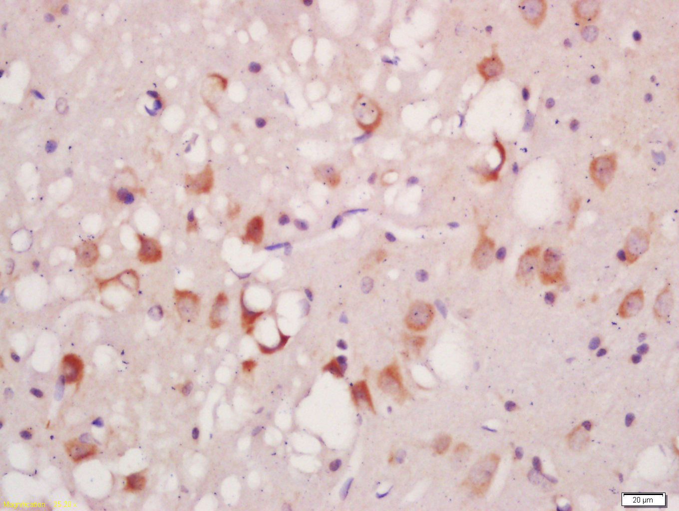
Tissue/cell: rat brain tissue; 4% Paraformaldehyde-fixed and paraffin-embedded;
Antigen retrieval: citrate buffer ( 0.01M, pH 6.0 ), Boiling bathing for 15min; Block endogenous peroxidase by 3% Hydrogen peroxide for 30min; Blocking buffer (normal goat serum,C-0005) at 37℃ for 20 min;
Incubation: Anti-Phospho-ATG1(Ser556) Polyclonal Antibody, Unconjugated(bs-3464R) 1:200, overnight at 4°C, followed by conjugation to the secondary antibody(SP-0023) and DAB(C-0010) staining
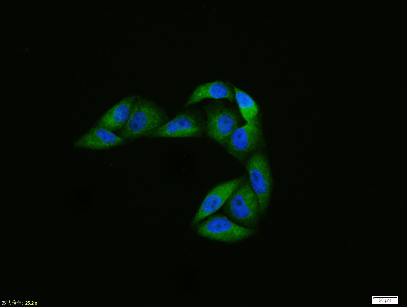
Hela cell; 4% Paraformaldehyde-fixed; Triton X-100 at room temperature for 20 min; Blocking buffer (normal goat serum, C-0005) at 37°C for 20 min; Antibody incubation with (Phospho-ATG1 (Ser556)) polyclonal Antibody, Unconjugated (bs-3464R) 1:100, 90 minutes at 37°C; followed by a conjugated Goat Anti-Rabbit IgG antibody at 37°C for 90 minutes, DAPI (blue, C02-04002) was used to stain the cell nuclei.
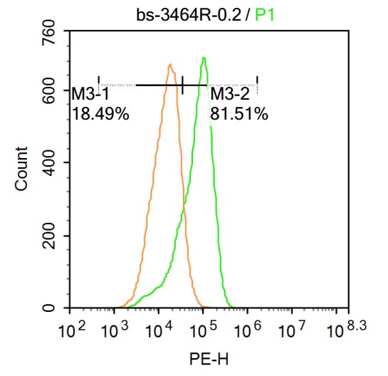
Blank control:A549.
Primary Antibody (green line): Rabbit Anti-Phospho-ATG1(Ser556) antibody (bs-3464R)
Dilution: 1μg /10^6 cells;
Isotype Control Antibody (orange line): Rabbit IgG .
Secondary Antibody : Goat anti-rabbit IgG-PE
Dilution: 3μg /test.
Protocol
The cells were fixed with 4% PFA (10min at room temperature)and then permeabilized with 20% PBST for 20 min at room temperature. The cells were then incubated in 5% BSA to block non-specific protein-protein interactions for 30 min at at room temperature .Cells stained with Primary Antibody for 30 min at room temperature. The secondary antibody used for 40 min at room temperature. Acquisition of 20,000 events was performed.
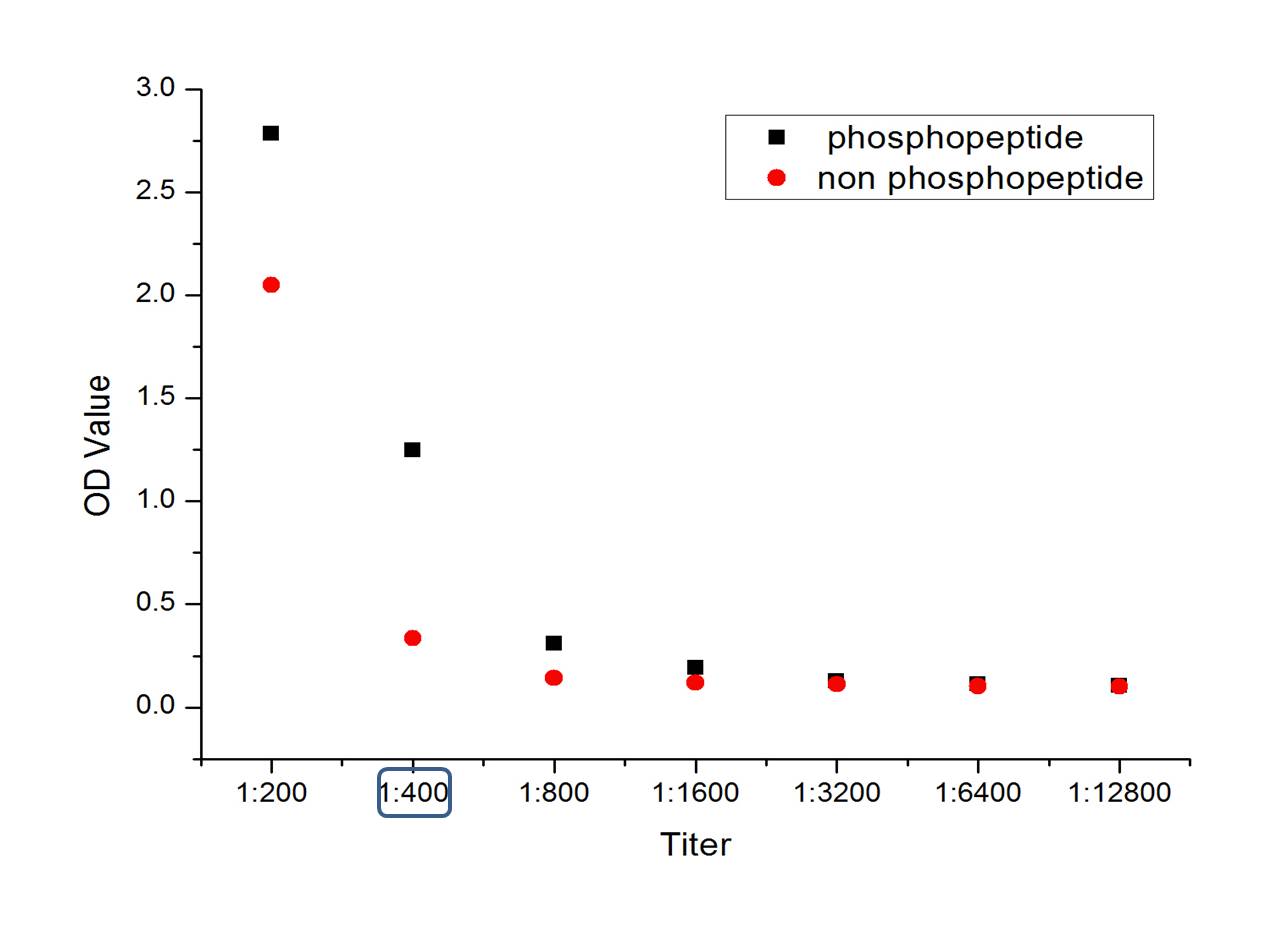
phosphopeptide
non phosphopeptide
|
| 1、抗体溶解方法 | |
| 2、抗体修复方式 | |
| 3、常用试剂的配制 | |
| 4、免疫组化操作步骤 | |
| 5、免疫组化问题解答 | |
| 6、Western Blotting 操作步骤 | |
| 7、Western Blotting 问题解答 | |
| 8、关于肽链的设计 | |
| 9、多肽的溶解与保存 | |
| 10、酶标抗体效价测定程序 | |


