

| 产品编号 | bs-3633R |
| 英文名称 | Synaptopodin Rabbit pAb |
| 中文名称 | 突触足蛋白抗体 |
| 别 名 | SYNPO1; 9030217H17Rik; 9130229N11; 9330140I15Rik; SYNPO_HUMAN; SYNPO; KIAA1029; SYNPO_MOUSE; SYNPO_RAT; |
 |
Specific References (2) | bs-3633R has been referenced in 2 publications.
[IF=3.73] Zhang, Xueming, et al. "Resolvin D1 Protects Podocytes in Adriamycin-Induced Nephropathy through Modulation of 14-3-3β Acetylation." PLOS ONE 8.6 (2013): e67471. IP ; Mouse.
[IF=0] Garovic, Vesna D., and Muthuvel Jayachandran. "DETECTING PODOCYTE INJURY IN DIABETIC NEPHROPATHY AND GLOMERULONEPHRITIS." U.S. Patent No. 20,170,003,299. 5 Jan. 2017. Human.
|
| 研究领域 | 肿瘤 细胞生物 免疫学 神经生物学 信号转导 细胞凋亡 转录调节因子 |
| 抗体来源 | Rabbit |
| 克隆类型 | Polyclonal |
| 克 隆 号 | |
| 交叉反应 | Human,Mouse,Rat |
| 产品应用 | WB=1:500-2000,IHC-P=1:100-500,IHC-F=1:100-500,IF=1:100-500,Flow-Cyt=1μg /test
not yet tested in other applications. optimal dilutions/concentrations should be determined by the end user. |
| 理论分子量 | 102 kDa |
| 检测分子量 | 76/102 |
| 细胞定位 | 细胞浆 细胞膜 |
| 性 状 | Liquid |
| 浓 度 | 1mg/ml |
| 免 疫 原 | KLH conjugated synthetic peptide derived from human Synaptopodin: 601-700/903 |
| 亚 型 | IgG |
| 纯化方法 | affinity purified by Protein A |
| 缓 冲 液 | 0.01M TBS (pH7.4) with 1% BSA, 0.02% Proclin300 and 50% Glycerol. |
| 保存条件 | Shipped at 4℃. Store at -20℃ for one year. Avoid repeated freeze/thaw cycles. |
| 注意事项 | This product as supplied is intended for research use only, not for use in human, therapeutic or diagnostic applications. |
| PubMed | PubMed |
| 产品介绍 |
Synaptopodin is an actin-associated protein that may play a role in actin-based cell shape and motility. May be essential for the formation of spine apparatuses in spines of telencephalic neurons, involved in synaptic plasticity. The name synaptopodin derives from the protein's associations with postsynaptic densities and dendritic spines and with renal podocytes. Subunit: Interacts with BAIAP1. Interacts with actin (By similarity). Interacts (via PPxY motifs) with WWC1 (via WW domains). Subcellular Location: Cytoplasm, cytoskeleton (By similarity). Cell junction, tight junction (By similarity). Perikaryon (By similarity). Cell projection, dendritic spine (By similarity). Cell junction, synapse, postsynaptic cell membrane, postsynaptic density (By similarity). Cell junction, synapse (By similarity). Note=Localized at the tight junction of cells. In brain, localized to the postsynaptic densities and in the perikarya. Asssociated with dendritic spines of a subset of synapses (By similarity). Tissue Specificity: Expressed in brain, namely in the olfactory bulb, cerebral cortex, striatum, and hippocampus, but not in the cerebellum. Also expressed in the podocytes of kidney glomeruli. In the hippocampus, mainly expressed in the principal cell layer of the dentate gyrus and Ammon's horn. Similarity: Belongs to the synaptopodin family. SWISS: Q8N3V7 Gene ID: 11346 Database links: Entrez Gene: 11346 Human Entrez Gene: 104027 Mouse Omim: 608155 Human SwissProt: Q8N3V7 Human SwissProt: Q8CC35 Mouse Unigene: 435228 Human synaptopodin是一种与肌动蛋白微丝偶联的蛋白,其富含脯氨酸,为线状蛋白质,在动物体内表达于肾小球的足细胞和后脑的突触内,synaptopodin被认为是足细胞特异的分化成熟标志物。 |
| 产品图片 |
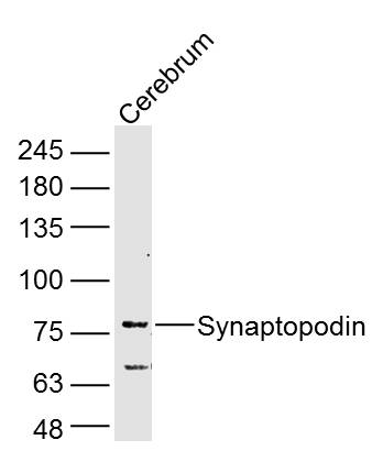
Sample: Cerebrum (Rat) Lysate at 40 ug
Primary: Anti-Synaptopodin (bs-3633R) at 1/300 dilution
Secondary: IRDye800CW Goat Anti-Rabbit IgG at 1/20000 dilution
Predicted band size: 102 kD
Observed band size: 76 kD
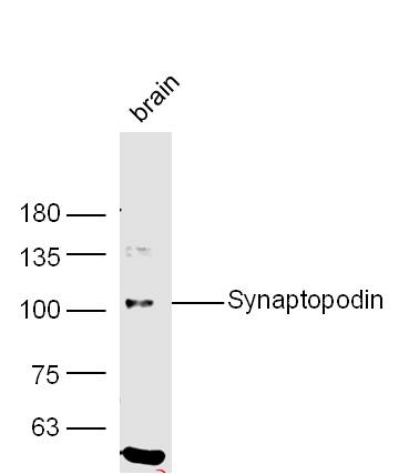
Sample: Brain (Mouse) Lysate at 40 ug
Primary: Anti-Synaptopodin (bs-3633R) at 1/300 dilution
Secondary: IRDye800CW Goat Anti-Rabbit IgG at 1/20000 dilution
Predicted band size: 102 kD
Observed band size: 102 kD
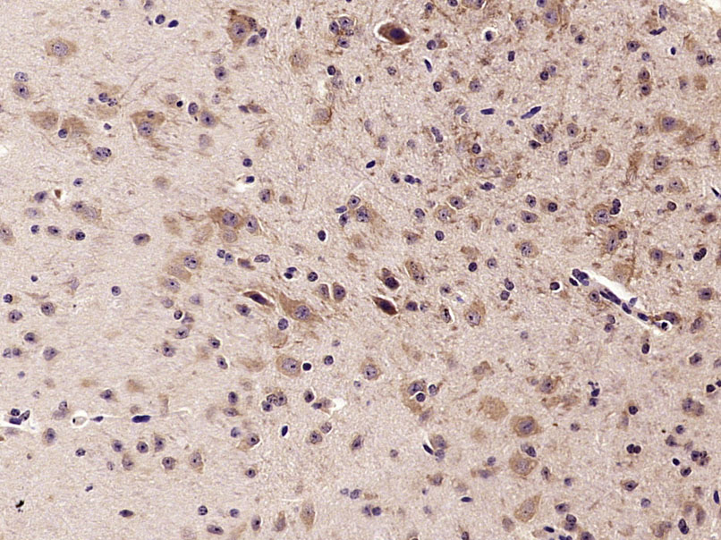
Paraformaldehyde-fixed, paraffin embedded (Mouse brain); Antigen retrieval by microwave in sodium citrate buffer (pH6.0) ; Block endogenous peroxidase by 3% hydrogen peroxide for 30 minutes; Blocking buffer (3% BSA) at RT for 30min; Antibody incubation with (Synaptopodin) Polyclonal Antibody, Unconjugated (bs-3633R) at 1:400 overnight at 4°C, followed by conjugation to the secondary antibody (labeled with HRP)and DAB staining.
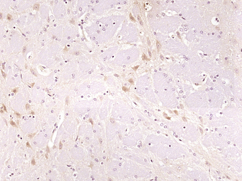
Paraformaldehyde-fixed, paraffin embedded (Rat brain); Antigen retrieval by boiling in sodium citrate buffer (pH6.0) for 15min; Block endogenous peroxidase by 3% hydrogen peroxide for 20 minutes; Blocking buffer (normal goat serum) at 37°C for 30min; Antibody incubation with (Synaptopodin) Polyclonal Antibody, Unconjugated (bs-3633R) at 1:400 overnight at 4°C, followed by operating according to SP Kit(Rabbit) (sp-0023) instructionsand DAB staining.
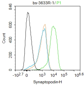
Blank control(black line):SH-SY5Y.
Primary Antibody (green line): Rabbit Anti-Synaptopodin antibody (bs-3633R)
Dilution:1ug/Test;
Secondary Antibody(white blue line): Goat anti-rabbit IgG-AF488
Dilution: 0.5ug/Test.
Isotype control(orange line): Normal Rabbit IgG
Protocol
The cells were fixed with 4% PFA (10min at room temperature)and then permeabilized with 90% ice-cold methanol for 20 min at -20℃, The cells were then incubated in 5%BSA to block non-specific protein-protein interactions for 30 min at room temperature .Cells stained with Primary Antibody for 30 min at room temperature. The secondary antibody used for 40 min at room temperature. Acquisition of 20,000 events was performed.
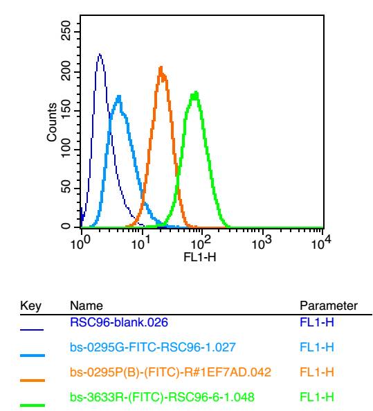
Positive control: RSC96
Isotype Control Antibody: Rabbit IgG;
Secondary Antibody: Goat anti-rabbit IgG-FITC;
Dilution: 1:200 in 1 X PBS containing 0.5% BSA
Primary Antibody catalog number: bs-3633R;
Dilution: 1μg in 100 μl 1X PBS containing 0.5% BSA
|
| 1、抗体溶解方法 | |
| 2、抗体修复方式 | |
| 3、常用试剂的配制 | |
| 4、免疫组化操作步骤 | |
| 5、免疫组化问题解答 | |
| 6、Western Blotting 操作步骤 | |
| 7、Western Blotting 问题解答 | |
| 8、关于肽链的设计 | |
| 9、多肽的溶解与保存 | |
| 10、酶标抗体效价测定程序 | |


