

| 产品编号 | bs-1497R |
| 英文名称 | MUC1 Rabbit pAb |
| 中文名称 | 粘蛋白-1抗体 |
| 别 名 | ADMCKD; ADMCKD1; ADTKD2; CA 15-3; CD227; Ca15-3; EMA; H23AG; KL-6; MAM6; MCD; MCKD; MCKD1; MUC-1; MUC-1/SEC; MUC-1/X; MUC1/ZD; PEM; PEMT; PUM; MUC1_HUMAN; MUC1; Breast carcinoma-associated antigen DF3; Cancer antigen 15-3 (CA 15-3); Carcinoma-associated m |
 |
Specific References (3) | bs-1497R has been referenced in 3 publications.
[IF=8.46] Taki, K., et al. "GNASR201H and KrasG12D cooperate to promote murine pancreatic tumorigenesis recapitulating human intraductal papillary mucinous neoplasm." Oncogene 35.18 (2016): 2407-2412. IHC-P ; Mouse.
[IF=3.101] Cao Y et al. Detection of three tumor biomarkers in human lung cancer serum using single particle ICP-MS combined with magnetic immunoassay. Spectrochimica Acta Part B: Atomic Spectroscopy, 105797 (2020). Sandwich-type magnetic immunoassay ;
[IF=2.32] Chen ZZ et al. An improved class of fluorescent silica nanoparticles for indirect immunofluorescence detection of MCF-7 cells. Optical Materials 88 (2019) 147–154. ICF ; Human.
|
| 研究领域 | 肿瘤 免疫学 |
| 抗体来源 | Rabbit |
| 克隆类型 | Polyclonal |
| 克 隆 号 | |
| 交叉反应 | Human,Mouse,Rat |
| 产品应用 | IHC-P=1:100-500,IHC-F=1:100-500,IF=1:100-500,ICC/IF=1:100-500
not yet tested in other applications. optimal dilutions/concentrations should be determined by the end user. |
| 理论分子量 | 20/138 kDa |
| 细胞定位 | 细胞核 细胞浆 细胞膜 分泌型蛋白 |
| 性 状 | Liquid |
| 浓 度 | 1mg/ml |
| 免 疫 原 | KLH conjugated synthetic peptide derived from human MUC1: 1101-1255/1255 <Cytoplasmic> |
| 亚 型 | IgG2b |
| 纯化方法 | affinity purified by Protein A |
| 缓 冲 液 | 0.01M TBS (pH7.4) with 1% BSA, 0.02% Proclin300 and 50% Glycerol. |
| 保存条件 | Shipped at 4℃. Store at -20℃ for one year. Avoid repeated freeze/thaw cycles. |
| 注意事项 | This product as supplied is intended for research use only, not for use in human, therapeutic or diagnostic applications. |
| PubMed | PubMed |
| 产品介绍 |
MUC1 is a large cell surface mucin glycoprotein expressed by most glandular and ductal epithelial cells and some hematopoietic cell lineages. It is expressed on most secretory epithelium, including mammary gland and some hematopoietic cells. It is expressed abundantly in lactating mammary glands and overexpressed abundantly in >90% breast carcinomas and metastases. Transgenic MUC1 has been shown to associate with all four cebB receptors and localize with erbB1 (EGFR) in lactating glands. The MUC1 gene contains seven exons and produces several different alternatively spliced variants. The major expressed form of MUC1 uses all seven exons and is a type 1 transmembrane protein with a large extracellular tandem repeat domain. The tandem repeat domain is highly O glycosylated and alterations in glycosylation have been shown in epithelial cancer cells. Function: The alpha subunit has cell adhesive properties. Can act both as an adhesion and an anti-adhesion protein. May provide a protective layer on epithelial cells against bacterial and enzyme attack. The beta subunit contains a C-terminal domain which is involved in cell signaling, through phosphorylations and protein-protein interactions. Modulates signaling in ERK, SRC and NF-kappa-B pathways. In activated T-cells, influences directly or indirectly the Ras/MAPK pathway. Promotes tumor progression. Regulates TP53-mediated transcription and determines cell fate in the genotoxic stress response. Binds, together with KLF4, the PE21 promoter element of TP53 and represses TP53 activity. Subunit: The alpha subunit forms a tight, non-covalent heterodimeric complex with the proteolytically-released beta-subunit. Subcellular Location: Apical cell membrane; Single-pass type I membrane protein. Isoform 5: Secreted. Isoform 7: Secreted. Isoform 9: Secreted. Mucin-1 subunit beta: Cell membrane. Cytoplasm. Nucleus. Tissue Specificity: Expressed on the apical surface of epithelial cells, especially of airway passages, breast and uterus. Also expressed in activated and unactivated T-cells. Overexpressed in epithelial tumors, such as breast or ovarian cancer and also in non-epithelial tumor cells. Isoform 7 is expressed in tumor cells only. Post-translational modifications: Highly glycosylated (N- and O-linked carbohydrates and sialic acid). O-glycosylated to a varying degree on serine and threonine residues within each tandem repeat, ranging from mono- to penta-glycosylation. The average density ranges from about 50% in human milk to over 90% in T47D breast cancer cells. Further sialylation occurs during recycling. Membrane-shed glycoproteins from kidney and breast cancer cells have preferentially sialyated core 1 structures, while secreted forms from the same tissues display mainly core 2 structures. The O-glycosylated content is overlapping in both these tissues with terminal fucose and galactose, 2- and 3-linked galactose, 3- and 3,6-linked GalNAc-ol and 4-linked GlcNAc predominating. Differentially O-glycosylated in breast carcinomas with 3,4-linked GlcNAc. N-glycosylation consists of high-mannose, acidic complex-type and hybrid glycans in the secreted form MUC1/SEC, and neutral complex-type in the transmembrane form, MUC1/TM. Proteolytic cleavage in the SEA domain occurs in the endoplasmic reticulum by an autoproteolytic mechanism and requires the full-length SEA domain as well as requiring a Ser, Thr or Cys residue at the P + 1 site. Cleavage at this site also occurs on isoform MUC1/X but not on isoform MUC1/Y. Ectodomain shedding is mediated by ADAM17. Dual palmitoylation on cysteine residues in the CQC motif is required for recycling from endosomes back to the plasma membrane. Phosphorylated on tyrosines and serine residues in the C-terminal. Phosphorylation on tyrosines in the C-terminal increases the nuclear location of MUC1 and beta-catenin. Phosphorylation by PKC delta induces binding of MUC1 to beta-catenin/CTNNB1 and thus decreases the formation of the beta-catenin/E-cadherin complex. Src-mediated phosphorylation inhibits interaction with GSK3B. Src-and EGFR-mediated phosphorylation on Tyr-1229 increases binding to beta-catenin/CTNNB1. GSK3B-mediated phosphorylation on Ser-1227 decreases this interaction but restores the formation of the beta-cadherin/E-cadherin complex. On T-cell receptor activation, phosphorylated by LCK. PDGFR-mediated phosphorylation increases nuclear colocalization of MUC1CT and CTNNB1. The N-terminal sequence has been shown to begin at position 24 or 28 (PubMed:11341784). Similarity: Contains 1 SEA domain. SWISS: P15941 Gene ID: 4582 Database links: Entrez Gene: 4582 Human Entrez Gene: 17829 Mouse Omim: 158340 Human SwissProt: P15941 Human SwissProt: Q02496 Mouse Unigene: 89603 Human Unigene: 16193 Mouse Unigene: 10779 Rat CA153为高分子糖蛋白,是乳腺癌标志物之一,其表达的量与乳腺癌的分化程度及雌激素受体高低有关联。近年来,很多学者研究认为:在人的很多种恶性肿瘤中都有CA15-3的不同表达。 |
| 产品图片 |
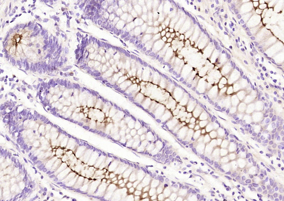
Paraformaldehyde-fixed, paraffin embedded (human colon carcinoma); Antigen retrieval by boiling in sodium citrate buffer (pH6.0) for 15min; Block endogenous peroxidase by 3% hydrogen peroxide for 20 minutes; Blocking buffer (normal goat serum) at 37°C for 30min; Antibody incubation with (MUC1) Polyclonal Antibody, Unconjugated (bs-1497R) at 1:200 overnight at 4°C, followed by operating according to SP Kit(Rabbit) (sp-0023) instructionsand DAB staining.
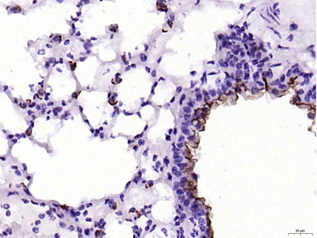
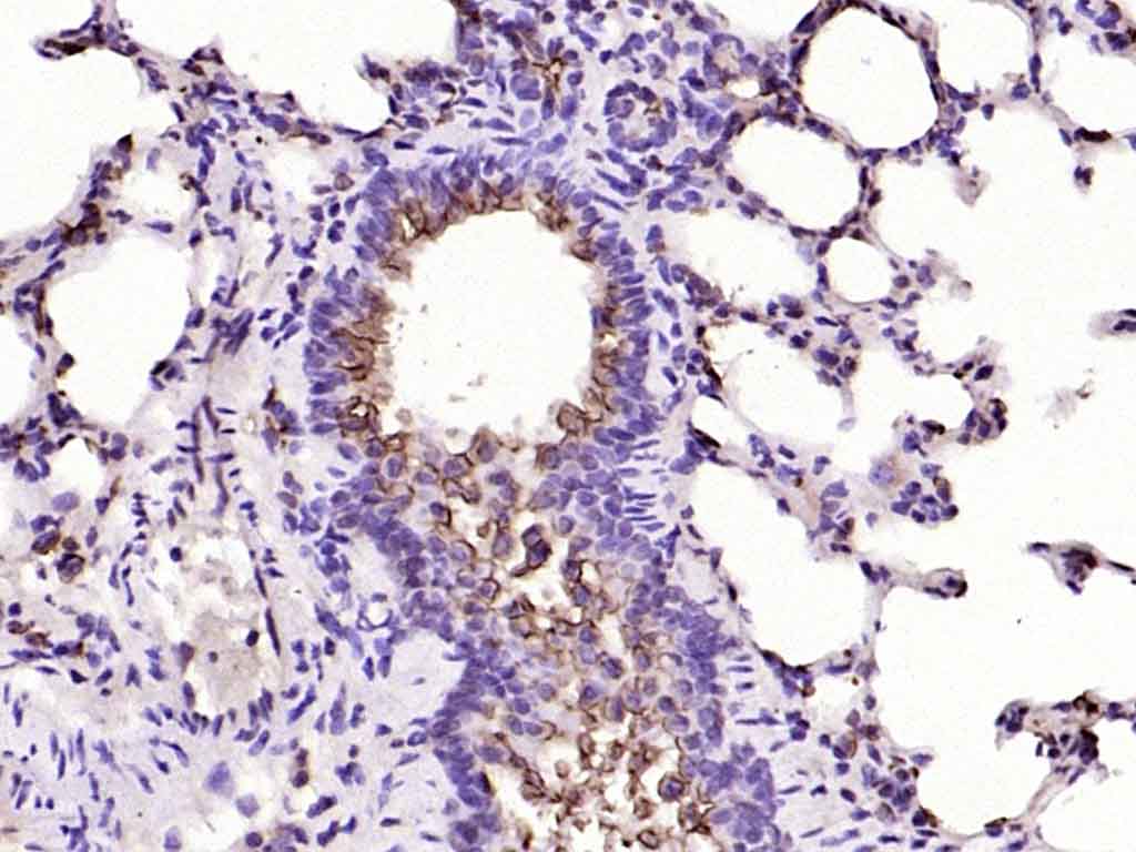
Paraformaldehyde-fixed, paraffin embedded (mouse lung); Antigen retrieval by boiling in sodium citrate buffer (pH6.0) for 15min; Block endogenous peroxidase by 3% hydrogen peroxide for 20 minutes; Blocking buffer (normal goat serum) at 37°C for 30min; Antibody incubation with (MUC1) Polyclonal Antibody, Unconjugated (bs-1497R) at 1:200 overnight at 4°C, followed by operating according to SP Kit(Rabbit) (sp-0023) instructionsand DAB staining.
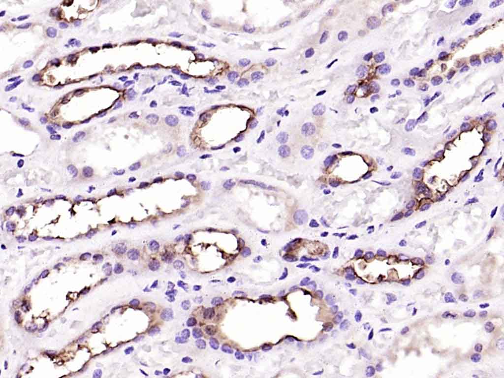
Paraformaldehyde-fixed, paraffin embedded (Human kidney); Antigen retrieval by boiling in sodium citrate buffer (pH6.0) for 15min; Block endogenous peroxidase by 3% hydrogen peroxide for 20 minutes; Blocking buffer (normal goat serum) at 37°C for 30min; Antibody incubation with (MUC1) Polyclonal Antibody, Unconjugated (bs-1497R) at 1:200 overnight at 4°C, followed by operating according to SP Kit(Rabbit) (sp-0023) instructionsand DAB staining.
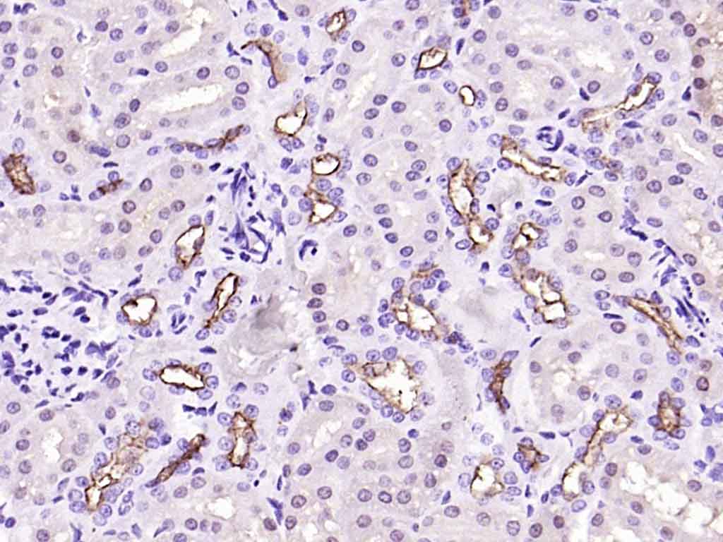
Paraformaldehyde-fixed, paraffin embedded (rat kidney); Antigen retrieval by boiling in sodium citrate buffer (pH6.0) for 15min; Block endogenous peroxidase by 3% hydrogen peroxide for 20 minutes; Blocking buffer (normal goat serum) at 37°C for 30min; Antibody incubation with (MUC1) Polyclonal Antibody, Unconjugated (bs-1497R) at 1:200 overnight at 4°C, followed by operating according to SP Kit(Rabbit) (sp-0023) instructionsand DAB staining.
Paraformaldehyde-fixed, paraffin embedded (rat lung); Antigen retrieval by boiling in sodium citrate buffer (pH6.0) for 15min; Block endogenous peroxidase by 3% hydrogen peroxide for 20 minutes; Blocking buffer (normal goat serum) at 37°C for 30min; Antibody incubation with (MUC1) Polyclonal Antibody, Unconjugated (bs-1497R) at 1:200 overnight at 4°C, followed by operating according to SP Kit(Rabbit) (sp-0023) instructionsand DAB staining.
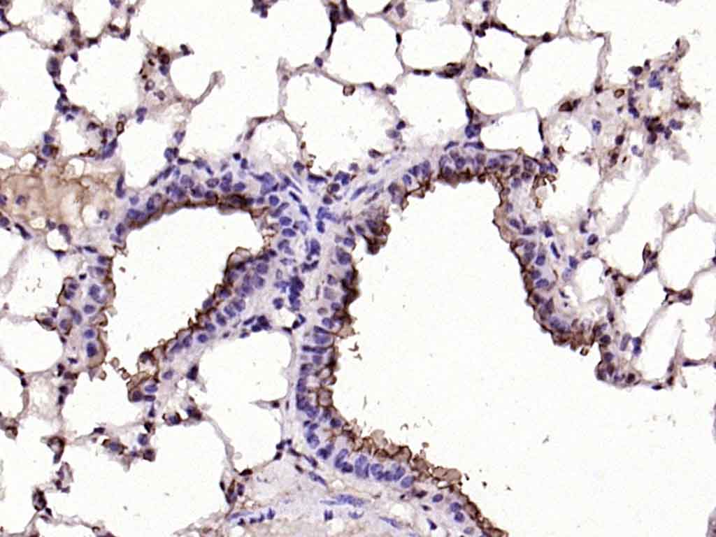
Paraformaldehyde-fixed, paraffin embedded (mouse lung); Antigen retrieval by boiling in sodium citrate buffer (pH6.0) for 15min; Block endogenous peroxidase by 3% hydrogen peroxide for 20 minutes; Blocking buffer (normal goat serum) at 37°C for 30min; Antibody incubation with (MUC1) Polyclonal Antibody, Unconjugated (bs-1497R) at 1:200 overnight at 4°C, followed by operating according to SP Kit(Rabbit) (sp-0023) instructionsand DAB staining.
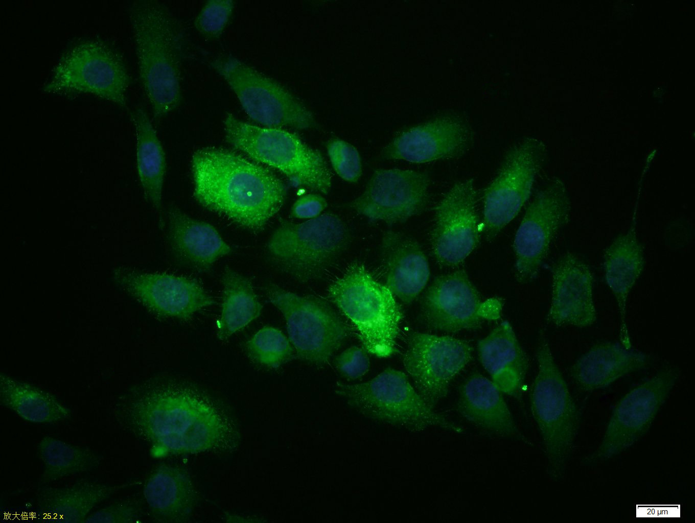
Tissue/cell: MCF-7 cell; 4% Paraformaldehyde-fixed; Triton X-100 at room temperature for 20 min; Blocking buffer (normal goat serum, C-0005) at 37°C for 20 min; Antibody incubation with (MUC1) Polyclonal Antibody, Unconjugated (bs-1497R) 1:100, 90 minutes at 37°C; followed by a conjugated Goat Anti-Rabbit IgG antibody (bs-0295G-FITC) at 37°C for 90 minutes, DAPI (5ug/ml, blue, C-0033) was used to stain the cell nuclei.
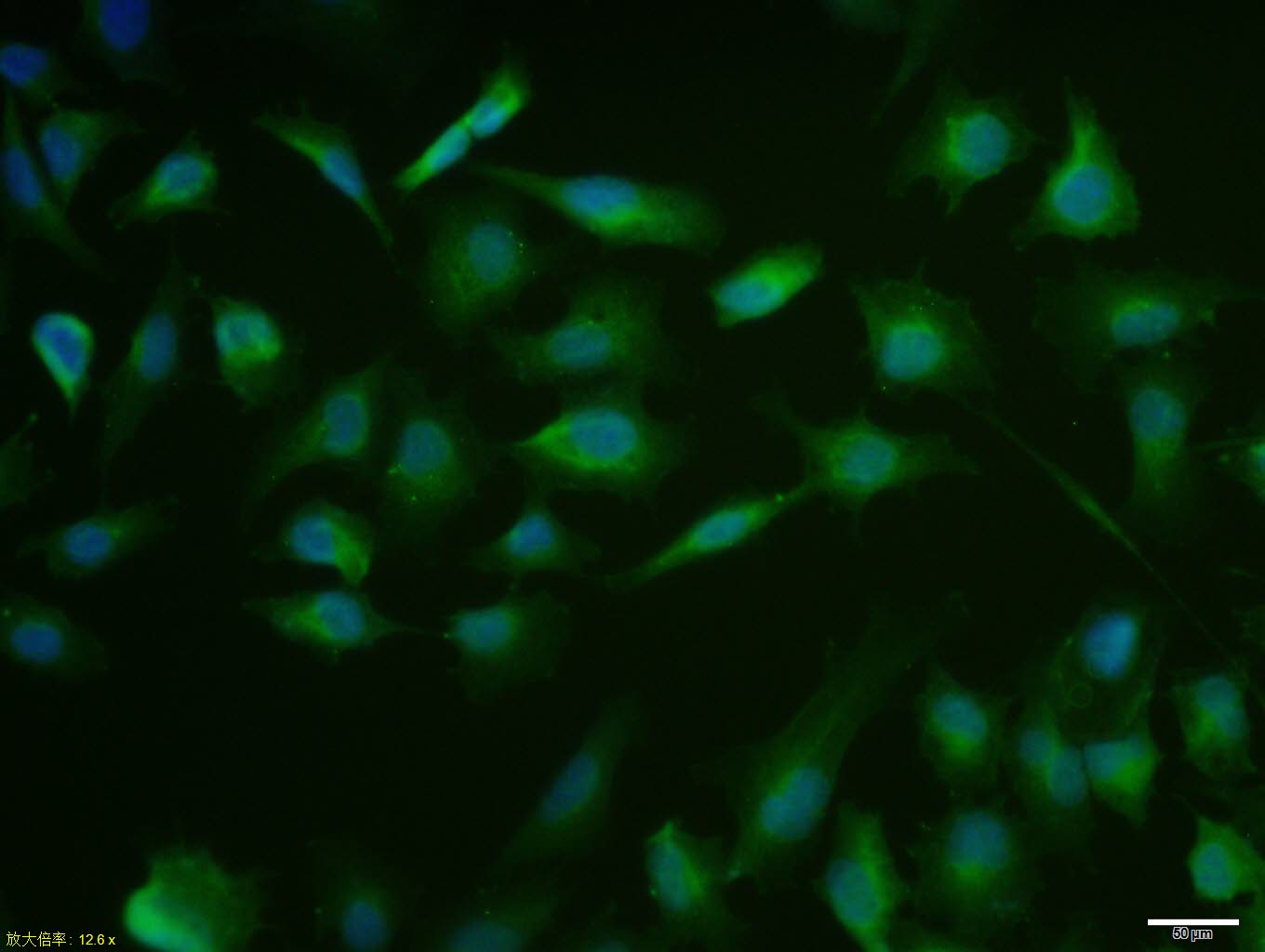
MCF7 cell; 4% Paraformaldehyde-fixed; Triton X-100 at room temperature for 20 min; Blocking buffer (normal goat serum, C-0005) at 37°C for 20 min; Antibody incubation with (MUC1) polyclonal Antibody, Unconjugated (bs-1497R) 1:100, 90 minutes at 37°C; followed by a conjugated Goat Anti-Rabbit IgG antibody at 37°C for 90 minutes, DAPI (blue, C02-04002) was used to stain the cell nuclei.
|
| 1、抗体溶解方法 | |
| 2、抗体修复方式 | |
| 3、常用试剂的配制 | |
| 4、免疫组化操作步骤 | |
| 5、免疫组化问题解答 | |
| 6、Western Blotting 操作步骤 | |
| 7、Western Blotting 问题解答 | |
| 8、关于肽链的设计 | |
| 9、多肽的溶解与保存 | |
| 10、酶标抗体效价测定程序 | |


