

| 产品编号 | bs-5039R |
| 英文名称 | Apolipoprotein E3 Rabbit pAb |
| 中文名称 | 载脂蛋白E3抗体 |
| 别 名 | AD2; APO-E; ApoE4; LDLCQ5; LPG; APOE_HUMAN; APOE; APOE_MOUSE; apolipoprotein E; Alzheimer disease 2 (APOE*E4-associated, late onset) |
 |
Specific References (1) | bs-5039R has been referenced in 1 publications.
[IF=5.108] Yuan C et al. OAB-14, a bexarotene derivative, improves Alzheimer's disease-related pathologies and cognitive impairments by increasing β-amyloid clearance in APP/PS1 mice.(2019) Biochim Biophys Acta Mol Basis Dis.Jan;1865(1):161-180. WB ; Mouse.
|
| 研究领域 | 肿瘤 细胞生物 免疫学 神经生物学 信号转导 细胞凋亡 转录调节因子 |
| 抗体来源 | Rabbit |
| 克隆类型 | Polyclonal |
| 交叉反应 | Human,Mouse,Rat (predicted: Pig,Cow) |
| 产品应用 | WB=1:500-2000,IHC-P=1:100-500,IHC-F=1:100-500,IF=1:100-500
not yet tested in other applications. optimal dilutions/concentrations should be determined by the end user. |
| 理论分子量 | 34kDa |
| 检测分子量 | 35 |
| 细胞定位 | 分泌型蛋白 |
| 性 状 | Liquid |
| 浓 度 | 1mg/ml |
| 免 疫 原 | KLH conjugated synthetic peptide derived from human APOE3: 101-180/191 |
| 亚 型 | IgG |
| 纯化方法 | affinity purified by Protein A |
| 缓 冲 液 | 0.01M TBS (pH7.4) with 1% BSA, 0.02% Proclin300 and 50% Glycerol. |
| 保存条件 | Shipped at 4℃. Store at -20℃ for one year. Avoid repeated freeze/thaw cycles. |
| 注意事项 | This product as supplied is intended for research use only, not for use in human, therapeutic or diagnostic applications. |
| PubMed | PubMed |
| 产品介绍 |
Apolipoprotein E, a main apoprotein of the chylomicron, binds to a specific receptor on liver cells and peripheral cells and is essential for the normal catabolism of triglyceride-rich lipoprotein constituents.
ApoE exists in three major isoforms; E2, E3, and E4, which differ from one another by a single amino-acid substitution. Compared with E3 and E4, E2 exhibits the lowest receptor binding affinity. Defects in ApoE are a cause of hyperlipoproteinemia type III due to increased plasma cholesterol and triglycerides levels which are the consequence of impaired clearance of chylomicron and VLDL remnants. Summary: Chylomicron remnants and very low density lipoprotein (VLDL) remnants are rapidly removed from the circulation by receptor-mediated endocytosis in the liver. Apolipoprotein E, a main apoprotein of the chylomicron, binds to a specific receptor on liver cells and peripheral cells. ApoE is essential for the normal catabolism of triglyceride-rich lipoprotein constituents. The APOE gene is mapped to chromosome 19 in a cluster with APOC1 and APOC2. Defects in apolipoprotein E result in familial dysbetalipoproteinemia, or type III hyperlipoproteinemia (HLP III), in which increased plasma cholesterol and triglycerides are the consequence of impaired clearance of chylomicron and VLDL remnants. [provided by RefSeq, Jul 2008]. Function: Mediates the binding, internalization, and catabolism of lipoprotein particles. It can serve as a ligand for the LDL (apo B/E) receptor and for the specific apo-E receptor (chylomicron remnant) of hepatic tissues. Subcellular Location: Secreted. Tissue Specificity: Occurs in all lipoprotein fractions in plasma. It constitutes 10-20% of very low density lipoproteins (VLDL) and 1-2% of high density lipoproteins (HDL). APOE is produced in most organs. Significant quantities are produced in liver, brain, spleen, lung, adrenal, ovary, kidney and muscle. Post-translational modifications: Synthesized with the sialic acid attached by O-glycosidic linkage and is subsequently desialylated in plasma. O-glycosylated with core 1 or possibly core 8 glycans. Thr-307 is a minor glycosylation site compared to Ser-308. Glycated in plasma VLDL of normal subjects, and of hyperglycemic diabetic patients at a higher level (2-3 fold). Phosphorylation sites are present in the extracellular medium. DISEASE: Defects in APOE are a cause of hyperlipoproteinemia type 3 (HLPP3) [MIM:107741]; also known as familial dysbetalipoproteinemia. Individuals with HLPP3 are clinically characterized by xanthomas, yellowish lipid deposits in the palmar crease, or less specific on tendons and on elbows. The disorder rarely manifests before the third decade in men. In women, it is usually expressed only after the menopause. The vast majority of the patients are homozygous for APOE*2 alleles. More severe cases of HLPP3 have also been observed in individuals heterozygous for rare APOE variants. The influence of APOE on lipid levels is often suggested to have major implications for the risk of coronary artery disease (CAD). Individuals carrying the common APOE*4 variant are at higher risk of CAD. Genetic variations in APOE are associated with Alzheimer disease type 2 (AD2) [MIM:104310]. It is a late-onset neurodegenerative disorder characterized by progressive dementia, loss of cognitive abilities, and deposition of fibrillar amyloid proteins as intraneuronal neurofibrillary tangles, extracellular amyloid plaques and vascular amyloid deposits. The major constituent of these plaques is the neurotoxic amyloid-beta-APP 40-42 peptide (s), derived proteolytically from the transmembrane precursor protein APP by sequential secretase processing. The cytotoxic C-terminal fragments (CTFs) and the caspase-cleaved products such as C31 derived from APP, are also implicated in neuronal death. Note=The APOE*4 allele is genetically associated with the common late onset familial and sporadic forms of Alzheimer disease. Risk for AD increased from 20% to 90% and mean age at onset decreased from 84 to 68 years with increasing number of APOE*4 alleles in 42 families with late onset AD. Thus APOE*4 gene dose is a major risk factor for late onset AD and, in these families, homozygosity for APOE*4 was virtually sufficient to cause AD by age 80. The mechanism by which APOE*4 participates in pathogenesis is not known. Defects in APOE are a cause of sea-blue histiocyte disease (SBHD) [MIM:269600]; also known as sea-blue histiocytosis. This disorder is characterized by splenomegaly, mild thrombocytopenia and, in the bone marrow, numerous histiocytes containing cytoplasmic granules which stain bright blue with the usual hematologic stains. The syndrome is the consequence of an inherited metabolic defect analogous to Gaucher disease and other sphingolipidoses. Defects in APOE are a cause of lipoprotein glomerulopathy (LPG) [MIM:611771]. LPG is an uncommon kidney disease characterized by proteinuria, progressive kidney failure, and distinctive lipoprotein thrombi in glomerular capillaries. It mainly affects people of Japanese and Chinese origin. The disorder has rarely been described in Caucasians. Similarity: Belongs to the apolipoprotein A1/A4/E family. SWISS: P02649 Gene ID: 348 Database links: Entrez Gene: 348 Human Entrez Gene: 11816 Mouse Omim: 107741 Human SwissProt: P02649 Human SwissProt: P08226 Mouse Unigene: 654439 Human Unigene: 305152 Mouse |
| 产品图片 |
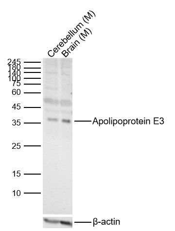
Sample:
Lane 1: Mouse Cerebellum Lysates
Lane 2: Mouse Brain Lysates
Primary: Anti-Apolipoprotein E3 (bs-5039R) at 1/1000 dilution
Secondary: IRDye800CW Goat Anti-Rabbit IgG at 1/20000 dilution
Predicted band size: 34kDa
Observed band size: 35kDa
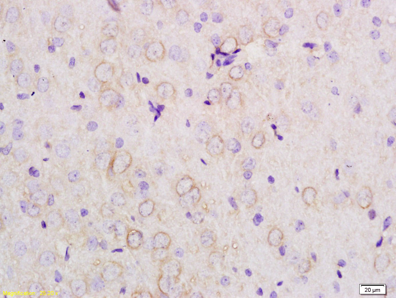
Tissue/cell: rat brain tissue; 4% Paraformaldehyde-fixed and paraffin-embedded;
Antigen retrieval: citrate buffer ( 0.01M, pH 6.0 ), Boiling bathing for 15min; Block endogenous peroxidase by 3% Hydrogen peroxide for 30min; Blocking buffer (normal goat serum,C-0005) at 37℃ for 20 min;
Incubation: Anti-APOE3 Polyclonal Antibody, Unconjugated(bs-5039R) 1:200, overnight at 4°C, followed by conjugation to the secondary antibody(SP-0023) and DAB(C-0010) staining
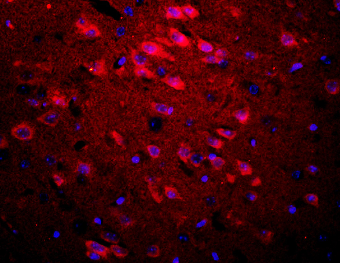
Tissue/cell: rat brain tissue;4% Paraformaldehyde-fixed and paraffin-embedded;
Antigen retrieval: citrate buffer ( 0.01M, pH 6.0 ), Boiling bathing for 15min; Blocking buffer (normal goat serum,C-0005) at 37℃ for 20 min;
Incubation: Anti-APOE3 Polyclonal Antibody, Unconjugated(bs-5039R) 1:200, overnight at 4°C; The secondary antibody was Goat Anti-Rabbit IgG, Cy3 conjugated(bs-0295G-Cy3)used at 1:200 dilution for 40 minutes at 37°C. DAPI(5ug/ml,blue,C-0033) was used to stain the cell nuclei
|
| 1、抗体溶解方法 | |
| 2、抗体修复方式 | |
| 3、常用试剂的配制 | |
| 4、免疫组化操作步骤 | |
| 5、免疫组化问题解答 | |
| 6、Western Blotting 操作步骤 | |
| 7、Western Blotting 问题解答 | |
| 8、关于肽链的设计 | |
| 9、多肽的溶解与保存 | |
| 10、酶标抗体效价测定程序 | |


