

| 产品编号 | bs-5305R |
| 英文名称 | phospho-E2F1 (Ser337) Rabbit pAb |
| 中文名称 | 磷酸化转录因子E2F-1抗体 |
| 别 名 | E2F1 (phospho-S337); p-E2F1; phospho-E2F1; E2F1 (phospho-Ser337); E2F-1; RBAP1; RBBP3; RBP3; Tg(Wnt1-cre)2Sor; mKIAA4009; E2F1_HUMAN; E2F1; PBR3; Retinoblastoma-associated protein 1 (RBAP-1); Retinoblastoma-binding protein 3 (RBBP-3); pRB-binding protein |
| 产品类型 | 磷酸化抗体 |
| 研究领域 | 肿瘤 免疫学 转录调节因子 |
| 抗体来源 | Rabbit |
| 克隆类型 | Polyclonal |
| 克 隆 号 | |
| 交叉反应 | Human,Mouse (predicted: Horse) |
| 产品应用 | WB=1:500-2000,IHC-P=1:100-500,IHC-F=1:100-500,IF=1:100-500,Flow-Cyt=1ug/Test,ICC/IF=1:100-500
not yet tested in other applications. optimal dilutions/concentrations should be determined by the end user. |
| 理论分子量 | 46 kDa |
| 检测分子量 | 53 |
| 细胞定位 | 细胞核 |
| 性 状 | Liquid |
| 浓 度 | 1mg/ml |
| 免 疫 原 | KLH conjugated Synthesised phosphopeptide derived from human E2F1 around the phosphorylation site of Ser337: PS(p-S)PP |
| 亚 型 | IgG |
| 纯化方法 | affinity purified by Protein A |
| 缓 冲 液 | 0.01M TBS (pH7.4) with 1% BSA, 0.02% Proclin300 and 50% Glycerol. |
| 保存条件 | Shipped at 4℃. Store at -20℃ for one year. Avoid repeated freeze/thaw cycles. |
| 注意事项 | This product as supplied is intended for research use only, not for use in human, therapeutic or diagnostic applications. |
| PubMed | PubMed |
| 产品介绍 |
E2F's are DNA binding proteins, which associate with negative regulators, such as the retinoblastoma p107 protein, resulting in an altered rate of gene transcription. The E2F proteins contain several evolutionally conserved domains found in most members of the family. These domains include a DNA binding domain, a dimerization domain which determines interaction with the differentiation regulated transcription factor proteins (DP), a transactivation domain enriched in acidic amino acids, and a tumor suppressor protein association domain which is embedded within the transactivation domain. This protein and another 2 members, E2F2 and E2F3, have an additional cyclin binding domain. E2F1 is proposed to be involved in several cellular processes that range from tumor suppressor, cell progression and oncogenesis. E2F1 overexpression can also drive cells into apoptosis. Function: Transcription activator that binds DNA cooperatively with dp proteins through the E2 recognition site, 5'-TTTC[CG]CGC-3' found in the promoter region of a number of genes whose products are involved in cell cycle regulation or in DNA replication. The DRTF1/E2F complex functions in the control of cell-cycle progression from G1 to S phase. E2F-1 binds preferentially RB1 protein, in a cell-cycle dependent manner. It can mediate both cell proliferation and p53-dependent apoptosis. Subunit: Component of the DRTF1/E2F transcription factor complex. Forms heterodimers with DP family members. The E2F-1 complex binds specifically hypophosphorylated retinoblastoma protein RB1. During the cell cycle, RB1 becomes phosphorylated in mid-to-late G1 phase, detaches from the DRTF1/E2F complex, rendering E2F transcriptionally active. Interacts with TRRAP, which probably mediates its interaction with histone acetyltransferase complexes, leading to transcription activation. Binds TOPBP1. Interacts with ARID3A (By similarity). Binds EAPP. Subcellular Location: Nucleus. Post-translational modifications: Phosphorylated by CDK2 and cyclin A-CDK2 in the S-phase (By similarity). Similarity: Belongs to the E2F/DP family. SWISS: Q01094 Gene ID: 1869 Database links: Entrez Gene: 1869 Human Omim: 189971 Human SwissProt: Q01094 Human Unigene: 654393 Human E2F1—属于调节性转录因子E2F家族。有学者认为:E2F-1既可作为癌基因起作用,又可作为抑癌基因起作用。其不同可能由细胞中其他生长促进或抑制性蛋白质水平和(或)活性决定,同时与细胞所处环境及器官特异性有关。在控制细胞周期和肿瘤抑制基因蛋白的活性方面起关键作用。 |
| 产品图片 |
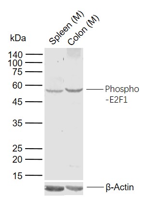
Sample:
Lane 1: Mouse Spleen tissue lysates
Lane 2: Mouse Colon tissue lysates
Primary: Anti-phospho-E2F1 (Ser337) (bs-5305R) at 1/1000 dilution
Secondary: IRDye800CW Goat Anti-Rabbit IgG at 1/20000 dilution
Predicted band size: 46 kDa
Observed band size: 53 kDa
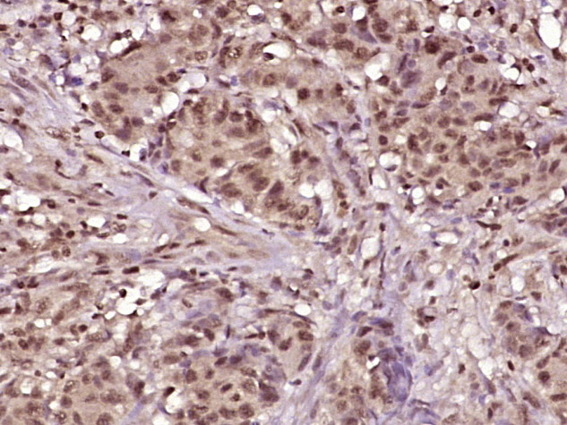
Paraformaldehyde-fixed, paraffin embedded (human colon carcinoma); Antigen retrieval by boiling in sodium citrate buffer (pH6.0) for 15min; Block endogenous peroxidase by 3% hydrogen peroxide for 20 minutes; Blocking buffer (normal goat serum) at 37°C for 30min; Antibody incubation with (E2F1 (Ser337)) Polyclonal Antibody, Unconjugated (bs-5305R) at 1:400 overnight at 4°C, followed by operating according to SP Kit(Rabbit) (sp-0023) instructionsand DAB staining.
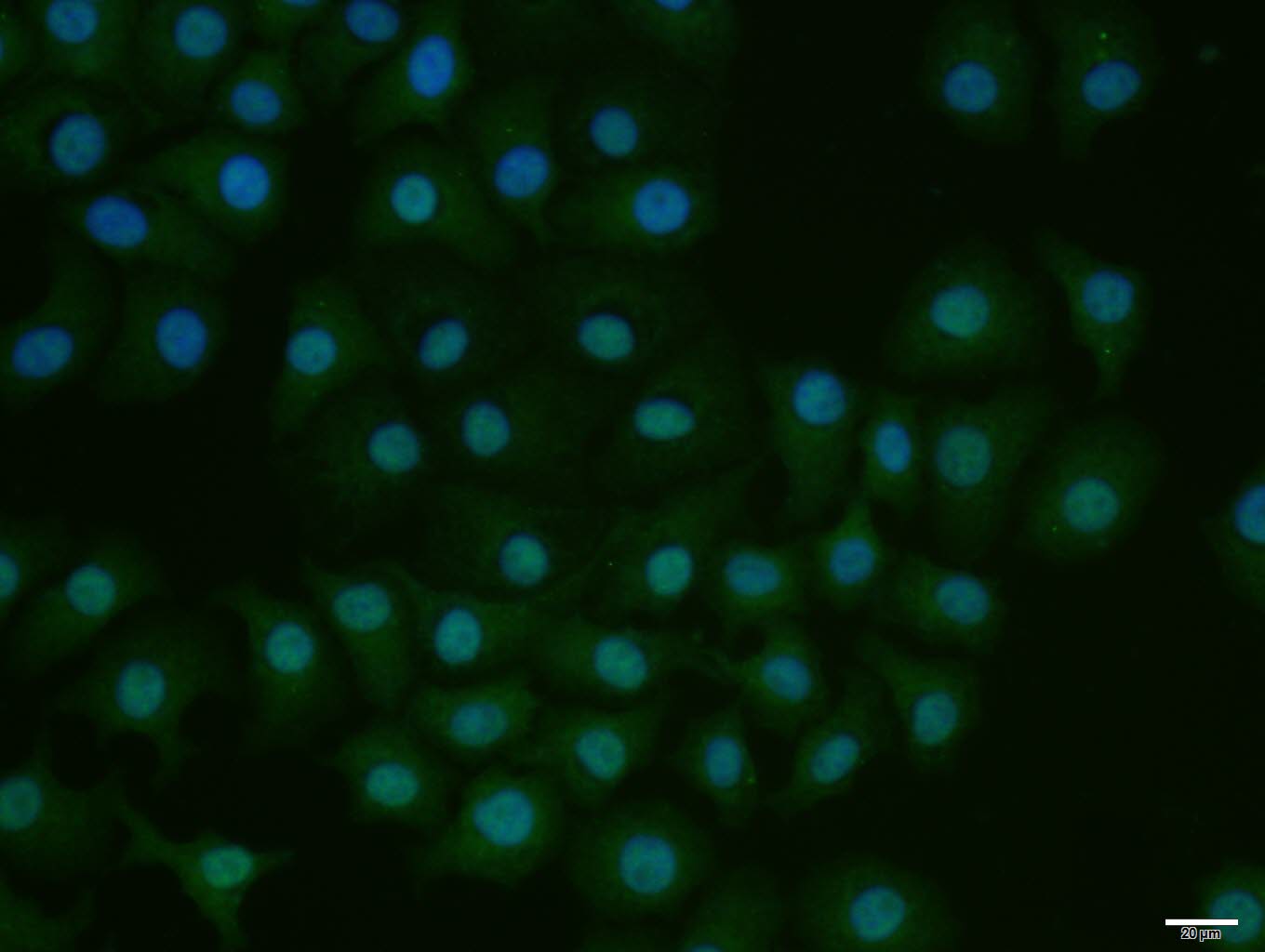
HepG2 cell; 4% Paraformaldehyde-fixed; Triton X-100 at room temperature for 20 min; Blocking buffer (normal goat serum, C-0005) at 37°C for 20 min; Antibody incubation with (phospho-E2F1 (Ser337)) polyclonal Antibody, Unconjugated (bs-5305R) 1:100, 90 minutes at 37°C; followed by a conjugated Goat Anti-Rabbit IgG antibody at 37°C for 90 minutes, DAPI (blue, C02-04002) was used to stain the cell nuclei.
Blank control(blue): TM4 cells(fixed with 2% paraformaldehyde (10 min) , then permeabilized with 90% ice-cold methanol for 30 min on ice). Primary Antibody:Rabbit Anti-E2F 1 antibody(bs-5305R), Dilution: 1μg in 100 μL 1X PBS containing 0.5% BSA; Isotype Control Antibody: Rabbit IgG(orange) ,used under the same conditions ); Secondary Antibody: Goat anti-rabbit IgG-PE(white blue), Dilution: 1:200 in 1 X PBS containing 0.5% BSA.
Protocol
The cells were . Primary antibody (bs-5305R, 1μg /1x10^6 cells) were incubated for 30 min on the ice, followed by 1 X PBS containing 0.5% BSA + 1 0% goat serum (15 min) to block non-specific protein-protein interactions. Then the Goat Anti-rabbit IgG/PE antibody was added into the blocking buffer mentioned above to react with the primary antibody at 1/200 dilution for 30 min on ice. Acquisition of 20,000 events was performed.
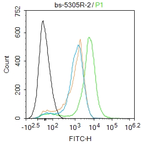
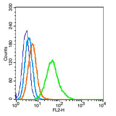
Blank control:Mosue spleen.
Primary Antibody (green line): Rabbit Anti-phospho-E2F1 (Ser337) antibody (bs-5305R)
Dilution: 2μg /10^6 cells;
Isotype Control Antibody (orange line): Rabbit IgG .
Secondary Antibody : Goat anti-rabbit IgG-FITC
Dilution: 1μg /test.
Protocol
The cells were fixed with 4% PFA (10min at room temperature)and then permeabilized with 90% ice-cold methanol for 20 min at -20℃. The cells were then incubated in 5%BSA to block non-specific protein-protein interactions for 30 min at room temperature .Cells stained with Primary Antibody for 30 min at room temperature. The secondary antibody used for 40 min at room temperature. Acquisition of 20,000 events was performed.
|
| 1、抗体溶解方法 | |
| 2、抗体修复方式 | |
| 3、常用试剂的配制 | |
| 4、免疫组化操作步骤 | |
| 5、免疫组化问题解答 | |
| 6、Western Blotting 操作步骤 | |
| 7、Western Blotting 问题解答 | |
| 8、关于肽链的设计 | |
| 9、多肽的溶解与保存 | |
| 10、酶标抗体效价测定程序 | |


