

| 产品编号 | bs-4258R |
| 英文名称 | Vesicle docking protein p115 Rabbit pAb |
| 中文名称 | 囊泡对接蛋白p115抗体 |
| 别 名 | VDP; P115; TAP; 115kDa; USO1_BOVIN; USO1; Protein USO1 homolog; Transcytosis-associated protein (TAP); Vesicle-docking protein; USO1_HUMAN; USO1_MOUSE; USO1_RAT; USO1 vesicle transport factor; USO1 homolog, vesicle docking protein (yeast); USO1 vesicle docking protein homolog (yeast); vesicle docking protein; transcytosis associated protein |
| 研究领域 | 免疫学 信号转导 结合蛋白 G蛋白偶联受体 |
| 抗体来源 | Rabbit |
| 克隆类型 | Polyclonal |
| 交叉反应 | Human,Mouse (predicted: Rat,Pig,Sheep,Cow,Chicken,Horse) |
| 产品应用 | WB=1:500-2000,IHC-P=1:100-500,IHC-F=1:100-500,IF=1:100-500
not yet tested in other applications. optimal dilutions/concentrations should be determined by the end user. |
| 理论分子量 | 108kDa |
| 检测分子量 | 140 |
| 细胞定位 | 细胞浆 细胞膜 |
| 性 状 | Liquid |
| 浓 度 | 1mg/ml |
| 免 疫 原 | KLH conjugated synthetic peptide derived from human Vesicle docking protein p115: 501-600/962 |
| 亚 型 | IgG |
| 纯化方法 | affinity purified by Protein A |
| 缓 冲 液 | 0.01M TBS (pH7.4) with 1% BSA, 0.02% Proclin300 and 50% Glycerol. |
| 保存条件 | Shipped at 4℃. Store at -20℃ for one year. Avoid repeated freeze/thaw cycles. |
| 注意事项 | This product as supplied is intended for research use only, not for use in human, therapeutic or diagnostic applications. |
| PubMed | PubMed |
| 产品介绍 |
p115 (Vesicle docking protein p115) is a peripheral membrane protein that is located on the Golgi complex. p115 exists as a homodimer with two globular heads, an extended coiled-coil tail, followed by an acidic domain at the extreme C terminus. p115 is homologous to a yeast protein, Uso1p, which is required for ER to Golgi transport. p115 likely plays an important role in vesicle transportation from the ER to the cis-Golgi comparments. Function: General vesicular transport factor required for intercisternal transport in the Golgi stack; it is required for transcytotic fusion and/or subsequent binding of the vesicles to the target membrane. May well act as a vesicular anchor by interacting with the target membrane and holding the vesicular and target membranes in proximity. Subunit: Homodimer. Dimerizes by parallel association of the tails, resulting in an elongated structure with two globular head domains side by side, and a long rod-like tail structure (Probable). Interacts with MIF. Subcellular Location: Cytoplasm; cytosol. Golgi apparatus membrane. Recycles between the cytosol and the Golgi apparatus during interphase. During interphase, the phosphorylated form is found exclusively in cytosol; the unphosphorylated form is associated with Golgi apparatus membranes. Post-translational modifications: Phosphorylated in a cell cycle-specific manner; phosphorylated in interphase but not in mitotic cells. Dephosphorylated protein associates with the Golgi membrane; phosphorylation promotes dissociation. Similarity: Belongs to the VDP/USO1/EDE1 family. Contains 10 ARM repeats. SWISS: O60763 Gene ID: 8615 Database links: Entrez Gene: 8615 Human Entrez Gene: 56041 Mouse Omim: 603344 Human SwissProt: O60763 Human SwissProt: Q9Z1Z0 Mouse Unigene: 292689 Human Unigene: 15868 Mouse Unigene: 4746 Rat |
| 产品图片 |
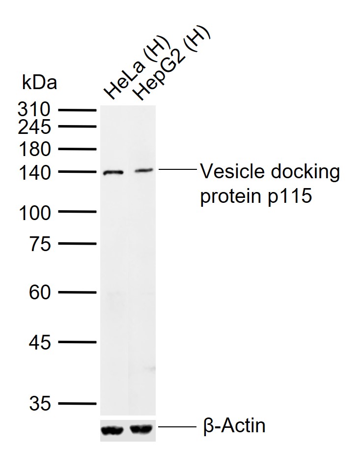
Sample:
Lane 1: Human HeLa cell lysates
Lane 2: Human HepG2 cell lysates
Primary: Anti-Vesicle docking protein p115 (bs-4258R) at 1/1000 dilution
Secondary: IRDye800CW Goat Anti-Rabbit IgG at 1/20000 dilution
Predicted band size: 108 kDa
Observed band size: 140 kDa
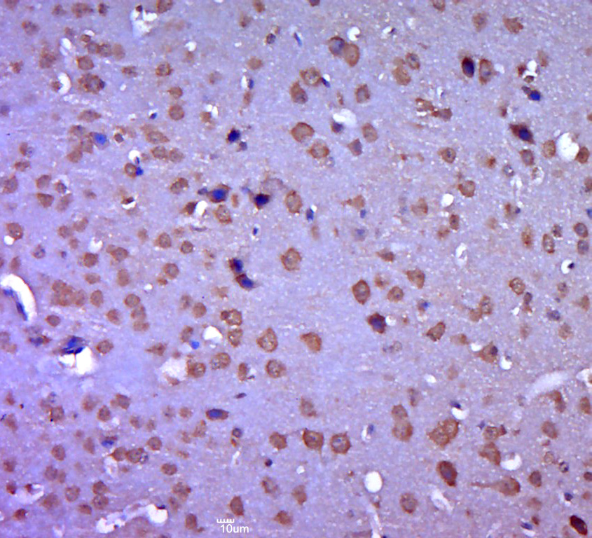
Paraformaldehyde-fixed, paraffin embedded (Mouse brain); Antigen retrieval by boiling in sodium citrate buffer (pH6.0) for 15min; Block endogenous peroxidase by 3% hydrogen peroxide for 20 minutes; Blocking buffer (normal goat serum) at 37°C for 30min; Antibody incubation with (Vesicle docking protein p115) Polyclonal Antibody, Unconjugated (bs-4258R) at 1:400 overnight at 4°C, followed by operating according to SP Kit(Rabbit) (sp-0023) instructionsand DAB staining.
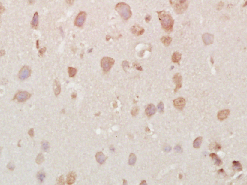
Paraformaldehyde-fixed, paraffin embedded (mouse brain tissue); Antigen retrieval by boiling in sodium citrate buffer (pH6.0) for 15min; Block endogenous peroxidase by 3% hydrogen peroxide for 20 minutes; Blocking buffer (normal goat serum) at 37°C for 30min; Antibody incubation with (Vesicle docking protein p115) Polyclonal Antibody, Unconjugated (bs-4258R) at 1:400 overnight at 4°C, followed by a conjugated secondary (sp-0023) for 20 minutes and DAB staining.
|
| 1、抗体溶解方法 | |
| 2、抗体修复方式 | |
| 3、常用试剂的配制 | |
| 4、免疫组化操作步骤 | |
| 5、免疫组化问题解答 | |
| 6、Western Blotting 操作步骤 | |
| 7、Western Blotting 问题解答 | |
| 8、关于肽链的设计 | |
| 9、多肽的溶解与保存 | |
| 10、酶标抗体效价测定程序 | |


