

| 产品编号 | bs-7048R |
| 英文名称 | INPPL1 Rabbit pAb |
| 中文名称 | 肌醇聚磷酸盐磷酸酶样蛋白1抗体 |
| 别 名 | OPSMD; SHIP2; 51C; SHIP2_HUMAN; INPPL1; Inositol polyphosphate phosphatase-like protein 1 (INPPL-1); Protein 51C; SH2 domain-containing inositol 5'-phosphatase 2 (SH2 domain-containing inositol phosphatase 2 | SHIP-2); 3.1.3.86; SHIP2_MOUSE; SHIP2_RAT; |
| 研究领域 | 肿瘤 心血管 免疫学 神经生物学 信号转导 生长因子和激素 激酶和磷酸酶 细胞骨架 新陈代谢 细胞外基质 |
| 抗体来源 | Rabbit |
| 克隆类型 | Polyclonal |
| 克 隆 号 | |
| 交叉反应 | Mouse,Rat (predicted: Human,Rabbit,Pig,Cow,Dog,Horse) |
| 产品应用 | IHC-P=1:100-500,IHC-F=1:100-500,IF=1:100-500
not yet tested in other applications. optimal dilutions/concentrations should be determined by the end user. |
| 理论分子量 | 139 kDa |
| 细胞定位 | 细胞浆 细胞膜 |
| 性 状 | Liquid |
| 浓 度 | 1mg/ml |
| 免 疫 原 | KLH conjugated synthetic peptide derived from human INPPL1: 701-800/1258 |
| 亚 型 | IgG |
| 纯化方法 | affinity purified by Protein A |
| 缓 冲 液 | 0.01M TBS (pH7.4) with 1% BSA, 0.02% Proclin300 and 50% Glycerol. |
| 保存条件 | Shipped at 4℃. Store at -20℃ for one year. Avoid repeated freeze/thaw cycles. |
| 注意事项 | This product as supplied is intended for research use only, not for use in human, therapeutic or diagnostic applications. |
| PubMed | PubMed |
| 产品介绍 |
INPPL1 is a phosphatidylinositol (PtdIns) phosphatase that specifically hydrolyzes the 5-phosphate of phosphatidylinositol-3,4,5-trisphosphate (PtdIns(3,4,5)P3) to produce PtdIns(3,4)P2, thereby negatively regulating the PI3K (phosphoinositide 3-kinase) pathways with wide reaching effects It plays a central role in regulation of PI3K-dependent insulin signaling and confers resistance to dietary obesity. It is part of a signaling pathway that regulates actin cytoskeleton remodeling. It regulates cell adhesion and cell spreading and acts as a regulator of neuritogenesis. It acts as a negative regulator of the FC-gamma-RIIA receptor (FCGR2A). and mediates signaling from the FC-gamma-RIIB receptor (FCGR2B), playing a central role in terminating signal transduction from activating immune/hematopoietic cell receptor systems. It is involved in the EGF signaling pathway. Cytoplasm: cytosol and cytoskeleton; actin patch. Membrane; Peripheral membrane protein. Note: Translocates to membrane ruffles when activated, translocation is probably due to different mechanisms depending on the stimulus and cell type. Function: Phosphatidylinositol (PtdIns) phosphatase that specifically hydrolyzes the 5-phosphate of phosphatidylinositol-3,4,5-trisphosphate (PtdIns(3,4,5)P3) to produce PtdIns(3,4)P2, thereby negatively regulating the PI3K (phosphoinositide 3-kinase) pathways. Plays a central role in regulation of PI3K-dependent insulin signaling, although the precise molecular mechanisms and signaling pathways remain unclear. While overexpression reduces both insulin-stimulated MAP kinase and Akt activation, its absence does not affect insulin signaling or GLUT4 trafficking. Confers resistance to dietary obesity. May act by regulating AKT2, but not AKT1, phosphorylation at the plasma membrane. Part of a signaling pathway that regulates actin cytoskeleton remodeling. Required for the maintenance and dynamic remodeling of actin structures as well as in endocytosis, having a major impact on ligand-induced EGFR internalization and degradation. Participates in regulation of cortical and submembraneous actin by hydrolyzing PtdIns(3,4,5)P3 thereby regulating membrane ruffling. Regulates cell adhesion and cell spreading. Required for HGF-mediated lamellipodium formation, cell scattering and spreading. Acts as a negative regulator of EPHA2 receptor endocytosis by inhibiting via PI3K-dependent Rac1 activation. Acts as a regulator of neuritogenesis by regulating PtdIns(3,4,5)P3 level and is required to form an initial protrusive pattern, and later, maintain proper neurite outgrowth. Acts as a negative regulator of the FC-gamma-RIIA receptor (FCGR2A). Mediates signaling from the FC-gamma-RIIB receptor (FCGR2B), playing a central role in terminating signal transduction from activating immune/hematopoietic cell receptor systems. Involved in EGF signaling pathway. Upon stimulation by EGF, it is recruited by EGFR and dephosphorylates PtdIns(3,4,5)P3. Plays a negative role in regulating the PI3K-PKB pathway, possibly by inhibiting PKB activity. Down-regulates Fc-gamma-R-mediated phagocytosis in macrophages independently of INPP5D/SHIP1. In macrophages, down-regulates NF-kappa-B-dependent gene transcription by regulating macrophage colony-stimulating factor (M-CSF)-induced signaling. May also hydrolyze PtdIns(1,3,4,5)P4, and could thus affect the levels of the higher inositol polyphosphates like InsP6 Subunit: Interacts with tyrosine phosphorylated form of SHC1, Interacts with EGFR. Upon stimulation by the EGF signaling pathway, it forms a complex with SHC1 and EGFR. Interacts with cytoskeletal protein SORBS3/vinexin, promoting its localization to the periphery of cells. Forms a complex with filamin (FLNA or FLNB), actin, GPIb (GP1BA or GP1BB) that regulates cortical and submembraneous actin. Interacts with c-Met/MET, when c-Met/MET is phosphorylated on 'Tyr-1356'. Interacts with p130Cas/BCAR1. Interacts with CENTD3/ARAP3 via its SAM domain. Interacts with c-Cbl/CBL and CAP/SORBS1. Interacts with activated EPHA2 receptor. Interacts with receptors FCGR2A and FCGR2B. Interacts with tyrosine kinases ABL1 and TEC. Interacts with CSF1R. Subcellular Location: Cytoplasm, cytosol. Cytoplasm, cytoskeleton, actin patch. Membrane; Peripheral membrane protein. Tissue Specificity: Widely expressed, most prominently in skeletal muscle, heart and brain. Present in platelets. Expressed in transformed myeloid cells and in primary macrophages, but not in peripheral blood monocytes. Post-translational modifications: Tyrosine phosphorylated by the members of the SRC family after exposure to a diverse array of extracellular stimuli such as insulin, growth factors such as EGF or PDGF, chemokines, integrin ligands and hypertonic and oxidative stress. May be phosphorylated upon IgG receptor FCGR2B-binding. Phosphorylated at Tyr-986 following cell attachment and spreading. Phosphorylated at Tyr-1162 following EGF signaling pathway stimulation. Phosphorylated at Thr-958 in response to PDGF. DISEASE: Defects in INPPL1 may be a cause of susceptibility to type 2 diabetes mellitus non-insulin dependent (NIDDM) [MIM:125853]. Note=Genetic variations in INPPL1 may be a cause of susceptibility to metabolic syndrome. Metabolic syndrome is characterized by diabetes, insulin resistance, hypertension, and hypertriglyceridemia is absent. Similarity: Belongs to the inositol 1,4,5-trisphosphate 5-phosphatase family. Contains 1 SAM (sterile alpha motif) domain. Contains 1 SH2 domain. SWISS: O15357 Gene ID: 3636 Database links: Entrez Gene: 3636 Human Entrez Gene: 16332 Mouse Omim: 600829 Human SwissProt: O15357 Human SwissProt: Q6P549 Mouse Unigene: 523875 Human Unigene: 476000 Mouse Unigene: 5028 Mouse Unigene: 42902 Rat |
| 产品图片 |
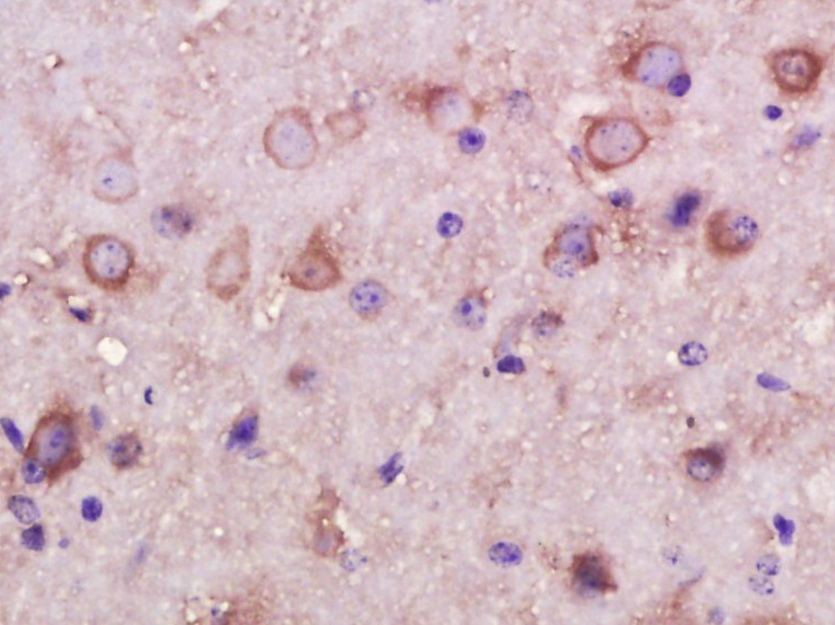
Paraformaldehyde-fixed, paraffin embedded (Mouse brain); Antigen retrieval by boiling in sodium citrate buffer (pH6.0) for 15min; Block endogenous peroxidase by 3% hydrogen peroxide for 20 minutes; Blocking buffer (normal goat serum) at 37°C for 30min; Antibody incubation with (INPPL1) Polyclonal Antibody, Unconjugated (bs-7048R) at 1:400 overnight at 4°C, followed by operating according to SP Kit(Rabbit) (sp-0023) instructionsand DAB staining.
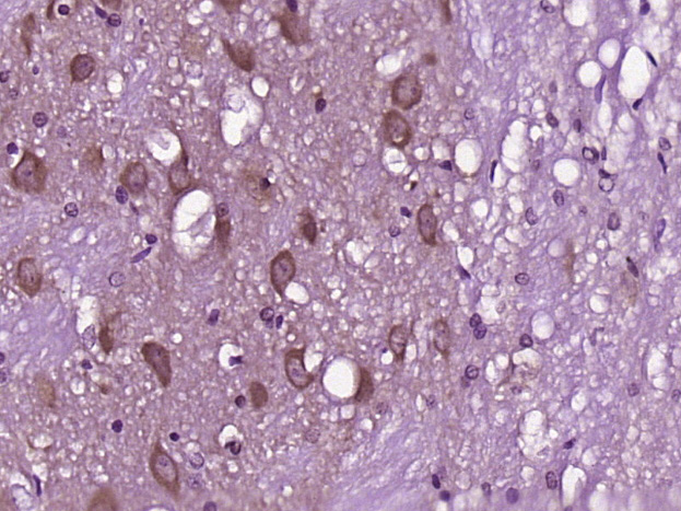
Paraformaldehyde-fixed, paraffin embedded (Rat brain); Antigen retrieval by boiling in sodium citrate buffer (pH6.0) for 15min; Block endogenous peroxidase by 3% hydrogen peroxide for 20 minutes; Blocking buffer (normal goat serum) at 37°C for 30min; Antibody incubation with (INPPL1) Polyclonal Antibody, Unconjugated (bs-7048R) at 1:400 overnight at 4°C, followed by operating according to SP Kit(Rabbit) (sp-0023) instructionsand DAB staining.
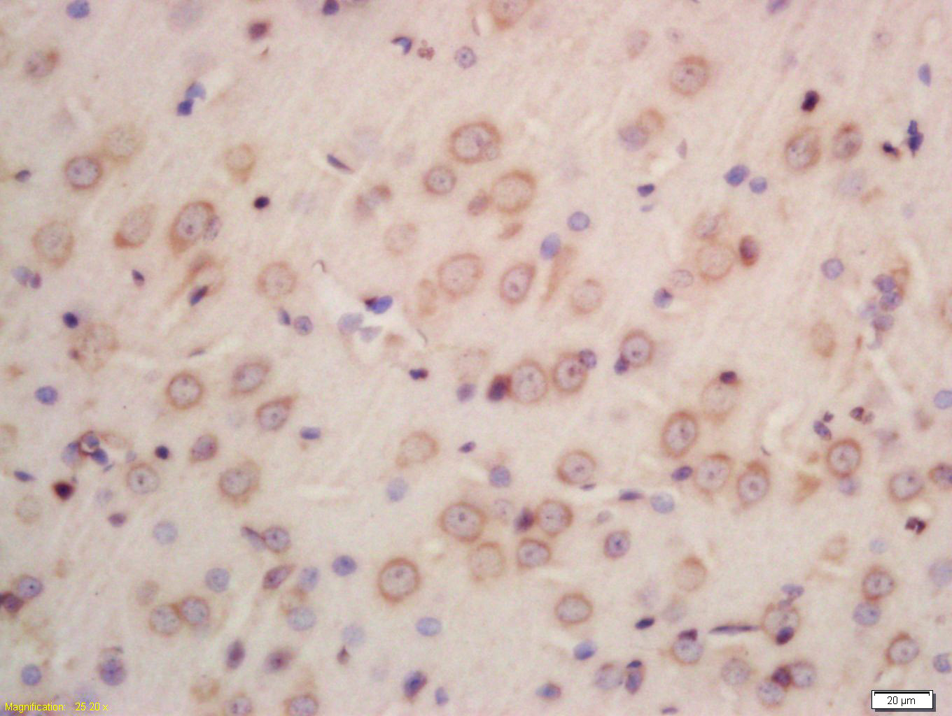
Tissue/cell: rat brain tissue; 4% Paraformaldehyde-fixed and paraffin-embedded;
Antigen retrieval: citrate buffer ( 0.01M, pH 6.0 ), Boiling bathing for 15min; Block endogenous peroxidase by 3% Hydrogen peroxide for 30min; Blocking buffer (normal goat serum,C-0005) at 37℃ for 20 min;
Incubation: Anti-INPPL1 Polyclonal Antibody, Unconjugated(bs-7048R) 1:200, overnight at 4°C, followed by conjugation to the secondary antibody(SP-0023) and DAB(C-0010) staining
|
| 1、抗体溶解方法 | |
| 2、抗体修复方式 | |
| 3、常用试剂的配制 | |
| 4、免疫组化操作步骤 | |
| 5、免疫组化问题解答 | |
| 6、Western Blotting 操作步骤 | |
| 7、Western Blotting 问题解答 | |
| 8、关于肽链的设计 | |
| 9、多肽的溶解与保存 | |
| 10、酶标抗体效价测定程序 | |


