

| 产品编号 | bs-6796R |
| 英文名称 | CIDEC Rabbit pAb |
| 中文名称 | 细胞死亡活化蛋白抗体 |
| 别 名 | CIDE-3; CIDE3; FPLD5; FSP27; CIDEC_HUMAN; CIDEC; Cell death activator CIDE-3; Cell death-inducing DFFA-like effector protein C; Fat-specific protein FSP27 homolog; CIDEC_MOUSE; Fat-specific protein FSP27; CIDEC_RAT; |
 |
Specific References (2) | bs-6796R has been referenced in 2 publications.
[IF=6.656] Qing-song Xia. et al. Ban-xia-xie-xin-tang ameliorates hepatic steatosis by regulating Cidea and Cidec expression in HFD-fed mice. PHYTOMEDICINE. 2022 Oct;105:154351 WB, IHC ; Mouse.
[IF=2.742] Liu, Yanrong. et al. Cinnamaldehyde affects lipid droplets metabolism after adipogenic differentiation of C2C12 cells. MOL BIOL REP. 2022 Dec;:1-7 WB ; Mouse.
|
| 研究领域 | 肿瘤 心血管 细胞生物 免疫学 信号转导 细胞凋亡 表观遗传学 |
| 抗体来源 | Rabbit |
| 克隆类型 | Polyclonal |
| 克 隆 号 | |
| 交叉反应 | Human,Mouse,Rat (predicted: Pig) |
| 产品应用 | WB=1:500-2000,IHC-P=1:100-500,IHC-F=1:100-500,IF=1:100-500,Flow-Cyt=1ug/Test,ICC/IF=1:50-200
not yet tested in other applications. optimal dilutions/concentrations should be determined by the end user. |
| 理论分子量 | 27 kDa |
| 检测分子量 | 27 |
| 细胞定位 | 细胞核 细胞浆 |
| 性 状 | Liquid |
| 浓 度 | 1mg/ml |
| 免 疫 原 | KLH conjugated synthetic peptide derived from human CIDEC: 101-200/238 |
| 亚 型 | IgG |
| 纯化方法 | affinity purified by Protein A |
| 缓 冲 液 | 0.01M TBS (pH7.4) with 1% BSA, 0.02% Proclin300 and 50% Glycerol. |
| 保存条件 | Shipped at 4℃. Store at -20℃ for one year. Avoid repeated freeze/thaw cycles. |
| 注意事项 | This product as supplied is intended for research use only, not for use in human, therapeutic or diagnostic applications. |
| PubMed | PubMed |
| 产品介绍 |
This gene encodes a member of the cell death-inducing DNA fragmentation factor-like effector family. Members of this family play important roles in apoptosis. The encoded protein promotes lipid droplet formation in adipocytes and may mediate adipocyte apoptosis. This gene is regulated by insulin and its expression is positively correlated with insulin sensitivity. Mutations in this gene may contribute to insulin resistant diabetes. A pseudogene of this gene is located on the short arm of chromosome 3. Alternatively spliced transcript variants that encode different isoforms have been observed for this gene. [provided by RefSeq, Dec 2010]. Tissue specificity: Expressed mainly in small intestine, heart, colon and stomach and, at lower levels, in brain, kidney and liver. Function: May act as a CEBPB coactivator in white adipose tissueto control the expression of a subset of CEBPB downstream targetgenes, including SOCS1, SOCS3, TGFB1, TGFBR1, ID2 and XDH (Bysimilarity). Binds to lipid droplets and regulates theirenlargement, thereby restricting lipolysis and favoring storage. Atfocal contact sites between lipid droplets, promotes directionalnet neutral lipid transfer from the smaller to larger lipiddroplets. The transfer direction may be driven by the internalpressure difference between the contacting lipid droplet pair. Whenoverexpressed in preadipocytes, induces apoptosis or increases cellsusceptibility to apoptosis induced by serum deprivation or TGFBtreatment. As mature adipocytes, that express high CIDEC levels,are quite resistant to apoptotic stimuli, the physiologicalsignificance of its role in apoptosis is unclear. Subunit: Interacts with CEBPB (By similarity). Interacts withCIDEA. Subcellular Location: Nucleus (By similarity). Endoplasmicreticulum (By similarity). Lipid droplet. Note=Diffuses quickly onlipid droplet surface, but becomes trapped and clustered at lipiddroplet contact sites, thereby enabling its rapid enrichment atlipid droplet contact sites. Tissue Specificity: Expressed mainly in adipose tissue, smallintestine, heart, colon and stomach and, at lower levels, in brain,kidney and liver. Post-translational modifications: Ubiquitinated and targeted to proteasomal degradation,resulting in a short half-life. Protein stability depends ontriaclyglycerol synthesis, fatty acid availability and lipiddroplet formation (By similarity). DISEASE: Note=In omental adipose tissue of obese patients matchedfor BMI, expression levels tend to correlate with insulinsensitivity. Expression is increased 2-3 fold in the group ofpatients with high insulin sensitivity, compared to theinsulin-resistant group. This observation is consistent with theidea that triglyceride storage in adipocytes plays an importantrole in sequestering triglycerides and fatty acids away from thecirculation and peripheral tissues, thus enhancing insulinsensitivity in liver and muscle. This effect is not significant insubcutaneous adipose tissue (PubMed:18509062). In subcutaneousadipose tissue of diabetic patients, tends to negatively correlatewith body mass index and total fat mass, independently of insulinsensitivity (PubMed:18334488). Similarity: Contains 1 CIDE-N domain. SWISS: Q96AQ7 Gene ID: 63924 Database links: Entrez Gene: 63924 Human Entrez Gene: 14311 Mouse Omim: 612120 Human SwissProt: Q96AQ7 Human SwissProt: P56198 Mouse Unigene: 567562 Human Unigene: 635072 Human Unigene: 10026 Mouse Unigene: 33794 Rat |
| 产品图片 |
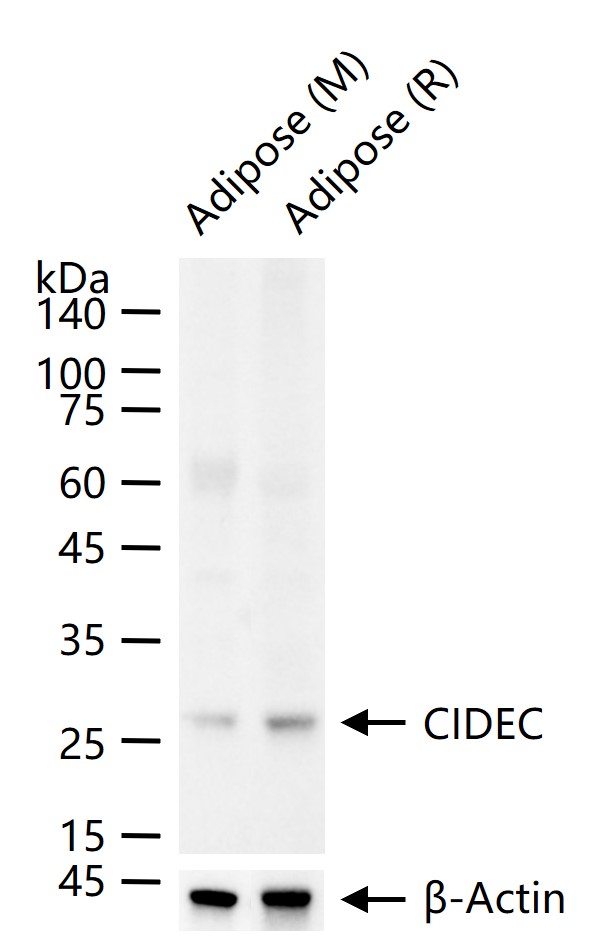
25 ug total protein per lane of various lysates (see on figure) probed with CIDEC polyclonal antibody, unconjugated (bs-6796R) at 1:1000 dilution and 4°C overnight incubation. Followed by conjugated secondary antibody incubation at r.t. for 60 min.
Paraformaldehyde-fixed, paraffin embedded (Rat brain); Antigen retrieval by boiling in sodium citrate buffer (pH6.0) for 15min; Block endogenous peroxidase by 3% hydrogen peroxide for 20 minutes; Blocking buffer (normal goat serum) at 37°C for 30min; Antibody incubation with (CIDEC) Polyclonal Antibody, Unconjugated (bs-6796R) at 1:400 overnight at 4°C, followed by operating according to SP Kit(Rabbit) (sp-0023) instructionsand DAB staining.
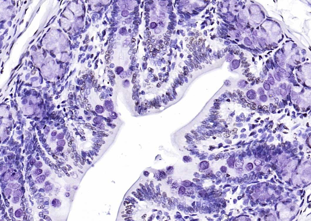
Paraformaldehyde-fixed, paraffin embedded Human Stomach; Antigen retrieval by boiling in sodium citrate buffer (pH6.0) for 15 min; The section was incubated with CIDEC Polyclonal Antibody, Unconjugated (bs-6796R) at 1:200 overnight at 4°C, followed by conjugation to the bs-0295G-HRP and DAB (C-0010) staining.
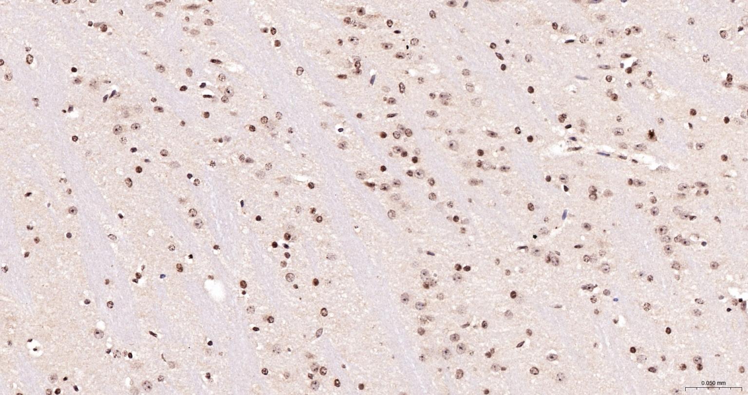
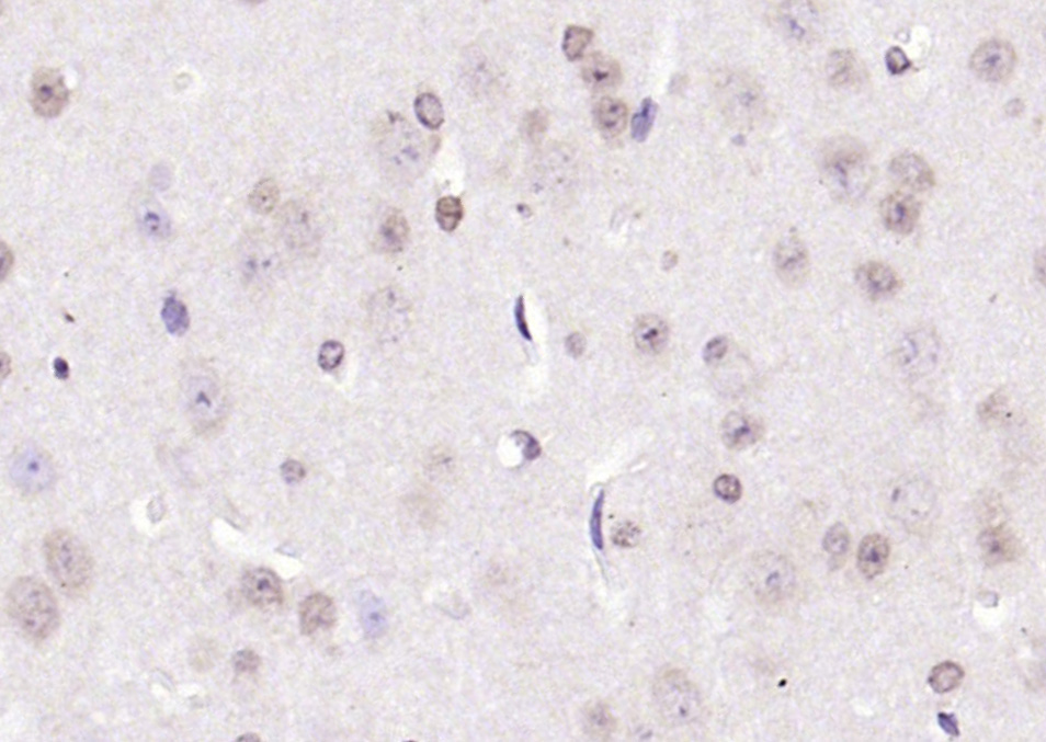
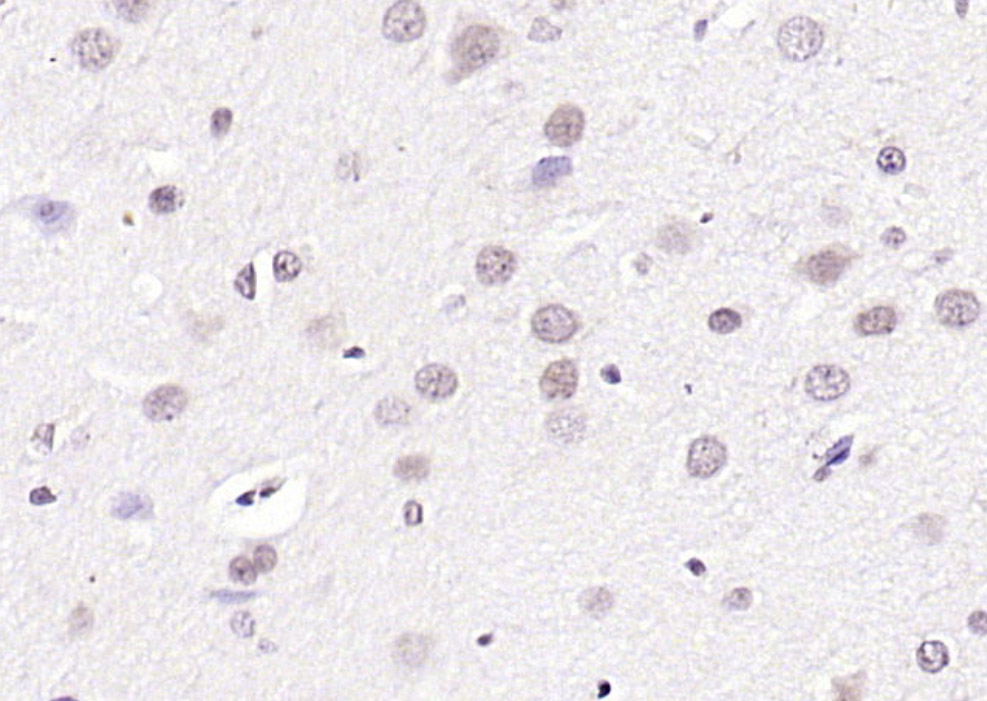
Paraformaldehyde-fixed, paraffin embedded Rat Cerebrum; Antigen retrieval by boiling in sodium citrate buffer (pH6.0) for 15 min; The section was incubated with CIDEC Polyclonal Antibody, Unconjugated (bs-6796R) at 1:200 overnight at 4°C, followed by conjugation to the bs-0295G-HRP and DAB (C-0010) staining.
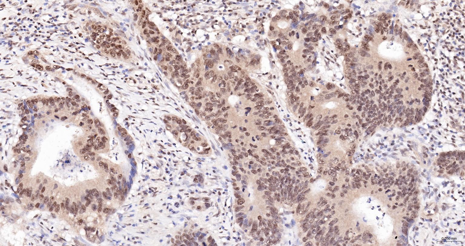
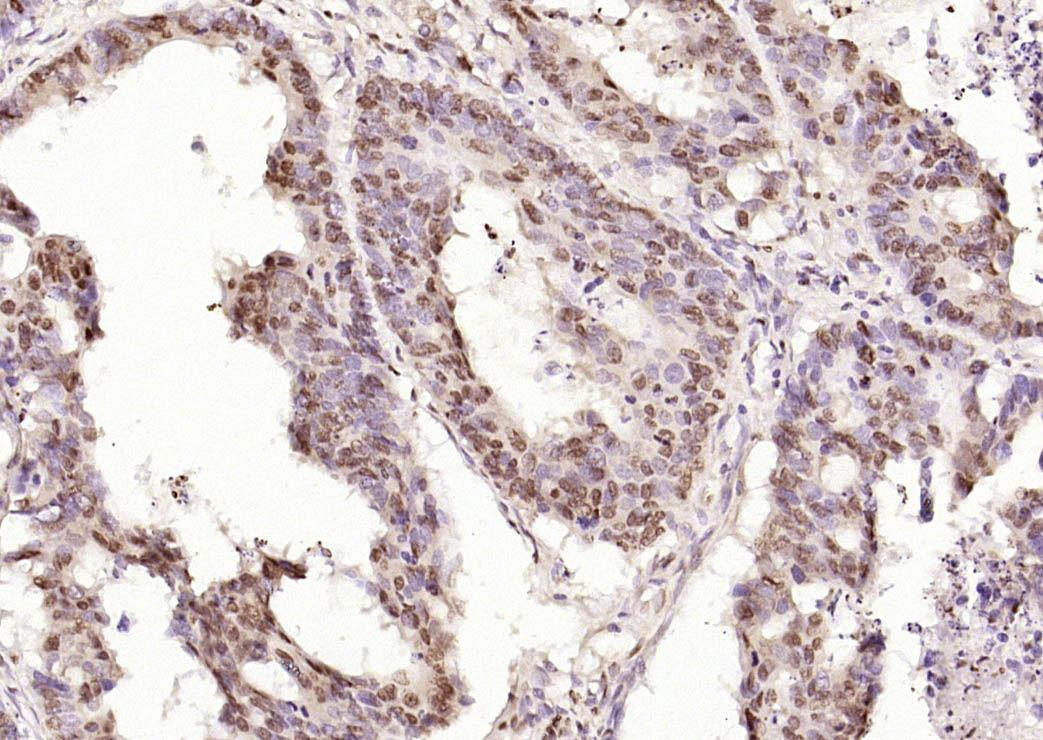
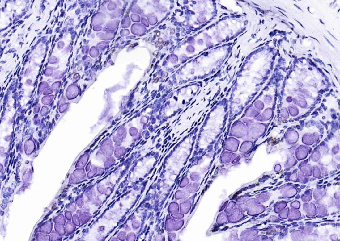
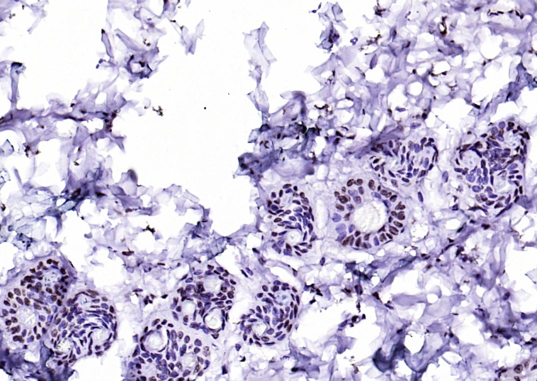
Paraformaldehyde-fixed, paraffin embedded Human Colon Cancer; Antigen retrieval by boiling in sodium citrate buffer (pH6.0) for 15 min; The section was incubated with CIDEC Polyclonal Antibody, Unconjugated (bs-6796R) at 1:200 overnight at 4°C, followed by conjugation to the bs-0295G-HRP and DAB (C-0010) staining.
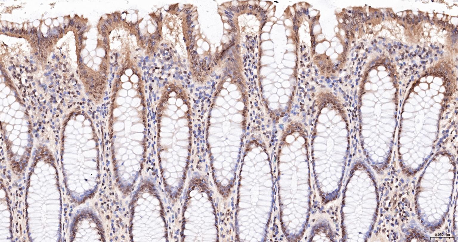
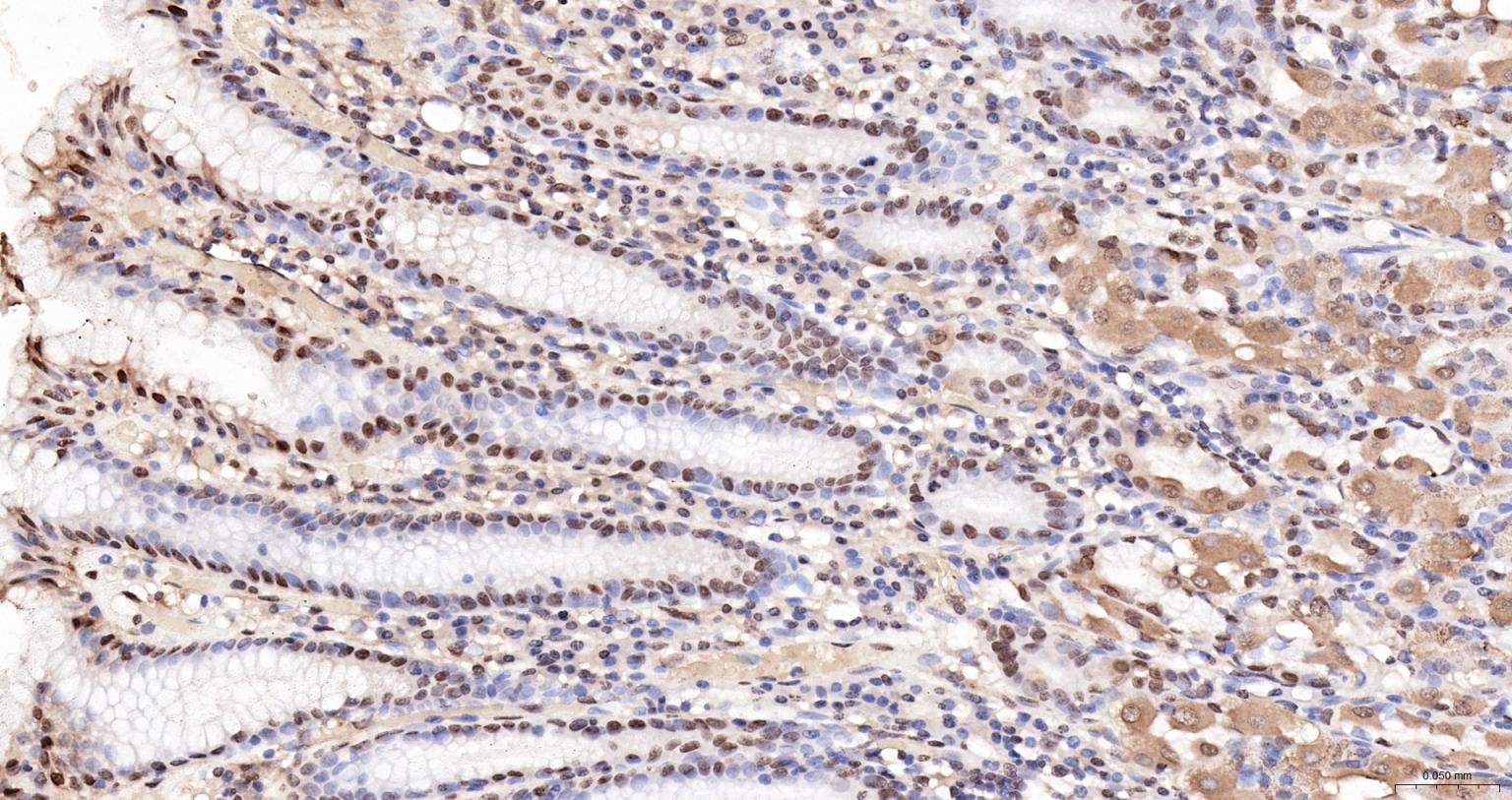
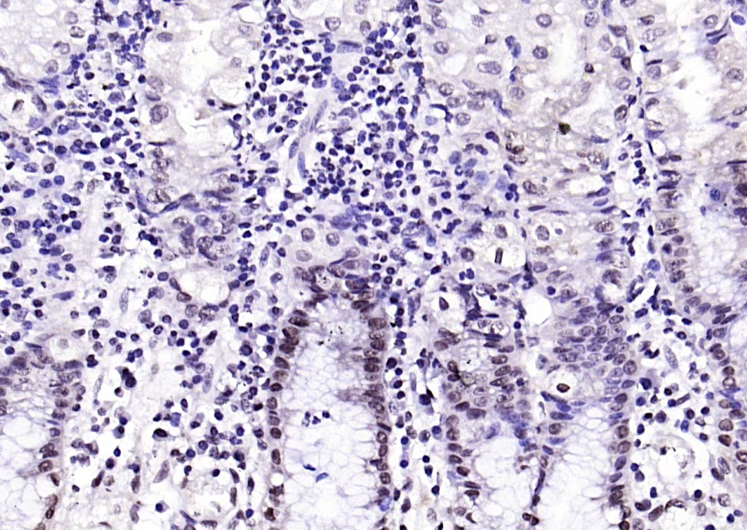
Paraformaldehyde-fixed, paraffin embedded Human Colon; Antigen retrieval by boiling in sodium citrate buffer (pH6.0) for 15 min; The section was incubated with CIDEC Polyclonal Antibody, Unconjugated (bs-6796R) at 1:200 overnight at 4°C, followed by conjugation to the bs-0295G-HRP and DAB (C-0010) staining.
Paraformaldehyde-fixed, paraffin embedded (Human colon carcinoma); Antigen retrieval by boiling in sodium citrate buffer (pH6.0) for 15min; Block endogenous peroxidase by 3% hydrogen peroxide for 20 minutes; Blocking buffer (normal goat serum) at 37°C for 30min; Antibody incubation with (CIDEC) Polyclonal Antibody, Unconjugated (bs-6796R) at 1:400 overnight at 4°C, followed by operating according to SP Kit(Rabbit) (sp-0023) instructionsand DAB staining.
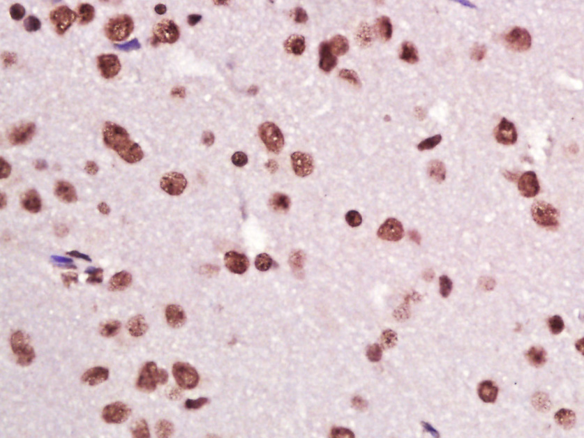
Paraformaldehyde-fixed, paraffin embedded (mouse brain); Antigen retrieval by boiling in sodium citrate buffer (pH6.0) for 15min; Block endogenous peroxidase by 3% hydrogen peroxide for 20 minutes; Blocking buffer (normal goat serum) at 37°C for 30min; Antibody incubation with (CIDEC) Polyclonal Antibody, Unconjugated (bs-6796R) at 1:200 overnight at 4°C, followed by operating according to SP Kit(Rabbit) (sp-0023) instructionsand DAB staining.
Paraformaldehyde-fixed, paraffin embedded (rat brain); Antigen retrieval by boiling in sodium citrate buffer (pH6.0) for 15min; Block endogenous peroxidase by 3% hydrogen peroxide for 20 minutes; Blocking buffer (normal goat serum) at 37°C for 30min; Antibody incubation with (CIDEC) Polyclonal Antibody, Unconjugated (bs-6796R) at 1:200 overnight at 4°C, followed by operating according to SP Kit(Rabbit) (sp-0023) instructionsand DAB staining.
Paraformaldehyde-fixed, paraffin embedded (human gastric); Antigen retrieval by boiling in sodium citrate buffer (pH6.0) for 15min; Block endogenous peroxidase by 3% hydrogen peroxide for 20 minutes; Blocking buffer (normal goat serum) at 37°C for 30min; Antibody incubation with (CIDEC) Polyclonal Antibody, Unconjugated (bs-6796R) at 1:200 overnight at 4°C, followed by operating according to SP Kit(Rabbit) (sp-0023) instructionsand DAB staining.
Paraformaldehyde-fixed, paraffin embedded (rat colon); Antigen retrieval by boiling in sodium citrate buffer (pH6.0) for 15min; Block endogenous peroxidase by 3% hydrogen peroxide for 20 minutes; Blocking buffer (normal goat serum) at 37°C for 30min; Antibody incubation with (CIDEC) Polyclonal Antibody, Unconjugated (bs-6796R) at 1:200 overnight at 4°C, followed by operating according to SP Kit(Rabbit) (sp-0023) instructionsand DAB staining.
Paraformaldehyde-fixed, paraffin embedded (Mouse colon); Antigen retrieval by boiling in sodium citrate buffer (pH6.0) for 15min; Block endogenous peroxidase by 3% hydrogen peroxide for 20 minutes; Blocking buffer (normal goat serum) at 37°C for 30min; Antibody incubation with (CIDEC) Polyclonal Antibody, Unconjugated (bs-6796R) at 1:200 overnight at 4°C, followed by operating according to SP Kit(Rabbit) (sp-0023) instructionsand DAB staining.
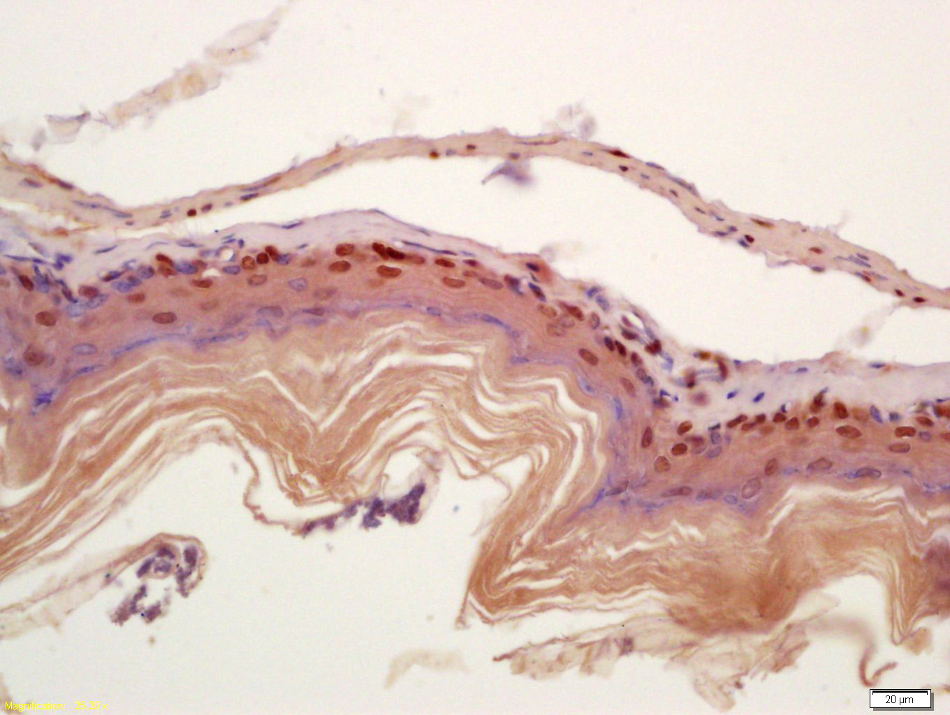
Paraformaldehyde-fixed, paraffin embedded (rat breast); Antigen retrieval by boiling in sodium citrate buffer (pH6.0) for 15min; Block endogenous peroxidase by 3% hydrogen peroxide for 20 minutes; Blocking buffer (normal goat serum) at 37°C for 30min; Antibody incubation with (CIDEC) Polyclonal Antibody, Unconjugated (bs-6796R) at 1:200 overnight at 4°C, followed by operating according to SP Kit(Rabbit) (sp-0023) instructionsand DAB staining.
Tissue/cell: mouse stomach wall; 4% Paraformaldehyde-fixed and paraffin-embedded;
Antigen retrieval: citrate buffer ( 0.01M, pH 6.0 ), Boiling bathing for 15min; Block endogenous peroxidase by 3% Hydrogen peroxide for 30min; Blocking buffer (normal goat serum,C-0005) at 37℃ for 20 min;
Incubation: Anti-CIDEC Polyclonal Antibody, Unconjugated(bs-6796R) 1:200, overnight at 4°C, followed by conjugation to the secondary antibody(SP-0023) and DAB(C-0010) staining
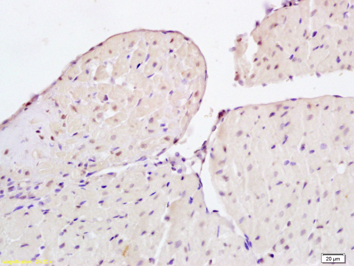
Tissue/cell: rat heart tissue; 4% Paraformaldehyde-fixed and paraffin-embedded;
Antigen retrieval: citrate buffer ( 0.01M, pH 6.0 ), Boiling bathing for 15min; Block endogenous peroxidase by 3% Hydrogen peroxide for 30min; Blocking buffer (normal goat serum,C-0005) at 37℃ for 20 min;
Incubation: Anti-CIDEC Polyclonal Antibody, Unconjugated(bs-6796R) 1:200, overnight at 4°C, followed by conjugation to the secondary antibody(SP-0023) and DAB(C-0010) staining
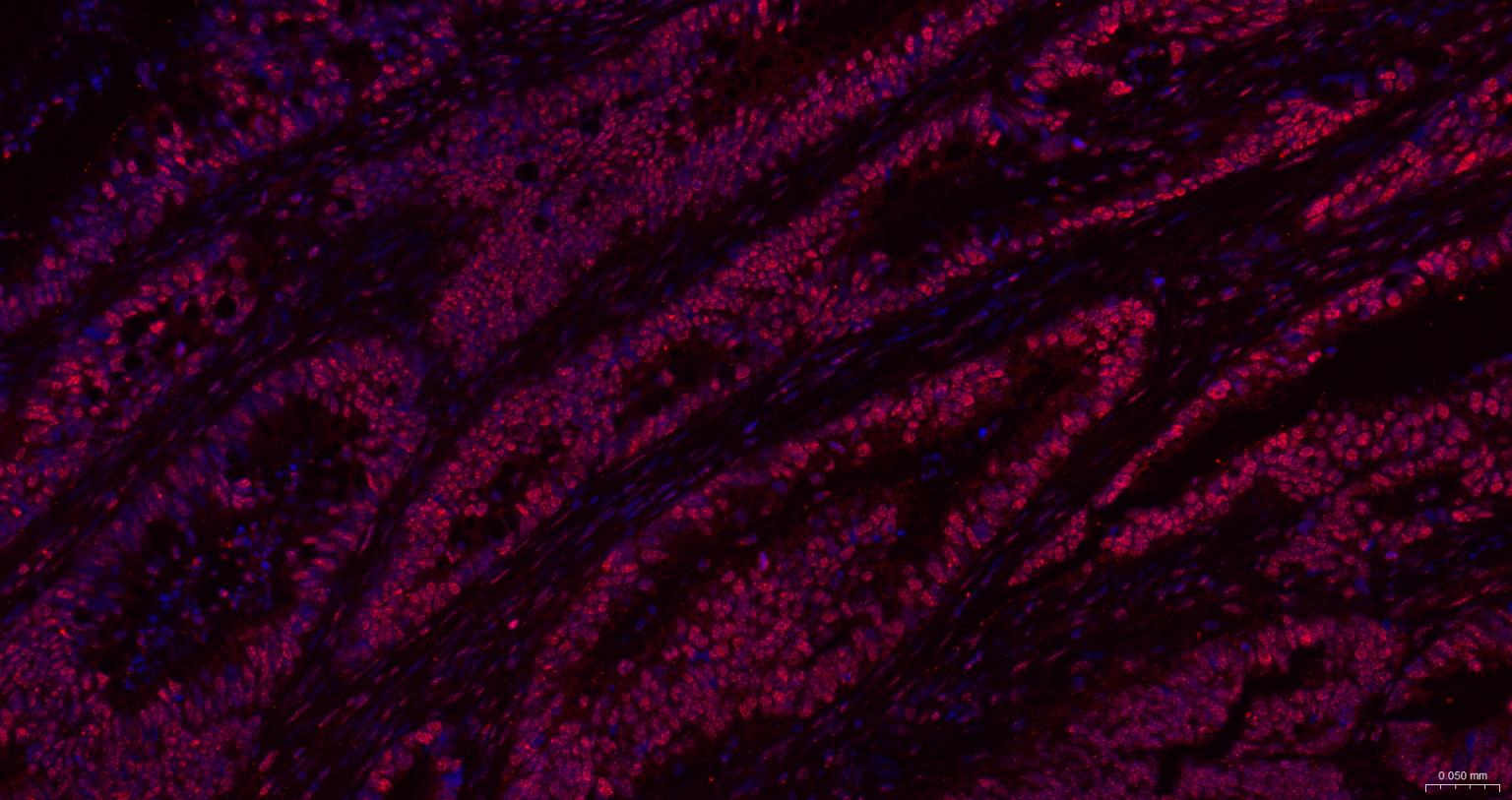
Paraformaldehyde-fixed, paraffin embedded Human Colon Cancer; Antigen retrieval by boiling in sodium citrate buffer (pH6.0) for 15 min; The section was incubated with CIDEC Polyclonal Antibody, Unconjugated (bs-6796R) at 1:200 overnight at 4°C. Followed by conjugated Goat Anti-Rabbit IgG antibody (Red, bs-0295G-BF594), DAPI (blue, C02-04002) was used to stain the cell nuclei.
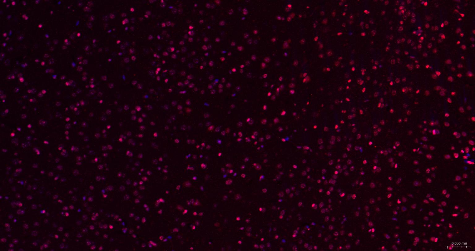
Paraformaldehyde-fixed, paraffin embedded Mouse Cerebrum; Antigen retrieval by boiling in sodium citrate buffer (pH6.0) for 15 min; The section was incubated with CIDEC Polyclonal Antibody, Unconjugated (bs-6796R) at 1:200 overnight at 4°C. Followed by conjugated Goat Anti-Rabbit IgG antibody (Red, bs-0295G-BF594), DAPI (blue, C02-04002) was used to stain the cell nuclei.
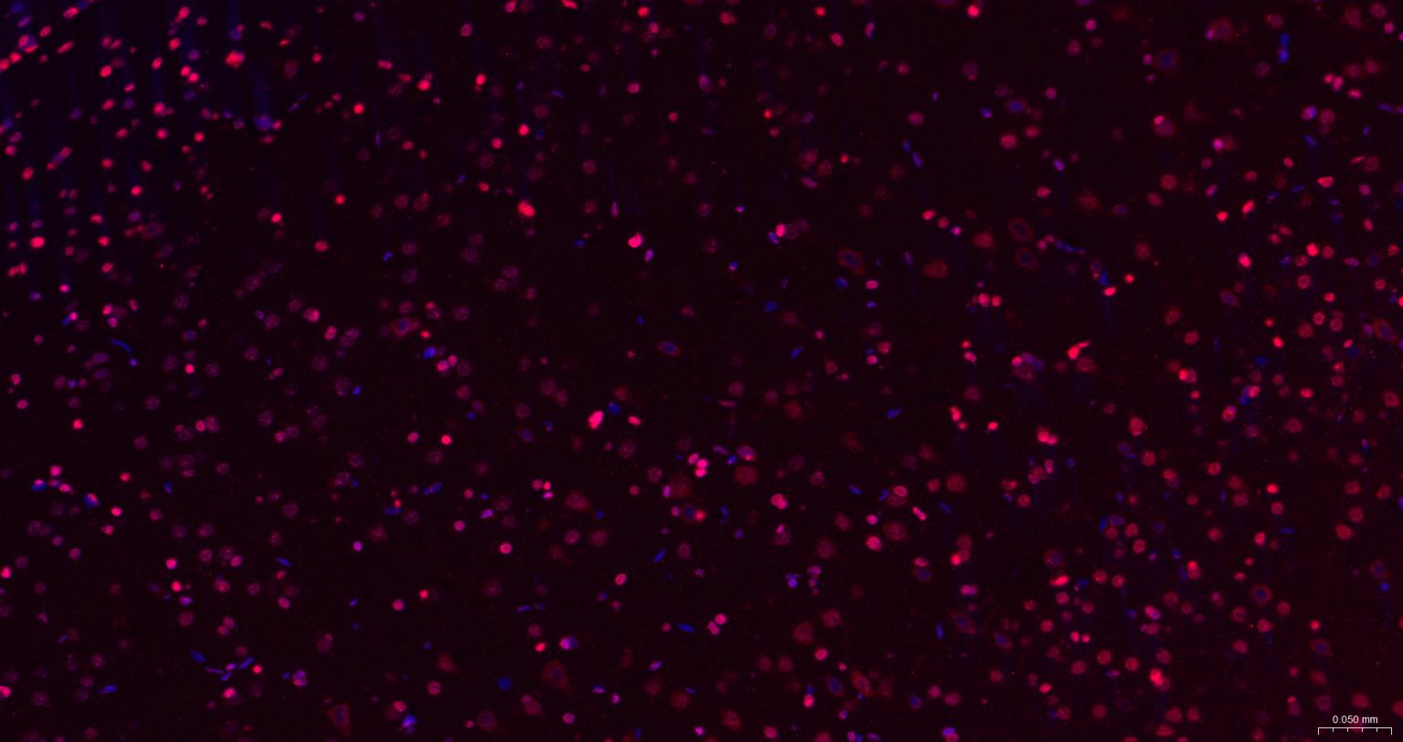
Paraformaldehyde-fixed, paraffin embedded Rat Cerebrum; Antigen retrieval by boiling in sodium citrate buffer (pH6.0) for 15 min; The section was incubated with CIDEC Polyclonal Antibody, Unconjugated (bs-6796R) at 1:200 overnight at 4°C. Followed by conjugated Goat Anti-Rabbit IgG antibody (Red, bs-0295G-BF594), DAPI (blue, C02-04002) was used to stain the cell nuclei.
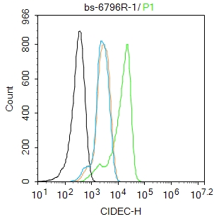
Blank control: A431.
Primary Antibody (green line): Rabbit Anti-CIDEC antibody (bs-6796R)
Dilution: 1ug/Test;
Secondary Antibody : Goat anti-rabbit IgG-FITC
Dilution: 0.5ug/Test.
Protocol
The cells were fixed with 4% PFA (10min at room temperature)and then permeabilized with 0.1% PBST for 20 min at room temperature.The cells were then incubated in 5%BSA to block non-specific protein-protein interactions for 30 min at room temperature .Cells stained with Primary Antibody for 30 min at room temperature. The secondary antibody used for 40 min at room temperature. Acquisition of 20,000 events was performed.
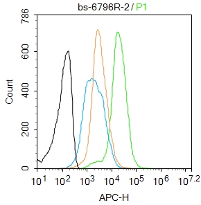
Blank control:Mouse spleen.
Primary Antibody (green line): Rabbit Anti-CIDEC antibody (bs-6796R)
Dilution: 2μg /10^6 cells;
Isotype Control Antibody (orange line): Rabbit IgG .
Secondary Antibody : Goat anti-rabbit IgG-AF647
Dilution: 1μg /test.
Protocol
The cells were fixed with 4% PFA (10min at room temperature)and then permeabilized with 90% ice-cold methanol for 20 min at-20℃. The cells were then incubated in 5%BSA to block non-specific protein-protein interactions for 30 min at room temperature .Cells stained with Primary Antibody for 30 min at room temperature. The secondary antibody used for 40 min at room temperature. Acquisition of 20,000 events was performed.
|
| 1、抗体溶解方法 | |
| 2、抗体修复方式 | |
| 3、常用试剂的配制 | |
| 4、免疫组化操作步骤 | |
| 5、免疫组化问题解答 | |
| 6、Western Blotting 操作步骤 | |
| 7、Western Blotting 问题解答 | |
| 8、关于肽链的设计 | |
| 9、多肽的溶解与保存 | |
| 10、酶标抗体效价测定程序 | |


