

| 产品编号 | bs-9439R |
| 英文名称 | FOXO1A Rabbit pAb |
| 中文名称 | 叉头蛋白O1抗体 |
| 别 名 | FKH1; FKHR; FOXO1A; Afxh; Fkhr1; FOXO1_HUMAN; FOXO1; Forkhead box protein O1A; Forkhead in rhabdomyosarcoma; FOXO1_MOUSE; FOXO1_RAT; forkhead box O1; forkhead homolog in rhabdomyosarcoma |
 |
Specific References (4) | bs-9439R has been referenced in 4 publications.
[IF=8.025] Long Chen. et al. Chondroitin sulfate stimulates the secretion of H2S by Desulfovibrio to improve insulin sensitivity in NAFLD mice. INT J BIOL MACROMOL. 2022 Jul;213:631 WB ; Mouse.
[IF=5.878] Xueying Zhang. et al. Scutellarin ameliorates hepatic lipid accumulation by enhancing autophagy and suppressing IRE1α/XBP1 pathway. 2021 Dec 02 WB ; Mouse ,Human.
[IF=2.81] Kinoshita et al. Associations between Forkhead Box O1 (FoxO1) Expression and Indicators of Hepatic Glucose Production in Transition Dairy Cows Supplemented with Dietary Nicotinic Acid. (2016) PLoS.On. 11:e0146670 WB ; Bovine.
[IF=0] Ahmed et al. Differential expression of mannose-6-phosphate receptor regulates T cell contraction. (2015) J.Leukoc.Biol. 98:313-8 other ; Mouse.
|
| 研究领域 | 细胞生物 免疫学 神经生物学 信号转导 表观遗传学 |
| 抗体来源 | Rabbit |
| 克隆类型 | Polyclonal |
| 交叉反应 | Human,Rat (predicted: Mouse,Rabbit,Pig,Cow,Chicken,Dog,Horse) |
| 产品应用 | WB=1:500-2000,IHC-P=1:100-500,IHC-F=1:100-500,IF=1:100-500,Flow-Cyt=2ug/Test
not yet tested in other applications. optimal dilutions/concentrations should be determined by the end user. |
| 理论分子量 | 72kDa |
| 检测分子量 | 72 |
| 细胞定位 | 细胞核 细胞浆 |
| 性 状 | Liquid |
| 浓 度 | 1mg/ml |
| 免 疫 原 | KLH conjugated synthetic peptide derived from human FKHR: 165-270/655 |
| 亚 型 | IgG |
| 纯化方法 | affinity purified by Protein A |
| 缓 冲 液 | 0.01M TBS (pH7.4) with 1% BSA, 0.02% Proclin300 and 50% Glycerol. |
| 保存条件 | Shipped at 4℃. Store at -20℃ for one year. Avoid repeated freeze/thaw cycles. |
| 注意事项 | This product as supplied is intended for research use only, not for use in human, therapeutic or diagnostic applications. |
| PubMed | PubMed |
| 产品介绍 |
FKHR (for forkhead in rhabdomyosarcoma) and FKHRL1 are members of the forkhead family of transcription factors. Transcriptional activation of FKHR proteins is regulated by the serine/threonine kinase Akt1, which phosphorylates FKHRL1 and results in FKHRL1 associating with 14-3-3 proteins and being retained in the cytoplasm. Induction of apoptosis or withdrawal of growth factors stimulates dephosphorylation and nuclear translocation of FKHR proteins, leading to FKHR-induced gene-specific transcriptional activation. FKHR, also designated forkhead box protein O1A (FOXO1), is a ubiquitously expressed protein that shuttles between the cytoplasm and nucleus. Genetic mutations in FKHR genes, including the t(2;13) and t(1;3) translocations, are commonly found in alveolar rhabdomyosarcomas. These translocations result in the fusion of the amino terminus of Pax-3 or Pax-7, including the paired box and homeodomain DNA-binding domains, with the carboxy-terminus of FKHR, which contains a transcriptional activation domain. The Pax-3/FKHR fusion protein appears to function as an oncogenic transcription factor that enhances the activation of normal Pax-3 target genes. Function: Transcription factor which acts as a regulator of cell responses to oxidative stress. In the presence of KIRT1, mediates down-regulation of cyclin D1 and up-regulation of CDKN1B levels which are required for cell transition from proliferative growth to quiescence. Triggers death of postmitotic neurons when phosphorylated by CDK1. Activates transcription of PMAIP1. Subunit: Interacts with LRPPRC. Interacts with SIRT1 and this interaction requires the presence of KRIT1. Interacts with NLK. Binds to CDK1 and 14-3-3 proteins. Subcellular Location: Cytoplasm. Nucleus. Note=Shuttles between cytoplasm and nucleus. Translocates to the nucleus upon oxidative stress induced phosphorylation at Ser-212 by STK4/MST1. Translocates to the nucleus upon phosphorylation of Thr-24, Ser-256 and Ser-322 by SGK1. Tissue Specificity: Ubiquitous. Post-translational modifications: Phosphorylated by AKT1; insulin-induced. Phosphorylated by NLK, which inhibits transcriptional activity and promotes nuclear export. IGF1 rapidly induces phosphorylation of Ser-256, Thr-24, and Ser-319. Phosphorylation of Ser-256 decreases DNA-binding activity and promotes the phosphorylation of Thr-24, and Ser-319, permitting phosphorylation of Ser-322 and Ser-325, probably by CK1, leading to nuclear exclusion and loss of function. Phosphorylation of Ser-329 is independent of IGF1 and leads to reduced function. Phosphorylated upon DNA damage, probably by ATM or ATR. Phosphorylation of Ser-249 by CDK1 disrupts 14-3-3 proteins binding and thereby promotes FOXO1 nuclear accumulation and subsequent transcription activation and cell death. Phosphorylated by STK4/MST1 on Ser-212 upon oxidative stress. Phosphorylated on Thr-24, Ser-256 and Ser-322 by SGK1 resulting in its translocation from the nucleus to the cytoplasm. DISEASE: Defects in FOXO1 are a cause of rhabdomyosarcoma type 2 (RMS2) [MIM:268220]. It is a form of rhabdomyosarcoma, a highly malignant tumor of striated muscle derived from primitive mesenchimal cells and exhibiting differentiation along rhabdomyoblastic lines. Rhabdomyosarcoma is one of the most frequently occurring soft tissue sarcomas and the most common in children. It occurs in four forms: alveolar, pleomorphic, embryonal and botryoidal rhabdomyosarcomas. Note=Chromosomal aberrations involving FOXO1 are found in rhabdomyosarcoma. Translocation (2;13)(q35;q14) with PAX3; translocation t(1;13)(p36;q14) with PAX7. The resulting protein is a transcriptional activator. Similarity: Contains 1 fork-head DNA-binding domain. SWISS: Q12778 Gene ID: 2308 Database links: Entrez Gene: 2308 Human Entrez Gene: 56458 Mouse Omim: 136533 Human SwissProt: Q12778 Human SwissProt: Q9R1E0 Mouse Unigene: 370666 Human Unigene: 29891 Mouse Unigene: 116108 Rat |
| 产品图片 |
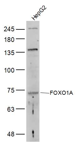
Sample:
HepG2(Human) Cell Lysate at 30 ug
Primary: Anti-FOXO1A (bs-9439R) at 1/300 dilution
Secondary: IRDye800CW Goat Anti-Rabbit IgG at 1/20000 dilution
Predicted band size: 72 kD
Observed band size: 72 kD
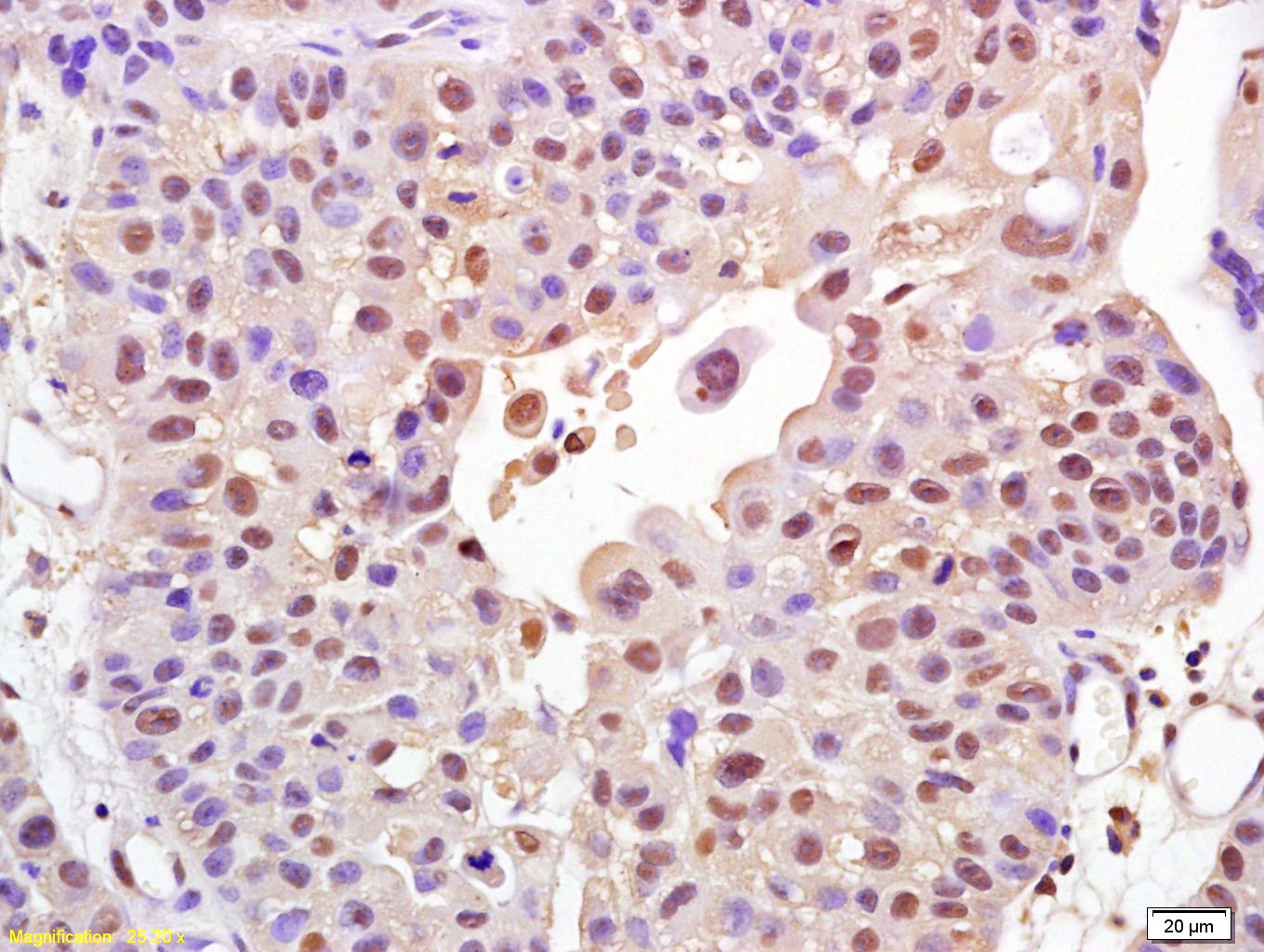
Tissue/cell: human bladder carcinoma; 4% Paraformaldehyde-fixed and paraffin-embedded;
Antigen retrieval: citrate buffer ( 0.01M, pH 6.0 ), Boiling bathing for 15min; Block endogenous peroxidase by 3% Hydrogen peroxide for 30min; Blocking buffer (normal goat serum,C-0005) at 37℃ for 20 min;
Incubation: Anti-FOXO1A Polyclonal Antibody, Unconjugated(bs-9439R) 1:200, overnight at 4°C, followed by conjugation to the secondary antibody(SP-0023) and DAB(C-0010) staining
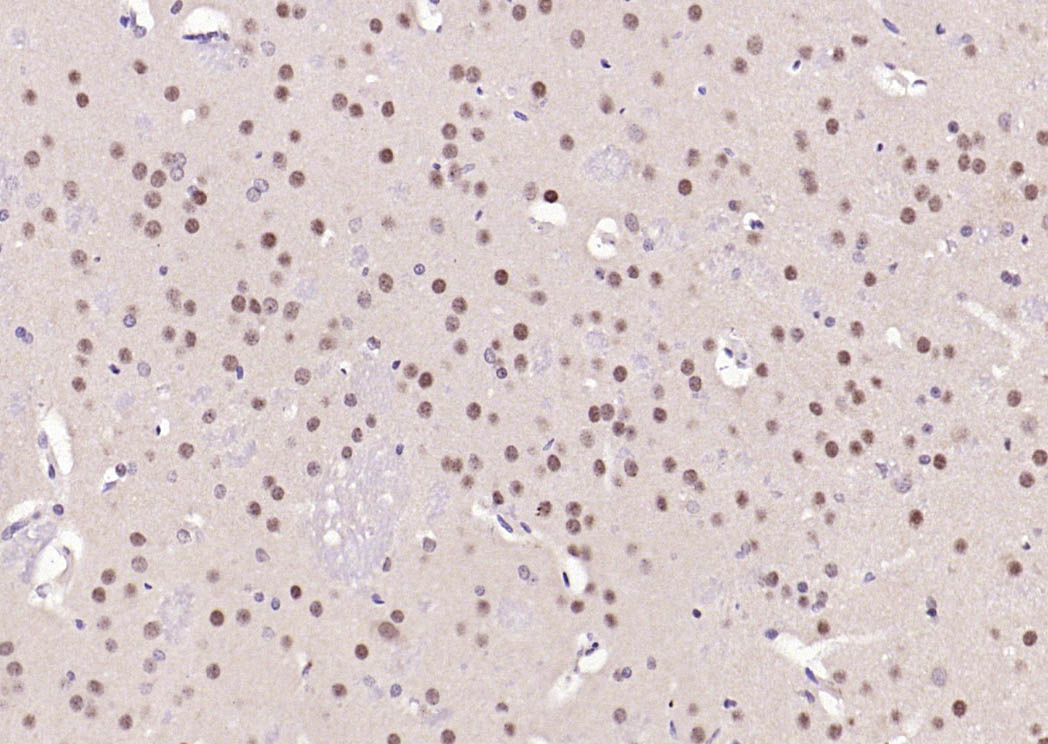
Paraformaldehyde-fixed, paraffin embedded (rat brain); Antigen retrieval by boiling in sodium citrate buffer (pH6.0) for 15min; Block endogenous peroxidase by 3% hydrogen peroxide for 20 minutes; Blocking buffer (normal goat serum) at 37°C for 30min; Antibody incubation with (FOXO1A) Polyclonal Antibody, Unconjugated (bs-9439R) at 1:200 overnight at 4°C, followed by operating according to SP Kit(Rabbit) (sp-0023) instructionsand DAB staining.
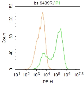
Blank control: HepG2.
Primary Antibody (green line): Rabbit Anti-FOXO1A antibody (bs-9439R)
Dilution: 1μg /10^6 cells;
Isotype Control Antibody (orange line): Rabbit IgG .
Secondary Antibody : Goat anti-rabbit IgG-PE
Dilution: 1μg /test.
Protocol
The cells were fixed with 4% PFA (10min at room temperature)and then permeabilized with 90% ice-cold methanol for 20 min at-20℃. The cells were then incubated in 5%BSA to block non-specific protein-protein interactions for 30 min at at room temperature .Cells stained with Primary Antibody for 30 min at room temperature. The secondary antibody used for 40 min at room temperature. Acquisition of 20,000 events was performed.
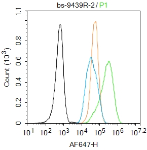
Blank control: HepG2.
Primary Antibody (green line): Rabbit Anti-FOXO1A antibody (bs-9439R)
Dilution: 1μg /10^6 cells;
Isotype Control Antibody (orange line): Rabbit IgG .
Secondary Antibody : Goat anti-rabbit IgG-AF647
Dilution: 1μg /test.
Protocol
The cells were fixed with 4% PFA (10min at room temperature)and then permeabilized with 90% ice-cold methanol for 20 min at -20℃. The cells were then incubated in 5%BSA to block non-specific protein-protein interactions for 30 min at room temperature .Cells stained with Primary Antibody for 30 min at room temperature. The secondary antibody used for 40 min at room temperature. Acquisition of 20,000 events was performed.
|
| 1、抗体溶解方法 | |
| 2、抗体修复方式 | |
| 3、常用试剂的配制 | |
| 4、免疫组化操作步骤 | |
| 5、免疫组化问题解答 | |
| 6、Western Blotting 操作步骤 | |
| 7、Western Blotting 问题解答 | |
| 8、关于肽链的设计 | |
| 9、多肽的溶解与保存 | |
| 10、酶标抗体效价测定程序 | |


