

| 产品编号 | bs-11397R |
| 英文名称 | Arginase II Rabbit pAb |
| 中文名称 | 精氨酸酶2抗体 |
| 别 名 | AII; ARGI2_HUMAN; ARG2; Arginase II; Kidney-type arginase; Non-hepatic arginase; Type II arginase; 3.5.3.1; ARGI2_MOUSE; ARGI2_RAT; |
 |
Specific References (2) | bs-11397R has been referenced in 2 publications.
[IF=12.12] Zaytouni et al. Critical role for arginase 2 in obesity-associated pancreatic cancer. (2017) Nat.Commun. 8:242 WB, IHC-P ; Human.
[IF=1.632] Liu Y et al. Isolation and characterization of ovine monocyte-derived macrophages from peripheral blood.(2018)Vet Immunol Immunopathol. Nov;205:83-92. IF ; Sheep.
|
| 研究领域 | 肿瘤 细胞生物 信号转导 激酶和磷酸酶 |
| 抗体来源 | Rabbit |
| 克隆类型 | Polyclonal |
| 克 隆 号 | |
| 交叉反应 | Human,Mouse (predicted: Rat,Rabbit,Cow,Chicken,Dog) |
| 产品应用 | WB=1:500-2000,IHC-P=1:100-500,IHC-F=1:100-500,IF=1:100-500,Flow-Cyt=1ug/Test,ICC/IF=1:100-500
not yet tested in other applications. optimal dilutions/concentrations should be determined by the end user. |
| 理论分子量 | 36 kDa |
| 检测分子量 | 36 |
| 细胞定位 | 细胞浆 |
| 性 状 | Liquid |
| 浓 度 | 1mg/ml |
| 免 疫 原 | KLH conjugated synthetic peptide derived from human Arginase II: 181-290/354 |
| 亚 型 | IgG |
| 纯化方法 | affinity purified by Protein A |
| 缓 冲 液 | 0.01M TBS (pH7.4) with 1% BSA, 0.02% Proclin300 and 50% Glycerol. |
| 保存条件 | Shipped at 4℃. Store at -20℃ for one year. Avoid repeated freeze/thaw cycles. |
| 注意事项 | This product as supplied is intended for research use only, not for use in human, therapeutic or diagnostic applications. |
| PubMed | PubMed |
| 产品介绍 |
Arginase I (also designated liver-type arginase), which is expressed almost exclusively in the liver, catalyzes the conversion of arginine to ornithine and urea (1). The human arginase I gene, which maps to chromosome 6q23, encodes a 322 amino acid protein. Arginase I exists as a homotrimeric protein and contains a binuclear manganese cluster (2-4). Arginase II catalyzes the same reaction as arginase I, but differs in its tissue specificity and subcellular location (5,6). Specifically, arginase II localizes to the mitochondria (5,6). Arginase II is expressed in non-hepatic tissues, with the highest levels of expression in the kidneys, but, unlike arginase I, is not expressed in liver (5,6). The human arginase II gene, which maps to chromosome 14q24.1, encodes a 354 amino acid protein (3,5-7). In addition, arginase II contains a putative amino-terminal mitochondrial localization sequence (5,6). Function: May play a role in the regulation of extra-urea cycle arginine metabolism and also in down-regulation of nitric oxide synthesis. Extrahepatic arginase functions to regulate L-arginine bioavailability to NO synthase. Since NO synthase is found in the penile corpus cavernosum smooth muscle, the clitoral corpus cavernosum and the vagina, arginase II plays a role in both male and female sexual arousal. It is therefore a potential target for the treatment of male and female sexual arousal disorders. Subunit: Homotrimer. Subcellular Location: Cytoplasm. Tissue Specificity: Expressed most strongly in kidney and prostate, much less strongly in the brain, skeletal muscle, placenta, lung, mammary gland, macrophage, uterus, testis and gut, but apparently not in the liver, heart and pancreas. Similarity: Belongs to the arginase family. SWISS: P78540 Gene ID: 384 Database links: Entrez Gene: 384 Human Entrez Gene: 11847 Mouse Omim: 107830 Human SwissProt: P78540 Human SwissProt: O08691 Mouse Unigene: 226007 Human Unigene: 723133 Human Unigene: 3506 Mouse Unigene: 11055 Rat |
| 产品图片 |
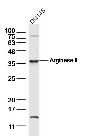
Sample:
DU145(human)cell Lysate at 30 ug
Primary: Anti-Arginase II (bs-11397R) at 1/300 dilution
Secondary: IRDye800CW Goat Anti-Rabbit IgG at 1/20000 dilution
Predicted band size: 36kD
Observed band size: 36 kD
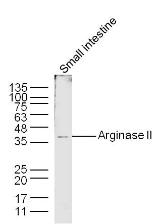
Sample: Small intestine (Mouse) Lysate at 40 ug
Primary: Anti-Arginase II (bs-11397R) at 1/300 dilution
Secondary: IRDye800CW Goat Anti-Rabbit IgG at 1/20000 dilution
Predicted band size: 36 kD
Observed band size: 36 kD

Sample:
Small intestine (Mouse) Lysate at 40 ug
Primary: Anti-Arginase II (Bs- 11397R) at 1/300 dilution
Secondary: IRDye800CW Goat Anti-Rabbit IgG at 1/20000 dilution
Predicted band size: 36 kD
Observed band size: 36 kD
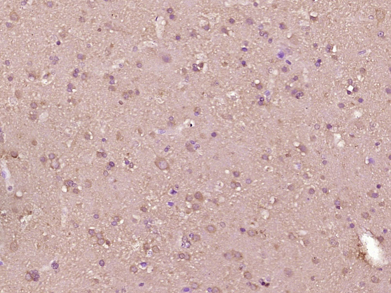
Paraformaldehyde-fixed, paraffin embedded (Human brain glioma); Antigen retrieval by boiling in sodium citrate buffer (pH6.0) for 15min; Block endogenous peroxidase by 3% hydrogen peroxide for 20 minutes; Blocking buffer (normal goat serum) at 37°C for 30min; Antibody incubation with (Arginase II) Polyclonal Antibody, Unconjugated (bs-11397R) at 1:400 overnight at 4°C, followed by operating according to SP Kit(Rabbit) (sp-0023) instructionsand DAB staining.
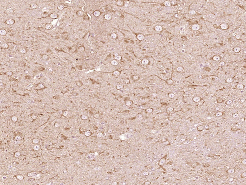
Paraformaldehyde-fixed, paraffin embedded (Mouse brain); Antigen retrieval by boiling in sodium citrate buffer (pH6.0) for 15min; Block endogenous peroxidase by 3% hydrogen peroxide for 20 minutes; Blocking buffer (normal goat serum) at 37°C for 30min; Antibody incubation with (Arginase II) Polyclonal Antibody, Unconjugated (bs-11397R) at 1:400 overnight at 4°C, followed by operating according to SP Kit(Rabbit) (sp-0023) instructionsand DAB staining.
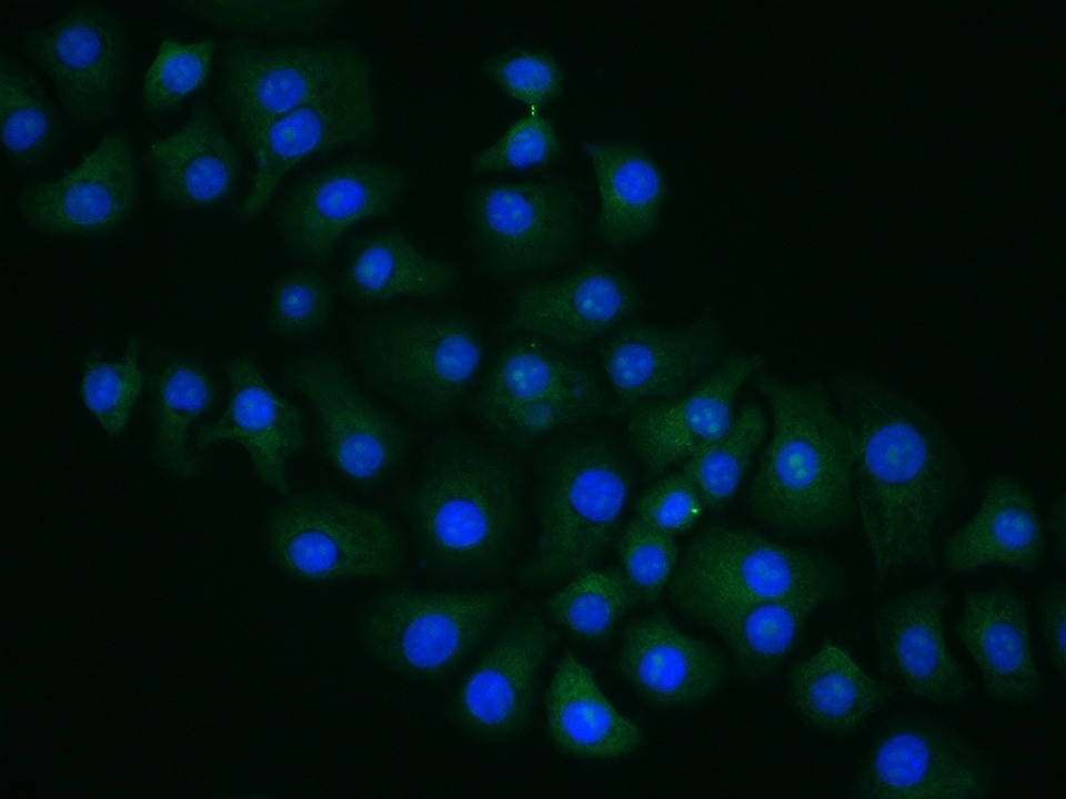
HepG2 cell; 4% Paraformaldehyde-fixed; Triton X-100 at room temperature for 20 min; Blocking buffer (normal goat serum, C-0005) at 37°C for 20 min; Antibody incubation with (Arginase II) polyclonal Antibody, Unconjugated (bs-11397R) 1:100, 90 minutes at 37°C; followed by a conjugated Goat Anti-Rabbit IgG antibody at 37°C for 90 minutes, DAPI (blue, C02-04002) was used to stain the cell nuclei.
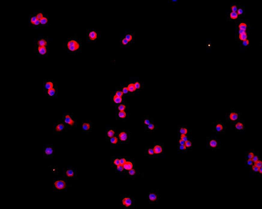
Tissue/cell: human MCF-7 cells;4% Paraformaldehyde-fixed and paraffin-embedded;
Antigen retrieval: citrate buffer ( 0.01M, pH 6.0 ), Boiling bathing for 15min; Blocking buffer (normal goat serum,C-0005) at 37℃ for 20 min;
Incubation: Anti-Arginase II Polyclonal Antibody, Unconjugated(bs-11397R) 1:200, overnight at 4°C; The secondary antibody was Goat Anti-Rabbit IgG, Cy3 conjugated(bs-0295G-Cy3)used at 1:200 dilution for 40 minutes at 37°C. DAPI(5ug/ml,blue,C-0033) was used to stain the cell nuclei
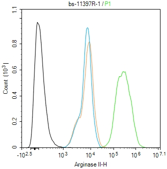
Blank control(black line):HepG2.
Primary Antibody (green line): Rabbit Anti-Arginase II antibody (bs-11397R)
Dilution:1ug/Test;
Secondary Antibody(white blue line): Goat anti-rabbit IgG-AF488
Dilution: 0.5ug/Test.
Isotype control(orange line): Normal Rabbit IgG
Protocol
The cells were fixed with 4% PFA (10min at room temperature)and then permeabilized with 90% ice-cold methanol for 20 min at -20℃, The cells were then incubated in 5%BSA to block non-specific protein-protein interactions for 30 min at room temperature .Cells stained with Primary Antibody for 30 min at room temperature. The secondary antibody used for 40 min at room temperature. Acquisition of 20,000 events was performed.
|
| 1、抗体溶解方法 | |
| 2、抗体修复方式 | |
| 3、常用试剂的配制 | |
| 4、免疫组化操作步骤 | |
| 5、免疫组化问题解答 | |
| 6、Western Blotting 操作步骤 | |
| 7、Western Blotting 问题解答 | |
| 8、关于肽链的设计 | |
| 9、多肽的溶解与保存 | |
| 10、酶标抗体效价测定程序 | |


