

| дә§е“Ғзј–еҸ· | bs-10106R |
| иӢұж–ҮеҗҚз§° | phospho-Tau (Thr548) Rabbit pAb |
| дёӯж–ҮеҗҚз§° | зЈ·й…ёеҢ–еҫ®з®Ўзӣёе…іиӣӢзҷҪжҠ—дҪ“ |
| еҲ« еҗҚ | MAPT | Tau (p-T548); p-Tau; phospho-Tau; tau; DDPAC; FTD1; FTDP-17; MAPTL; MSTD; MTBT1; MTBT2; PPND; PPP1R103; Tau-PHF6; tau-40; Mtapt; PHF-tau; MAPT_0N4R; MAPT_1N4R; MAPT_2N4R; RNPTAU; pTau; TAU_BOVIN; MAPT; Neurofibrillary tangle protein; Paired helical |
| дә§е“Ғзұ»еһӢ | зЈ·й…ёеҢ–жҠ—дҪ“ |
| з ”з©¶йўҶеҹҹ | з»Ҷиғһз”ҹзү© е…Қз–«еӯҰ зҘһз»Ҹз”ҹзү©еӯҰ дҝЎеҸ·иҪ¬еҜј з»ҶиғһеҮӢдәЎ Alzheimer's |
| жҠ—дҪ“жқҘжәҗ | Rabbit |
| е…ӢйҡҶзұ»еһӢ | Polyclonal |
| е…Ӣ йҡҶ еҸ· | |
| дәӨеҸүеҸҚеә” | Mouse,Rat (predicted: Human,Rabbit,Cow,Dog,Horse) |
| дә§е“Ғеә”з”Ё | WB=1:500-2000,IHC-P=1:100-500,IHC-F=1:100-500,IF=1:100-500
not yet tested in other applications. optimal dilutions/concentrations should be determined by the end user. |
| зҗҶи®әеҲҶеӯҗйҮҸ | 52/79 kDa |
| з»Ҷиғһе®ҡдҪҚ | з»ҶиғһжөҶ з»ҶиғһиҶң |
| жҖ§ зҠ¶ | Liquid |
| жө“ еәҰ | 1mg/ml |
| е…Қ з–« еҺҹ | KLH conjugated Synthesised phosphopeptide derived from human Tau around the phosphorylation site of Thr548: VR(p-T)PP |
| дәҡ еһӢ | IgG |
| зәҜеҢ–ж–№жі• | affinity purified by Protein A |
| зј“ еҶІ ж¶І | 0.01M TBS (pH7.4) with 1% BSA, 0.02% Proclin300 and 50% Glycerol. |
| дҝқеӯҳжқЎд»¶ | Shipped at 4в„ғ. Store at -20в„ғ for one year. Avoid repeated freeze/thaw cycles. |
| жіЁж„ҸдәӢйЎ№ | This product as supplied is intended for research use only, not for use in human, therapeutic or diagnostic applications. |
| PubMed | PubMed |
| дә§е“Ғд»Ӣз»Қ |
Tau proteins are important Promotes microtubule assembly and stability, and might be involved in the establishment and maintenance of neuronal polarity. The C-terminus binds axonal microtubules while the N-terminus binds neural plasma membrane components, suggesting that tau functions as a linker protein between both. Axonal polarity is predetermined by tau localization (in the neuronal cell) in the domain of the cell body defined by the centrosome. The short isoforms allow plasticity of the cytoskeleton whereas the longer isoforms may preferentially play a role in its stabilization. Tau proteins subcellular located in the axons of neurons, in the cytoso l and in association with plasma membrane components. It expressed in neurons. PNS-tau is expressed in the peripheral nervous system while the others are expressed in the central nervous system. Function: Promotes microtubule assembly and stability, and might be involved in the establishment and maintenance of neuronal polarity. The C-terminus binds axonal microtubules while the N-terminus binds neural plasma membrane components, suggesting that tau functions as a linker protein between both. Axonal polarity is predetermined by TAU/MAPT localization (in the neuronal cell) in the domain of the cell body defined by the centrosome. The short isoforms allow plasticity of the cytoskeleton whereas the longer isoforms may preferentially play a role in its stabilization. Subunit: Interacts with PSMC2 through SQSTM1. Interacts with SQSTM1 when polyubiquitinated. Interacts with FKBP4. Binds to CSNK1D. Interacts with SGK1. Subcellular Location: Cytoplasm, cytosol. Cell membrane; Peripheral membrane protein; Cytoplasmic side. Cytoplasm, cytoskeleton. Cell projection, axon. Note=Mostly found in the axons of neurons, in the cytosol and in association with plasma membrane components. Tissue Specificity: Expressed in neurons. Isoform PNS-tau is expressed in the peripheral nervous system while the others are expressed in the central nervous system. Post-translational modifications: Phosphorylation at serine and threonine residues in S-P or T-P motifs by proline-directed protein kinases (PDPK1: CDK1, CDK5, GSK3, MAPK) (only 2-3 sites per protein in interphase, seven-fold increase in mitosis, and in the form associated with paired helical filaments (PHF-tau)), and at serine residues in K-X-G-S motifs by MAP/microtubule affinity-regulating kinase (MARK1 or MARK2), causing detachment from microtubules, and their disassembly. Phosphorylation decreases with age. Phosphorylation within tau/MAP's repeat domain or in flanking regions seems to reduce tAU/MAP's interaction with, respectively, microtubules or plasma membrane components. Phosphorylation on Ser-610, Ser-622, Ser-641 and Ser-673 in several isoforms during mitosis. Phosphorylation at Ser-548 by GSK3B reduces ability to bind and stabilize microtubules. Phosphorylation at Ser-579 by BRSK1 and BRSK2 in neurons affects ability to bind microtubules and plays a role in neuron polarization. Phosphorylated at Ser-554, Ser-579, Ser-602, Ser-606 and Ser-669 by PHK. Phosphorylation at Ser-214 by SGK1 mediates microtubule depolymerization and neurite formation in hippocampal neurons. There is a reciprocal down-regulation of phosphorylation and O-GlcNAcylation. Phosphorylation on Ser-717 completely abolishes the O-GlcNAcylation on this site, while phosphorylation on Ser-713 and Ser-721 reduces glycosylation by a factor of 2 and 4 respectively. Phosphorylation on Ser-721 is reduced by about 41.5% by GlcNAcylation on Ser-717. DISEASE: Defects in MAPT are a cause of Parkinson-dementia syndrome (PARDE) [MIM:260540]. A syndrome characterized by parkinsonism tremor, rigidity, dementia, ophthalmoparesis and pyramidal signs. Neurofibrillary degeneration occurs in the hippocampus, basal ganglia and brainstem nuclei. Similarity : Contains 4 Tau/MAP repeats. Similarity: Contains 4 Tau/MAP repeats. SWISS: P10636-2 Gene ID: 4137 Database links: Entrez Gene: 4137 Human Entrez Gene: 17762 Mouse Omim: 157140 Human SwissProt: P10636 Human SwissProt: P10637 Mouse Unigene: 101174 Human Unigene: 1287 Mouse Unigene: 2455 Rat P-tauиӣӢзҷҪжҳҜи„‘еҶ…зҘһз»Ҹе…ғз»Ҷиғһж”Ҝжһ¶иӣӢзҷҪд№ӢдёҖгҖӮе…¶жӯЈеёёеҠҹиғҪжҳҜдҝғиҝӣеҫ®з®ЎиӣӢзҷҪз»„жҲҗеҫ®з®ЎпјҢ并з»ҙжҢҒе·ІеҪўжҲҗеҫ®з®Ўзҡ„зЁіе®ҡжҖ§гҖӮеҸӮдёҺз»ҙжҢҒз»ҶиғһеҪўжҖҒгҖҒдҝЎжҒҜдј йҖ’гҖҒз»ҶиғһеҲҶиЈӮзӯүйҮҚиҰҒз”ҹзү©еӯҰиҝҮзЁӢ,жҳҜиҪҙзӘҒз”ҹй•ҝеҸ‘иӮІе’ҢзҘһз»Ҹе…ғжһҒжҖ§еҪўжҲҗзҡ„дёҚеҸҜзјәе°‘еӣ зҙ гҖӮиҝ‘е№ҙжқҘеҸ‘зҺ°tauиӣӢзҷҪдёҺдёҖдәӣдёӯжһўзҘһз»Ҹзі»з»ҹеҸҳжҖ§з–ҫз—…еҜҶеҲҮзӣёе…і,е°Өе…¶жҳҜзҘһз»ҸTauе…·жңүеҗҜеҠЁеҫ®з®Ўзі»з»ҹзҡ„иЈ…й…Қд»ҘеҸҠзЁіе®ҡеҫ®з®Ўзі»з»ҹзҡ„дҪңз”ЁпјҢиҜҘиӣӢзҷҪзҡ„й”ҷиҜҜжҠҳеҸ дёҺиҖҒе№ҙжҖ§з—ҙе‘ҶзӯүзҘһз»ҸйҖҖиЎҢжҖ§з–ҫз—…еҜҶеҲҮзӣёе…ігҖӮ |
| дә§е“ҒеӣҫзүҮ |
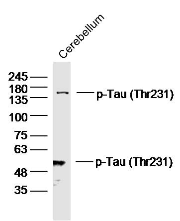
(mouse/tissue)Cerebellum lysates probed with p-Tau(Thr231) Polyclonal Antibody, Unconjugated (Catalog #bs-10106R) at 1:300 overnight at 4ЛҡC. Followed by a conjugated secondary antibody (Secondary Catalog #926-32211) at 1:10000 for 60 min at 37ЛҡC.
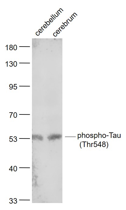
Sample:
Cerebellum (Mouse) Lysate at 40 ug
Cerebrum (Rat) Lysate at 40 ug
Primary: Anti- phospho-Tau (Thr548) (bs-10106R) at 1/1000 dilution
Secondary: IRDye800CW Goat Anti-Rabbit IgG at 1/20000 dilution
Predicted band size: 52/79 kD
Observed band size: 53 kD
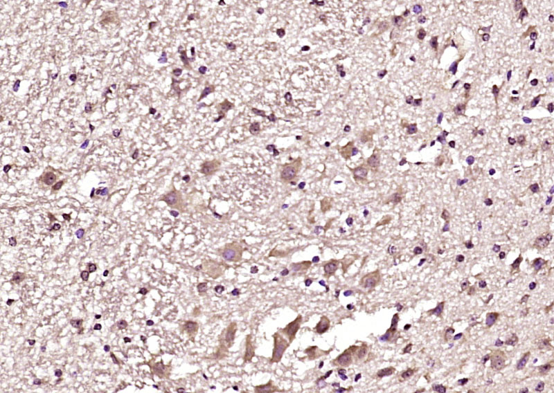
Paraformaldehyde-fixed, paraffin embedded (mouse cerebellum); Antigen retrieval by boiling in sodium citrate buffer (pH6.0) for 15min; Block endogenous peroxidase by 3% hydrogen peroxide for 20 minutes; Blocking buffer (normal goat serum) at 37В°C for 30min; Antibody incubation with (phospho-Tau (Thr548)) Polyclonal Antibody, Unconjugated (bs-10106R) at 1:200 overnight at 4В°C, followed by operating according to SP Kit(Rabbit) (sp-0023) instructionsand DAB staining.
|


