

| дә§е“Ғзј–еҸ· | bs-11575R |
| иӢұж–ҮеҗҚз§° | PDE7A Rabbit pAb |
| дёӯж–ҮеҗҚз§° | зЈ·й…ёдәҢй…Ҝй…¶7жҠ—дҪ“ |
| еҲ« еҗҚ | HCP1; PDE7; PDE7A_HUMAN; PDE7A; TM22; cAMP-specific phosphodiesterase 7A; 3.1.4.53; |
| з ”з©¶йўҶеҹҹ | еҝғиЎҖз®Ў з»Ҷиғһз”ҹзү© зҘһз»Ҹз”ҹзү©еӯҰ дҝЎеҸ·иҪ¬еҜј жҝҖй…¶е’ҢзЈ·й…ёй…¶ |
| жҠ—дҪ“жқҘжәҗ | Rabbit |
| е…ӢйҡҶзұ»еһӢ | Polyclonal |
| е…Ӣ йҡҶ еҸ· | |
| дәӨеҸүеҸҚеә” | Human,Mouse,Rat (predicted: Rabbit,Pig,Cow,Chicken,Dog,Horse) |
| дә§е“Ғеә”з”Ё | WB=1:500-2000,IHC-P=1:100-500,IHC-F=1:100-500,IF=1:100-500,Flow-Cyt=1Ојg/Test
not yet tested in other applications. optimal dilutions/concentrations should be determined by the end user. |
| зҗҶи®әеҲҶеӯҗйҮҸ | 55 kDa |
| жЈҖжөӢеҲҶеӯҗйҮҸ | 55 |
| з»Ҷиғһе®ҡдҪҚ | з»ҶиғһжөҶ |
| жҖ§ зҠ¶ | Liquid |
| жө“ еәҰ | 1mg/ml |
| е…Қ з–« еҺҹ | KLH conjugated synthetic peptide derived from human PDE7A: 341-440/482 |
| дәҡ еһӢ | IgG |
| зәҜеҢ–ж–№жі• | affinity purified by Protein A |
| зј“ еҶІ ж¶І | 0.01M TBS (pH7.4) with 1% BSA, 0.02% Proclin300 and 50% Glycerol. |
| дҝқеӯҳжқЎд»¶ | Shipped at 4в„ғ. Store at -20в„ғ for one year. Avoid repeated freeze/thaw cycles. |
| жіЁж„ҸдәӢйЎ№ | This product as supplied is intended for research use only, not for use in human, therapeutic or diagnostic applications. |
| PubMed | PubMed |
| дә§е“Ғд»Ӣз»Қ |
Phosphodiesterases (PDE, also designated cyclic nucleotide phosphodiesterase) are important for the downregulation of the intracellular level of the second messenger cyclic adenosine monophosphate (cAMP) by hydrolyzing cAMP to 5'AMP. Phosphodiesterase type 3 isoforms, PDE3A and 3B, are expressed primarily in cardiovascular tissue and adipose tissue, respectively. PDE3A, is found in myocardium and platelets and PDE3B is found in lymphocytes. The PDE7A1 (HCP1) isozyme and the PDE7A2 proteins, alternate splice products of PDE7A, are highly expressed in skeletal muscle. PDE7B is most highly expressed in pancreas. The PDE family contains proteins that serve tissue-specific roles in regulation of lipolysis, glycogenolysis, myocardial contractility, and smooth muscle relaxation. Function: Hydrolyzes the second messenger cAMP, which is a key regulator of many important physiological processes. May have a role in muscle signal transduction. Subunit: Interacts with CBFA2T3. Subcellular Location: Cytoplasm Tissue Specificity: PDE7A1 is found at high levels in skeletal muscle and at low levels in a variety of tissues including brain and heart. It is expressed as well in two T-cell lines. PDE7A2 is found abundantly in skeletal muscle and at low levels in heart. Similarity: Belongs to the cyclic nucleotide phosphodiesterase family. PDE7 subfamily. SWISS: Q13946 Gene ID: 5150 Database links: Entrez Gene: 5150 Human Omim: 171885 Human SwissProt: Q13946 Human Unigene: 527119 Human Unigene: 728847 Human |
| дә§е“ҒеӣҫзүҮ |
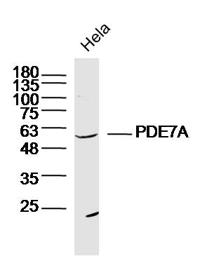
Sample: Hela Cell (Human) Lysate at 30 ug
Primary: Anti-PDE7A (bs-11575R) at 1/300 dilution
Secondary: IRDye800CW Goat Anti-Rabbit IgG at 1/20000 dilution
Predicted band size: 55kD
Observed band size: 55kD
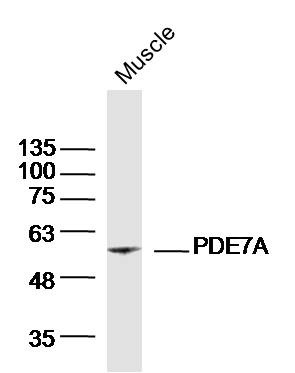
Sample: Muscle (Mouse)Lysate at 40 ug
Primary: Anti-PDE7A(bs-11575R)at 1/300 dilution
Secondary: IRDye800CW Goat Anti-RabbitIgG at 1/20000 dilution
Predicted band size: 55kD
Observed band size: 55kD
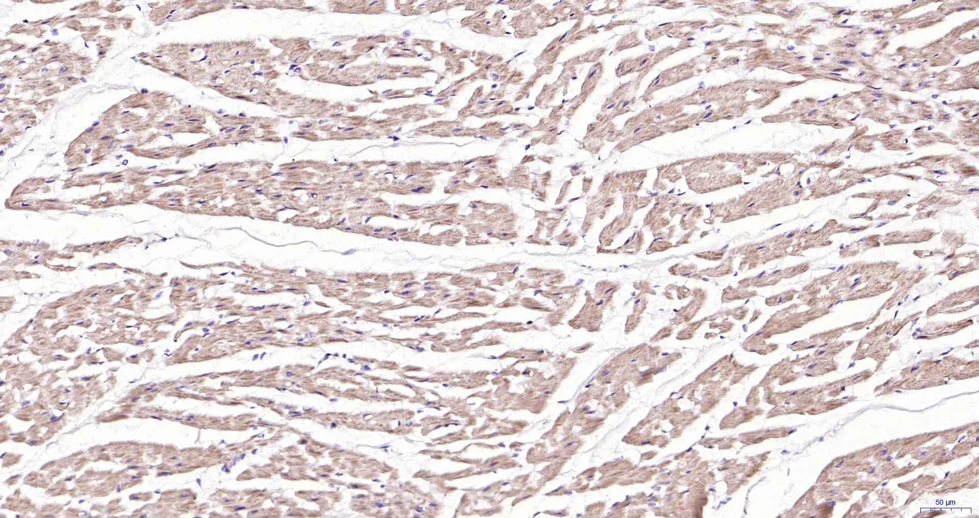
Paraformaldehyde-fixed, paraffin embedded Human Heart; Antigen retrieval by boiling in sodium citrate buffer (pH6.0) for 15 min; Antibody incubation with PDE7A Polyclonal Antibody, Unconjugated (bs-11575R) at 1:200 overnight at 4В°C, followed by conjugation to the SP Kit (Rabbit, SP-0023) and DAB (C-0010) staining.
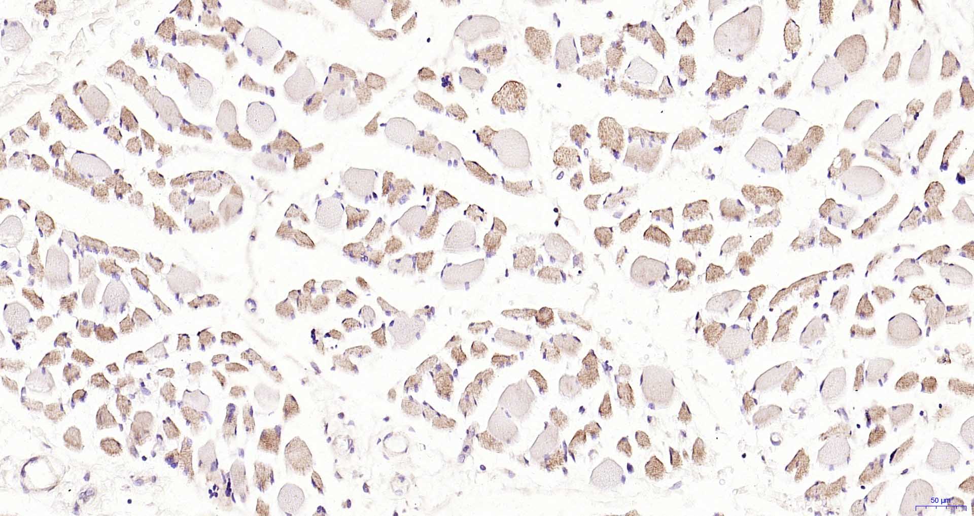
Paraformaldehyde-fixed, paraffin embedded Human Skeletal muscle; Antigen retrieval by boiling in sodium citrate buffer (pH6.0) for 15 min; Antibody incubation with PDE7A Polyclonal Antibody, Unconjugated (bs-11575R) at 1:200 overnight at 4В°C, followed by conjugation to the SP Kit (Rabbit, SP-0023) and DAB (C-0010) staining.
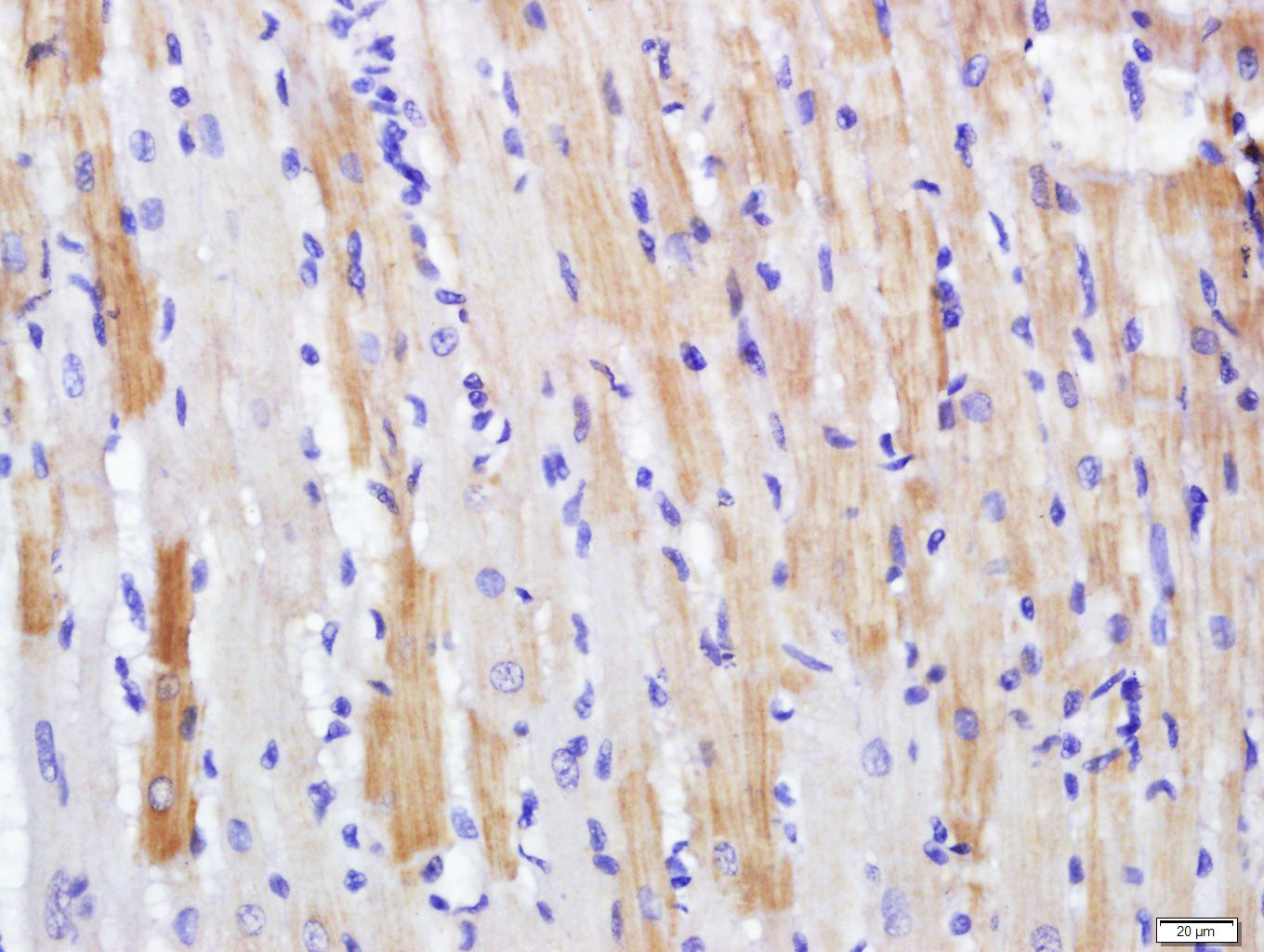
Paraformaldehyde-fixed, paraffin embedded (rat heart); Antigen retrieval by boiling in sodium citrate buffer (pH6.0) for 15min; Block endogenous peroxidase by 3% hydrogen peroxide for 20 minutes; Blocking buffer (normal goat serum) at 37В°C for 30min; Antibody incubation with (PDE7A) Polyclonal Antibody, Unconjugated (bs-11575R) at 1:500 overnight at 4В°C, followed by a conjugated secondary (sp-0023) for 20 minutes and DAB staining.
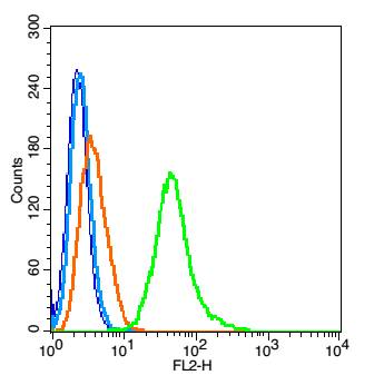
Blank control(blue): RSC96 cells(fixed with 2% paraformaldehyde (10 min) , then permeabilized with 90% ice-cold methanol for 30 min on ice). Primary Antibody:Rabbit Anti- PDE7A antibody(bs-11575R), Dilution: 1Ојg in 100 ОјL 1X PBS containing 0.5% BSA; Isotype Control Antibody: Rabbit IgG(orange) ,used under the same conditions ); Secondary Antibody: Goat anti-rabbit IgG-PE(white blue), Dilution: 1:200 in 1 X PBS containing 0.5% BSA.
|


