

| дә§е“Ғзј–еҸ· | bs-6951R |
| иӢұж–ҮеҗҚз§° | AKT1+2+3 Rabbit pAb |
| дёӯж–ҮеҗҚз§° | иӣӢзҷҪжҝҖй…¶AKT1,2,3жҠ—дҪ“ |
| еҲ« еҗҚ | AKT; PKB; PKB-ALPHA; PRKBA; RAC; RAC-ALPHA; LTR-akt; PKB/Akt; PKBalpha; O57513_CHICK; akt1; RAC-PK-alpha; 2.7.11.1; AKT1_HUMAN; Protein kinase B (PKB); Protein kinase B alpha (PKB alpha); Proto-oncogene c-Akt; AKT1_MOUSE; AKT1 kinase; Thymoma viral proto- Akt (pan); Akt (pan) (C67E7) ; |
 |
Specific References (24) | bs-6951R has been referenced in 24 publications.
|
| з ”з©¶йўҶеҹҹ | иӮҝзҳӨ з»Ҷиғһз”ҹзү© дҝЎеҸ·иҪ¬еҜј з»ҶиғһеҮӢдәЎ иҪ¬еҪ•и°ғиҠӮеӣ еӯҗ жҝҖй…¶е’ҢзЈ·й…ёй…¶ |
| жҠ—дҪ“жқҘжәҗ | Rabbit |
| е…ӢйҡҶзұ»еһӢ | Polyclonal |
| е…Ӣ йҡҶ еҸ· | |
| дәӨеҸүеҸҚеә” | Human,Mouse,Rat (predicted: Rabbit,Pig,Sheep,Cow,Chicken,Dog) |
| дә§е“Ғеә”з”Ё | WB=1:500-2000,IHC-P=1:100-500,IHC-F=1:100-500,IF=1:100-500,Flow-Cyt=1Ојg/Test
not yet tested in other applications. optimal dilutions/concentrations should be determined by the end user. |
| зҗҶи®әеҲҶеӯҗйҮҸ | 56 kDa |
| жЈҖжөӢеҲҶеӯҗйҮҸ | 56 |
| з»Ҷиғһе®ҡдҪҚ | з»ҶиғһжөҶ з»ҶиғһиҶң |
| жҖ§ зҠ¶ | Liquid |
| жө“ еәҰ | 1mg/ml |
| е…Қ з–« еҺҹ | KLH conjugated synthetic peptide derived from human AKT1/2/3: 401-480/480 |
| дәҡ еһӢ | IgG |
| зәҜеҢ–ж–№жі• | affinity purified by Protein A |
| зј“ еҶІ ж¶І | 0.01M TBS (pH7.4) with 1% BSA, 0.02% Proclin300 and 50% Glycerol. |
| дҝқеӯҳжқЎд»¶ | Shipped at 4в„ғ. Store at -20в„ғ for one year. Avoid repeated freeze/thaw cycles. |
| жіЁж„ҸдәӢйЎ№ | This product as supplied is intended for research use only, not for use in human, therapeutic or diagnostic applications. |
| PubMed | PubMed |
| дә§е“Ғд»Ӣз»Қ |
AKT, also known as protein kinase B (PKB), is a 57 kDa serine/threonine protein kinase. There are three mammalian isoforms of Akt: AKT1 (PKB alpha), AKT2 (PKB beta) and AKT3 (PKB gamma) with AKT2 and AKT3 being approximately 82% identical with the AKT1 isoform. Each isoform has a pleckstrin homology (PH)domain, a kinase domain and a carboxy terminal regulatory domain. AKT was originally cloned from the retrovirus AKT8, and is a key regulator of many signal transduction pathways. Its tight control over cell proliferation and cell viability are manifold; overexpression or inappropriate activation of AKT has been seen in many types of cancer. AKT mediates many of the downstream events of phosphatidylinositol 3 kinase (a lipid kinase activated by growth factors, cytokines and insulin). PI3 kinase recruits AKT to the membrane, where it is activated by PDK1 phosphorylation. Once phosphorylated, AKT dissociates from the membrane and phosphorylates targets in the cytoplasm and the cell nucleus. Function: IGF-1 leads to the activation of AKT3, which may play a role in regulating cell survival. Capable of phosphorylating several known proteins. Truncated isoform 2/PKB gamma 1 without the second serine phosphorylation site could still be stimulated but to a lesser extent. Subunit: Interacts (via the C-terminus) with CCDC88A (via its C-terminus). Interacts with GRB10; the interaction leads to GRB10 phosphorylation thus promoting YWHAE-binding. Interacts with AGAP2 (isoform 2/PIKE-A); the interaction occurs in the presence of guanine nucleotides. Interacts with AKTIP. Interacts (via PH domain) with MTCP1, TCL1A AND TCL1B. Interacts with CDKN1B; the interaction phosphorylates CDKN1B promoting 14-3-3 binding and cell-cycle progression. Interacts with MAP3K5 and TRAF6. Interacts with BAD, PPP2R5B, STK3 and STK4. Interacts (via PH domain) with SIRT1. Interacts with SRPK2 in a phosphorylation-dependent manner. Interacts with RAF1. Interacts with TRIM13; the interaction ubiquitinates AKT1 leading to its proteasomal degradation. Interacts with TNK2 and CLK2. Interacts (via the C-terminus) with THEM4 (via its C-terminus). Interacts with and phosphorylated by PDPK1. Subcellular Location: Cytoplasm. Nucleus. Cell membrane. Note=Nucleus after activation by integrin-linked protein kinase 1 (ILK1). Nuclear translocation is enhanced by interaction with TCL1A. Phosphorylation on Tyr-176 by TNK2 results in its localization to the cell membrane where it is targeted for further phosphorylations on Thr-308 and Ser-473 leading to its activation and the activated form translocates to the nucleus. Tissue Specificity: Expressed in prostate cancer and levels increase from the normal to the malignant state (at protein level). Expressed in all human cell types so far analyzed. The Tyr-176 phosphorylated form shows a significant increase in expression in breast cancers during the progressive stages i.e. normal to hyperplasia (ADH), ductal carcinoma in situ (DCIS), invasive ductal carcinoma (IDC) and lymph node metastatic (LNMM) stages. Post-translational modifications: O-GlcNAcylation at Thr-305 and Thr-312 inhibits activating phosphorylation at Thr-308 via disrupting the interaction between AKT1 and PDPK1. O-GlcNAcylation at Ser-473 also probably interferes with phosphorylation at this site. Phosphorylation on Thr-308, Ser-473 and Tyr-474 is required for full activity. Activated TNK2 phosphorylates it on Tyr-176 resulting in its binding to the anionic plasma membrane phospholipid PA. This phosphorylated form localizes to the cell membrane, where it is targeted by PDPK1 and PDPK2 for further phosphorylations on Thr-308 and Ser-473 leading to its activation. Ser-473 phosphorylation by mTORC2 favors Thr-308 phosphorylation by PDPK1. Ser-473 phosphorylation is enhanced by interaction with AGAP2 isoform 2 (PIKE-A). Ser-473 phosphorylation is enhanced in focal cortical dysplasias with Taylor-type balloon cells. Ser-473 phosphorylation is enhanced by signaling through activated FLT3. Dephosphorylated at Thr-308 and Ser-473 by PP2A phosphatase. The phosphorylated form of PPP2R5B is required for bridging AKT1 with PP2A phosphatase. Ubiquitinated via 'Lys-48'-linked polyubiquitination by ZNRF1, leading to its degradation by the proteasome. Ubiquitinated; undergoes both 'Lys-48'- and 'Lys-63'-linked polyubiquitination. TRAF6-induced 'Lys-63'-linked AKT1 ubiquitination is critical for phosphorylation and activation. When ubiquitinated, it translocates to the plasma membrane, where it becomes phosphorylated. When fully phosphorylated and translocated into the nucleus, undergoes 'Lys-48'-polyubiquitination catalyzed by TTC3, leading to its degradation by the proteasome. Also ubiquitinated by TRIM13 leading to its proteasomal degradation. Acetylated on Lys-14 and Lys-20 by the histone acetyltransferases EP300 and KAT2B. Acetylation results in reduced phosphorylation and inhibition of activity. Deacetylated at Lys-14 and Lys-20 by SIRT1. SIRT1-mediated deacetylation relieves the inhibition. DISEASE: Defects in AKT1 are a cause of susceptibility to breast cancer (BC) [MIM:114480]. A common malignancy originating from breast epithelial tissue. Breast neoplasms can be distinguished by their histologic pattern. Invasive ductal carcinoma is by far the most common type. Breast cancer is etiologically and genetically heterogeneous. Important genetic factors have been indicated by familial occurrence and bilateral involvement. Mutations at more than one locus can be involved in different families or even in the same case. Defects in AKT1 are associated with colorectal cancer (CRC) [MIM:114500]. Note=Genetic variations in AKT1 may play a role in susceptibility to ovarian cancer. Defects in AKT1 are a cause of Proteus syndrome (PROTEUSS) [MIM:176920]. A highly variable, severe disorder of asymmetric and disproportionate overgrowth of body parts, connective tissue nevi, epidermal nevi, dysregulated adipose tissue, and vascular malformations. Many features of Proteus syndrome overlap with other overgrowth syndromes. Similarity: Belongs to the protein kinase superfamily. AGC Ser/Thr protein kinase family. RAC subfamily. Contains 1 AGC-kinase C-terminal domain. Contains 1 PH domain. Contains 1 protein kinase domain. SWISS: P31749 Gene ID: 207 Database links: Entrez Gene: 207 Human Entrez Gene: 11651 Mouse Omim: 164730 Human SwissProt: O57513 Chicken SwissProt: P31749 Human SwissProt: P31750 Mouse Unigene: 525622 Human Unigene: 6645 Mouse Unigene: 11422 Rat |
| дә§е“ҒеӣҫзүҮ |
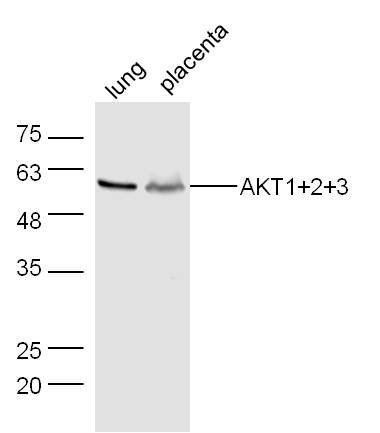
Sample:
Lung (Mouse) Lysate at 40 ug
Placenta (Mouse) Lysate at 40 ug
Primary: Anti-AKT1+2+3 (bs-6951R) at 1/300 dilution
Secondary: IRDye800CW Goat Anti-Rabbit IgG at 1/20000 dilution
Predicted band size: 56 kD
Observed band size: 56 kD
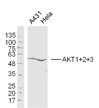
Sample:
A431Cell (Human) Lysate at 30 ug
Hela Cell(Human)Lysate at 30 ug
Primary: Anti-AKT1+2+3(bs-6951R)at 1/300 dilution
Secondary: IRDye800CW Goat Anti-Rabbit IgG at 1/20000 dilution
Predicted band size: 56 kD
Observed band size: 56 kD
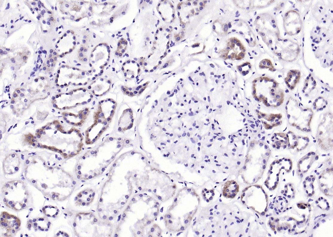
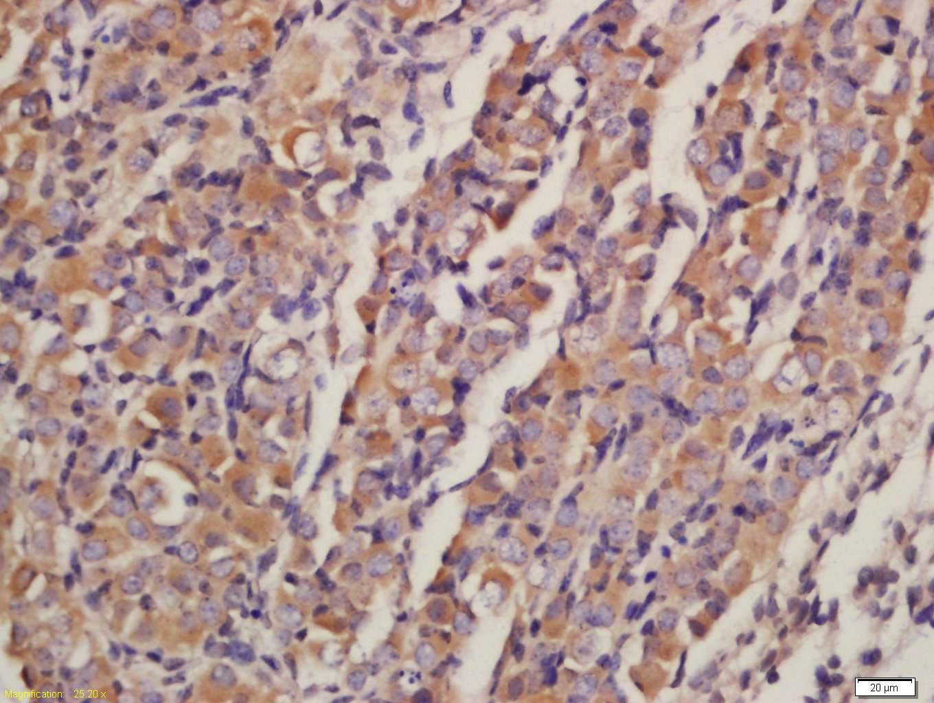
Paraformaldehyde-fixed, paraffin embedded (Human kidney); Antigen retrieval by boiling in sodium citrate buffer (pH6.0) for 15min; Block endogenous peroxidase by 3% hydrogen peroxide for 20 minutes; Blocking buffer (normal goat serum) at 37В°C for 30min; Antibody incubation with (AKT1+2+3) Polyclonal Antibody, Unconjugated (bs-6951R) at 1:200 overnight at 4В°C, followed by operating according to SP Kit(Rabbit) (sp-0023) instructionsand DAB staining.
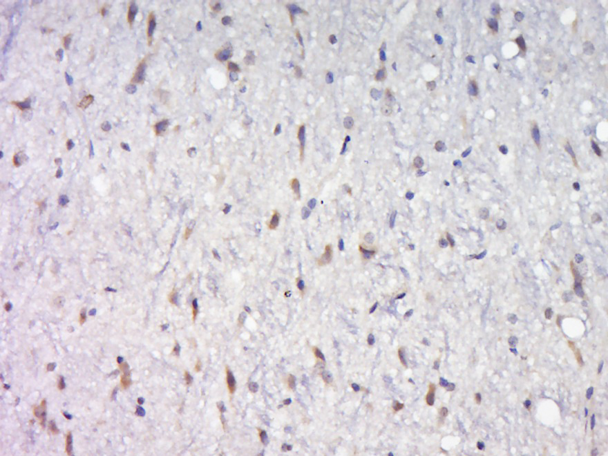
Paraformaldehyde-fixed, paraffin embedded (rat brain); Antigen retrieval by boiling in sodium citrate buffer (pH6.0) for 15min; Block endogenous peroxidase by 3% hydrogen peroxide for 20 minutes; Blocking buffer (normal goat serum) at 37В°C for 30min; Antibody incubation with (AKT1+2+3) Polyclonal Antibody, Unconjugated (bs-6951R) at 1:400 overnight at 4В°C, followed by a conjugated secondary (sp-0023) for 20 minutes and DAB staining.
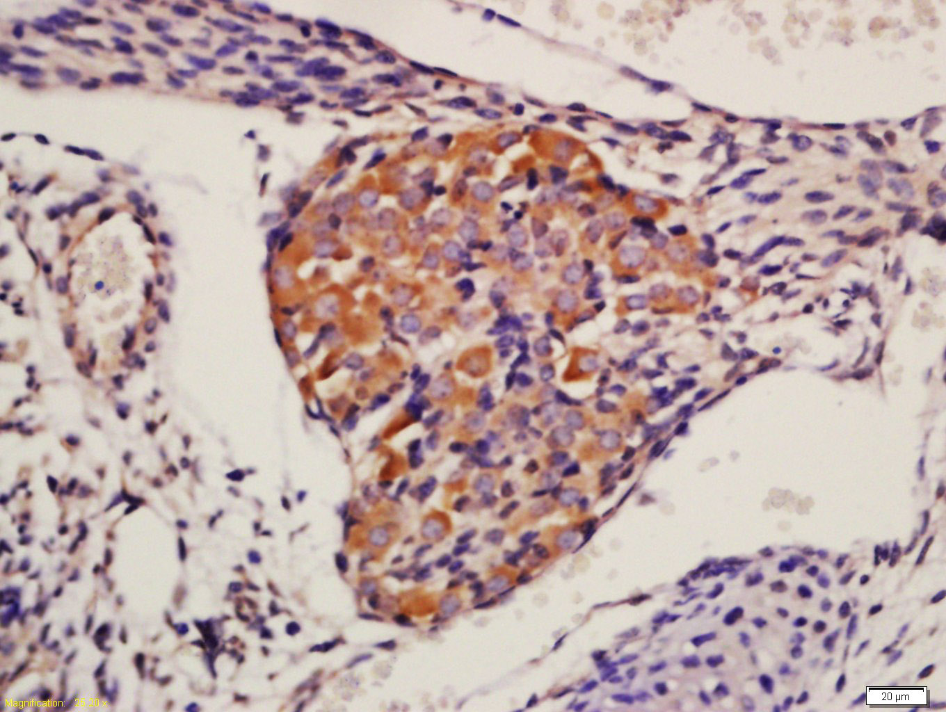
Tissue/cell: mouse embryo tissue; 4% Paraformaldehyde-fixed and paraffin-embedded;
Antigen retrieval: citrate buffer ( 0.01M, pH 6.0 ), Boiling bathing for 15min; Block endogenous peroxidase by 3% Hydrogen peroxide for 30min; Blocking buffer (normal goat serum,C-0005) at 37в„ғ for 20 min;
Incubation: Anti-AKT1+2+3 Polyclonal Antibody, Unconjugated(bs-6951R) 1:200, overnight at 4В°C, followed by conjugation to the secondary antibody(SP-0023) and DAB(C-0010) staining
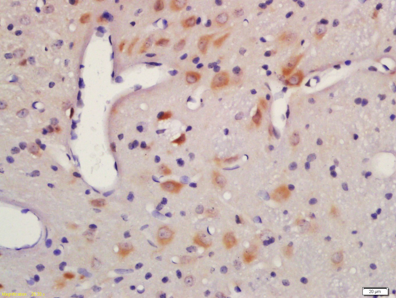
Tissue/cell: rat brain tissue; 4% Paraformaldehyde-fixed and paraffin-embedded;
Antigen retrieval: citrate buffer ( 0.01M, pH 6.0 ), Boiling bathing for 15min; Block endogenous peroxidase by 3% Hydrogen peroxide for 30min; Blocking buffer (normal goat serum,C-0005) at 37в„ғ for 20 min;
Incubation: Anti-AKT1+2+3 Polyclonal Antibody, Unconjugated(bs-6951R) 1:200, overnight at 4В°C, followed by conjugation to the secondary antibody(SP-0023) and DAB(C-0010) staining
Tissue/cell: Mouse embryos tissue; 4% Paraformaldehyde-fixed and paraffin-embedded;
Antigen retrieval: citrate buffer ( 0.01M, pH 6.0 ), Boiling bathing for 15min; Block endogenous peroxidase by 3% Hydrogen peroxide for 30min; Blocking buffer (normal goat serum,C-0005) at 37в„ғ for 20 min;
Incubation: Anti-AKT1 + 2 + 3 Polyclonal Antibody, Unconjugated(bs-6951R) 1:200, overnight at 4В°C, followed by conjugation to the secondary antibody(SP-0023) and DAB(C-0010) staining
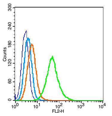
Blank control(blue): TM4 cells(fixed with 2% paraformaldehyde (10 min) , then permeabilized with 90% ice-cold methanol for 30 min on ice). Primary Antibody:Rabbit Anti-AKT1+2+3 antibody(bs-6951R), Dilution: 1Ојg in 100 ОјL 1X PBS containing 0.5% BSA; Isotype Control Antibody: Rabbit IgG(orange) ,used under the same conditions ); Secondary Antibody: Goat anti-rabbit IgG-PE(white blue), Dilution: 1:200 in 1 X PBS containing 0.5% BSA.
|


