

| 产品编号 | bs-11764R |
| 英文名称 | OPA1 Rabbit pAb |
| 中文名称 | 视神经萎缩相关蛋白1抗体 |
| 别 名 | Dynamin like 120 kDa protein; Dynamin like 120 kDa protein, mitochondrial; Dynamin-like 120 kDa protein; Dynamin-like 120 kDa protein, form S1; FLJ12460; Juvenile kjer type optic atrophy; Juvenile kjer-type optic atrophy; KIAA0567; KJER type; Large GTP binding protein; largeG; MGM1; Mitochondrial dynamin like 120 kDa protein; Mitochondrial dynamin like GTPase; NPG; NTG; OAK; OPA 1; OPA1; OPA1 gene; OPA1_HUMAN; Optic atrophy 1(autosomal dominant); OPTIC ATROPHY 1; Optic atrophy 1 gene protein; Optic atrophy 1 homolog(human); Optic atrophy protein 1; Optic atrophy protein 1 homolog. |
 |
Specific References (13) | bs-11764R has been referenced in 13 publications.
|
| 研究领域 | 心血管 细胞生物 神经生物学 |
| 抗体来源 | Rabbit |
| 克隆类型 | Polyclonal |
| 克 隆 号 | |
| 交叉反应 | Rat (predicted: Human,Mouse,Rabbit,Pig,Sheep,Cow,Dog,Horse) |
| 产品应用 | WB=1:500-2000,IHC-P=1:100-500,IHC-F=1:100-500,IF=1:100-500
not yet tested in other applications. optimal dilutions/concentrations should be determined by the end user. |
| 理论分子量 | 111 kDa |
| 检测分子量 | |
| 细胞定位 | 细胞浆 细胞膜 |
| 性 状 | Liquid |
| 浓 度 | 1mg/ml |
| 免 疫 原 | KLH conjugated synthetic peptide derived from human OPA1: 651-750/960 |
| 亚 型 | IgG |
| 纯化方法 | affinity purified by Protein A |
| 缓 冲 液 | 0.01M TBS (pH7.4) with 1% BSA, 0.02% Proclin300 and 50% Glycerol. |
| 保存条件 | Shipped at 4℃. Store at -20℃ for one year. Avoid repeated freeze/thaw cycles. |
| 注意事项 | This product as supplied is intended for research use only, not for use in human, therapeutic or diagnostic applications. |
| PubMed | PubMed |
| 产品介绍 |
OPA1 is a 120kDa protein belonging to the dynamin family. The OPA1 gene has been localized to 3q29. The gene is targeted to mitochondria and is involved in mitochondrial biogenesis. Defects in OPA1 are a cause of optic atrophy type 1. OPA1 is mostly expressed in retina but can also be expressed in brain, testis, heart and skeletal muscle. Function: Dynamin-related GTPase required for mitochondrial fusion and regulation of apoptosis. May form a diffusion barrier for proteins stored in mitochondrial cristae. Proteolytic processing in response to intrinsic apoptotic signals may lead to disassembly of OPA1 oligomers and release of the caspase activator cytochrome C (CYCS) into the mitochondrial intermembrane space. Subcellular Location: Mitochondrion inner membrane. Mitochondrion intermembrane space. Tissue Specificity: Highly expressed in retina. Also expressed in brain, testis, heart and skeletal muscle. Isoform 1 expressed in retina, skeletal muscle, heart, lung, ovary, colon, thyroid gland, leukocytes and fetal brain. Isoform 2 expressed in colon, liver, kidney, thyroid gland and leukocytes. Low levels of all isoforms expressed in a variety of tissues. Post-translational modifications: PARL-dependent proteolytic processing releases an antiapoptotic soluble form not required for mitochondrial fusion. DISEASE: Defects in OPA1 are a cause of optic atrophy type 1 (OPA1) [MIM:165500]. OPA1 is a dominantly inherited optic neuropathy occurring in 1 in 50,000 individuals that features progressive loss in visual acuity leading, in many cases, to legal blindness. Defects in OPA1 are the cause of optic atrophy 1 with deafness (OPA1D) [MIM:125250]. Some individuals with mutations in OPA1 manifest also ophthalmoplegia and myopathy. Similarity: Belongs to the dynamin family. SWISS: O60313 Gene ID: 4976 Database links: Entrez Gene: 424900 Chicken Entrez Gene: 4976 Human Entrez Gene: 74143 Mouse Omim: 605290 Human SwissProt: O60313 Human SwissProt: P58281 Mouse Unigene: 594504 Human Unigene: 274285 Mouse Unigene: 9783 Rat |
| 产品图片 |
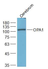
Sample:
Cerebrum (Rat) Lysate at 40 ug
Primary: Anti-OPA1 (bs-11764R) at 1/2000 dilution
Secondary: IRDye800CW Goat Anti-Rabbit IgG at 1/20000 dilution
Predicted band size: 111 kD
Observed band size: 111 kD
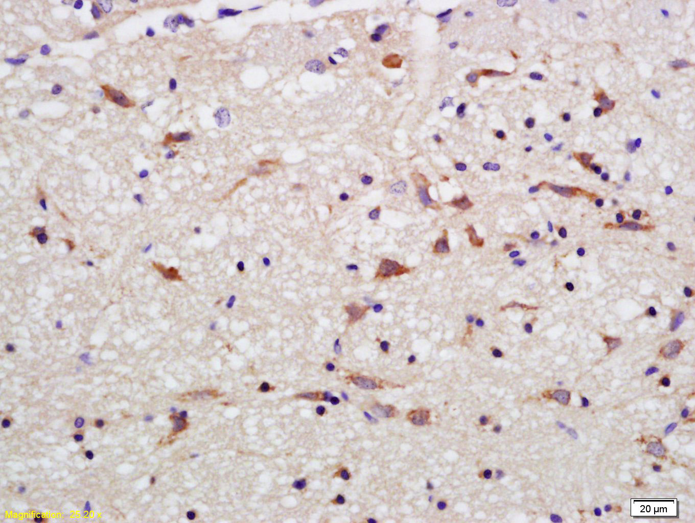
Tissue/cell: rat brain tissue; 4% Paraformaldehyde-fixed and paraffin-embedded;
Antigen retrieval: citrate buffer ( 0.01M, pH 6.0 ), Boiling bathing for 15min; Block endogenous peroxidase by 3% Hydrogen peroxide for 30min; Blocking buffer (normal goat serum,C-0005) at 37℃ for 20 min;
Incubation: Anti-OPA1 Polyclonal Antibody, Unconjugated(bs-11764R) 1:200, overnight at 4°C, followed by conjugation to the secondary antibody(SP-0023) and DAB(C-0010) staining
|
| 1、抗体溶解方法 | |
| 2、抗体修复方式 | |
| 3、常用试剂的配制 | |
| 4、免疫组化操作步骤 | |
| 5、免疫组化问题解答 | |
| 6、Western Blotting 操作步骤 | |
| 7、Western Blotting 问题解答 | |
| 8、关于肽链的设计 | |
| 9、多肽的溶解与保存 | |
| 10、酶标抗体效价测定程序 | |


