

| 产品编号 | bs-10196R |
| 英文名称 | alpha smooth muscle Actin Rabbit pAb |
| 中文名称 | 肌动蛋白α/α-SMA/α Actin抗体 |
| 别 名 | ACTSA; SMDYS; 0610041G09Rik; Actvs; SMAalpha; SMalphaA; a-SMA; alphaSMA; ACT-4; actin; ACTA_BOVIN; ACTA2; Alpha-actin-2; 3.6.4.-; ACTA_HUMAN; Cell growth-inhibiting gene 46 protein; ACTA_MOUSE; ACTA_RABIT; ACTA_RAT; α-Smooth Muscle Actin; α Smooth Muscle Actin; |
 |
Specific References (86) | bs-10196R has been referenced in 86 publications.
|
| 研究领域 | 肿瘤 细胞生物 免疫学 细胞骨架 |
| 抗体来源 | Rabbit |
| 克隆类型 | Polyclonal |
| 克 隆 号 | |
| 交叉反应 | Human,Mouse,Rat (predicted: Rabbit,Dog) |
| 产品应用 | WB=1:2000-10000,IHC-P=1:200-800,IHC-F=1:200-800,IF=1:200-800,Flow-Cyt=1μg/Test
not yet tested in other applications. optimal dilutions/concentrations should be determined by the end user. |
| 理论分子量 | 42 kDa |
| 检测分子量 | 42 |
| 细胞定位 | 细胞浆 |
| 性 状 | Liquid |
| 浓 度 | 1mg/ml |
| 免 疫 原 | KLH conjugated synthetic peptide derived from human Actin alpha: 165-260/377 |
| 亚 型 | IgG |
| 纯化方法 | affinity purified by Protein A |
| 缓 冲 液 | 0.01M TBS (pH7.4) with 1% BSA, 0.02% Proclin300 and 50% Glycerol. |
| 保存条件 | Shipped at 4℃. Store at -20℃ for one year. Avoid repeated freeze/thaw cycles. |
| 注意事项 | This product as supplied is intended for research use only, not for use in human, therapeutic or diagnostic applications. |
| PubMed | PubMed |
| 产品介绍 |
All eukaryotic cells express Actin, which often constitutes as much as 50% of total cellular protein. Actin filaments can form both stable and labile structures and are crucial components of microvilli and the contractile apparatus of muscle cells. While lower eukaryotes, such as yeast, have only one Actin gene, higher eukaryotes have several isoforms encoded by a family of genes. At least six types of Actin are present in mammalian tissues and fall into three classes. alpha-Actin expression is limited to various types of muscle, whereas beta- and gamma-Actin are the principle constituents of filaments in other tissues. Members of the small GTPase family regulate the organization of the Actin cytoskeleton. Rho controls the assembly of Actin stress fibers and focal adhesion. Rac regulates Actin filament accumulation at the plasma membrane. Cdc42 stimulates formation of filopodia. Function: Actins are highly conserved proteins that are involved in various types of cell motility and are ubiquitously expressed in all eukaryotic cells. Subunit: Polymerization of globular actin (G-actin) leads to a structural filament (F-actin) in the form of a two-stranded helix. Each actin can bind to 4 others. Subcellular Location: Cytoplasm, cytoskeleton. Post-translational modifications: Oxidation of Met-46 by MICALs (MICAL1, MICAL2 or MICAL3) to form methionine sulfoxide promotes actin filament depolymerization. Methionine sulfoxide is produced stereospecifically, but it is not known whether the (S)-S-oxide or the (R)-S-oxide is produced (By similarity). DISEASE: Defects in ACTA2 are the cause of aortic aneurysm familial thoracic type 6 (AAT6) [MIM:611788]. AATs are characterized by permanent dilation of the thoracic aorta usually due to degenerative changes in the aortic wall. They are primarily associated with a characteristic histologic appearance known as 'medial necrosis' or 'Erdheim cystic medial necrosis' in which there is degeneration and fragmentation of elastic fibers, loss of smooth muscle cells, and an accumulation of basophilic ground substance. Similarity: Belongs to the actin family. SWISS: P62736 Gene ID: 59 Database links: Entrez Gene: 101021287 Baboon Entrez Gene: 59 Human Entrez Gene: 11475 Mouse Entrez Gene: 100009271 Rabbit Omim: 102620 Human SwissProt: P62736 Human SwissProt: P62737 Mouse SwissProt: P62740 Rabbit Unigene: 500483 Human Unigene: 213025 Mouse Unigene: 195319 Rat Unigene: 3114 Rat Actin α/α-Actin 是一种具有收缩能力的微丝蛋白,a-SMA广泛分布于几乎所有的肌型细胞中。Actin-α蛋白主要用于检测骨骼肌、平滑肌、血管平滑肌、心肌和肌原性肿瘤 包括:平滑肌瘤、平滑肌肉瘤、横纹肌肉瘤以及肌上细胞和肌上皮瘤。Actin(肌动蛋白)是在所有真核细胞中都表达的高度保守的蛋白质。它们沿微管组成了细胞骨架的主要成分。肌动蛋白至少表达为6种异构形式。它在心脏、骨骼横纹肌组织和某些平滑肌组织中表达,调节其收缩功能。有报导说肌动蛋白在乳房瘤中是高度磷酸化的。肌动蛋白的功能失调也会导致某种类型的心脏病。平滑肌α肌动蛋白使人更感兴趣,因为编码它的基因是相对局限于在血管平滑肌细胞中表达的少数几个基因之一。肌动蛋白是标记平滑肌和肌上皮细胞肿瘤的有效工具。 |
| 产品图片 |
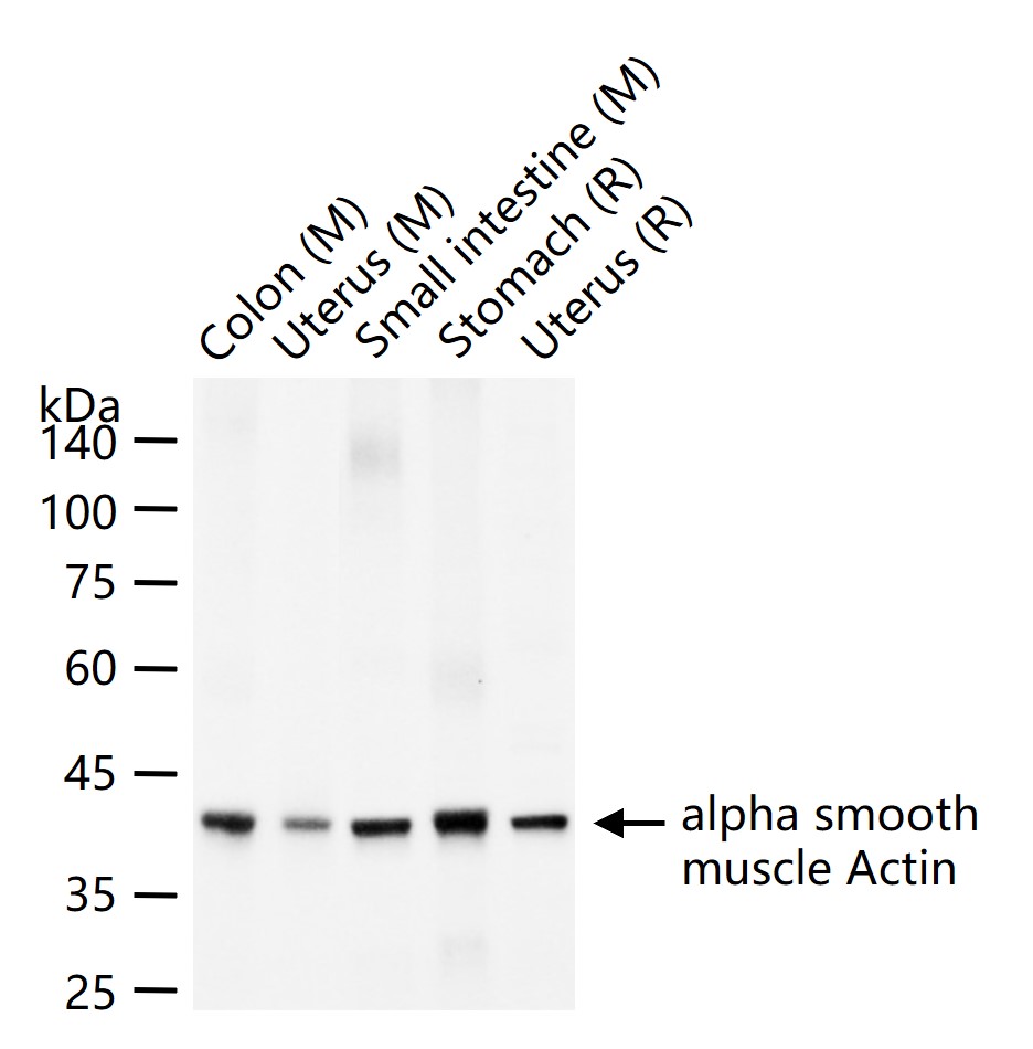
25 ug total protein per lane of various lysates (see on figure) probed with alpha smooth muscle Actin polyclonal antibody, unconjugated (bs-10196R) at 1:2000 dilution and 4°C overnight incubation. Followed by conjugated secondary antibody incubation at r.t. for 60 min.
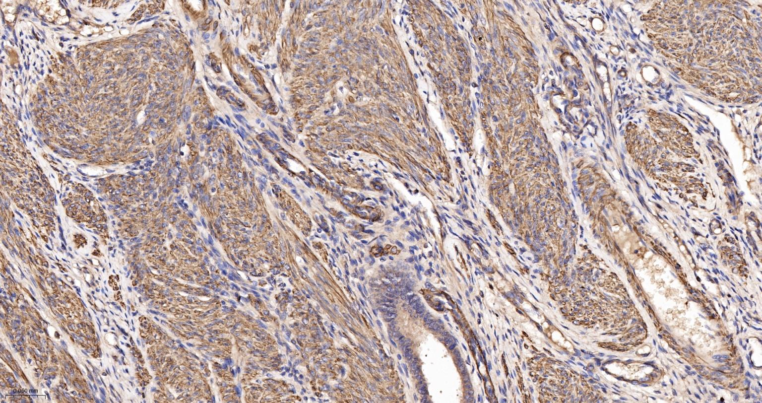
Paraformaldehyde-fixed, paraffin embedded Human Uterus; Antigen retrieval by boiling in sodium citrate buffer (pH6.0) for 15 min; The section was incubated with alpha smooth muscle Actin Polyclonal Antibody, Unconjugated (bs-10196R) at 1:200 overnight at 4°C, followed by conjugation to the bs-0295G-HRP and DAB (C-0010) staining.
Paraformaldehyde-fixed, paraffin embedded Rat Uterus; Antigen retrieval by boiling in sodium citrate buffer (pH6.0) for 15 min; The section was incubated with alpha smooth muscle Actin Polyclonal Antibody, Unconjugated (bs-10196R) at 1:200 overnight at 4°C, followed by conjugation to the bs-0295G-HRP and DAB (C-0010) staining.
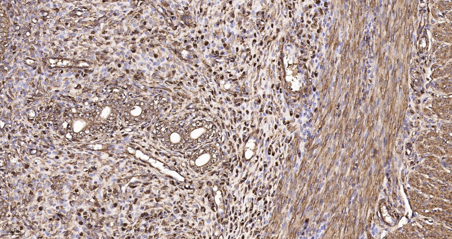
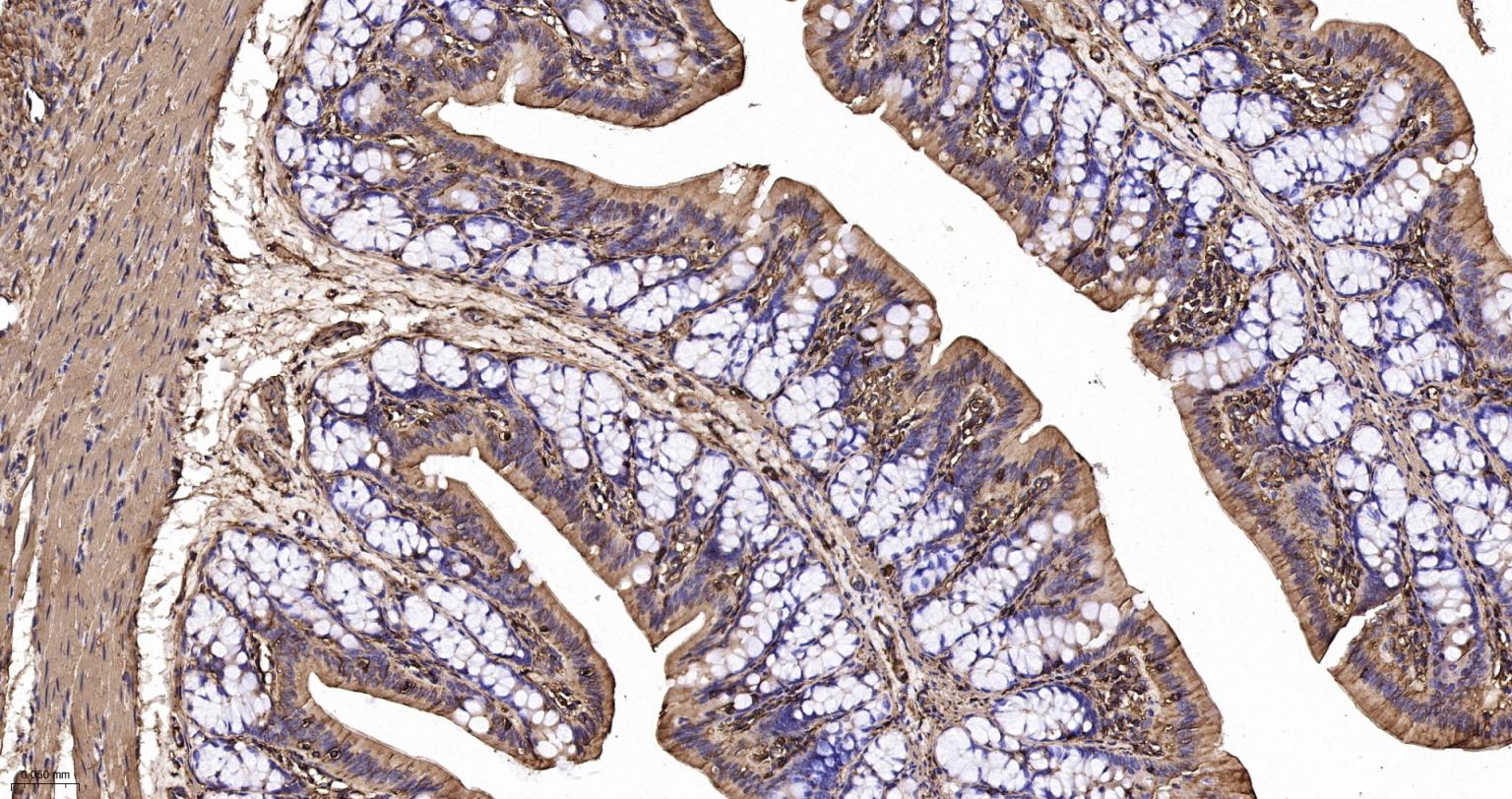
Paraformaldehyde-fixed, paraffin embedded Human Colon; Antigen retrieval by boiling in sodium citrate buffer (pH6.0) for 15 min; The section was incubated with alpha smooth muscle Actin Polyclonal Antibody, Unconjugated (bs-10196R) at 1:200 overnight at 4°C, followed by conjugation to the bs-0295G-HRP and DAB (C-0010) staining.
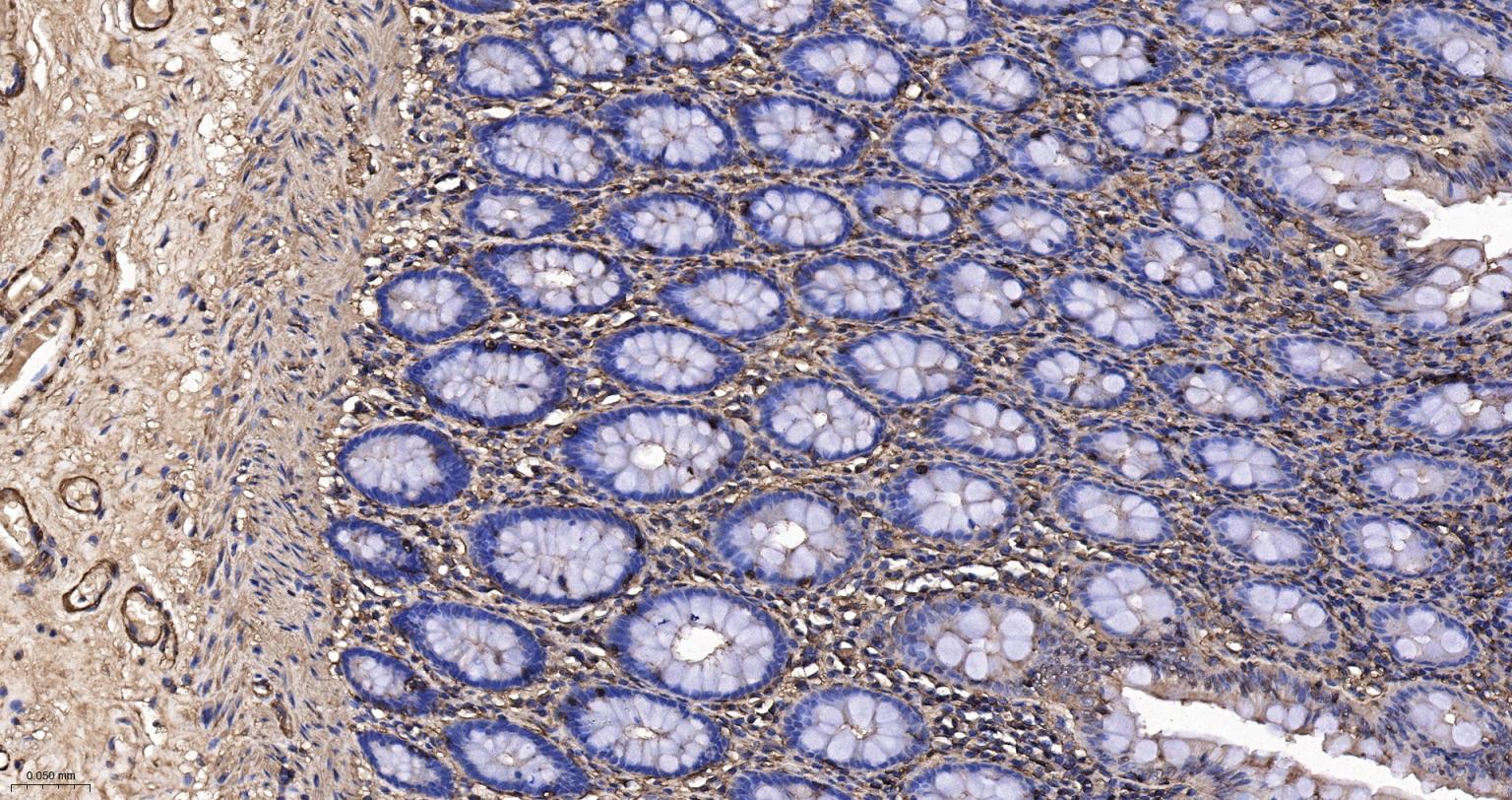
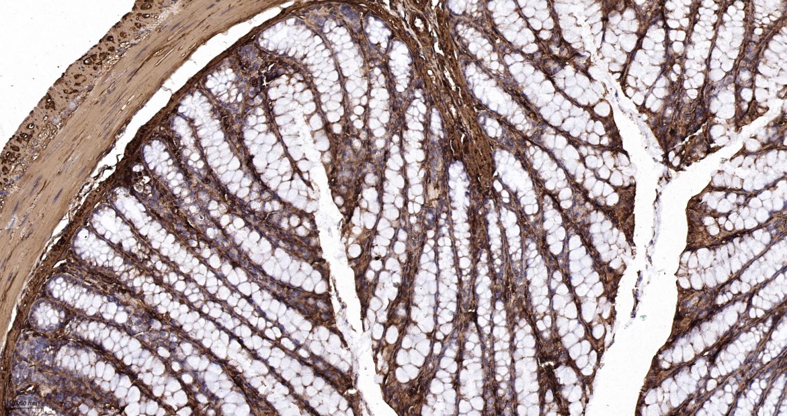
Paraformaldehyde-fixed, paraffin embedded Rat Colon; Antigen retrieval by boiling in sodium citrate buffer (pH6.0) for 15 min; The section was incubated with alpha smooth muscle Actin Polyclonal Antibody, Unconjugated (bs-10196R) at 1:200 overnight at 4°C, followed by conjugation to the bs-0295G-HRP and DAB (C-0010) staining.
Paraformaldehyde-fixed, paraffin embedded Mouse Colon; Antigen retrieval by boiling in sodium citrate buffer (pH6.0) for 15 min; The section was incubated with alpha smooth muscle Actin Polyclonal Antibody, Unconjugated (bs-10196R) at 1:200 overnight at 4°C, followed by conjugation to the bs-0295G-HRP and DAB (C-0010) staining.
|
| 1、抗体溶解方法 | |
| 2、抗体修复方式 | |
| 3、常用试剂的配制 | |
| 4、免疫组化操作步骤 | |
| 5、免疫组化问题解答 | |
| 6、Western Blotting 操作步骤 | |
| 7、Western Blotting 问题解答 | |
| 8、关于肽链的设计 | |
| 9、多肽的溶解与保存 | |
| 10、酶标抗体效价测定程序 | |


