

| 产品编号 | bs-10216R |
| 英文名称 | SOD1 Rabbit pAb |
| 中文名称 | 超氧化物歧化酶1(铜/锌过氧化物歧化酶SOD抗体 |
| 别 名 | ALS; ALS1; HEL-S-44; IPOA; SOD; STAHP; hSod1; homodimer; B430204E11Rik; Cu/Zn-SOD; CuZnSOD; Ipo-1; Ipo1; SODC; Sod-1; SODC_HUMAN; SOD1; Superoxide dismutase 1 (hSod1); 1.15.1.1; SODC_MOUSE; SODC_RAT; |
 |
Specific References (20) | bs-10216R has been referenced in 20 publications.
|
| 抗体来源 | Rabbit |
| 克隆类型 | Polyclonal |
| 克 隆 号 | |
| 交叉反应 | Human,Mouse,Rat (predicted: Pig,Cow,Horse) |
| 产品应用 | WB=1:500-2000,IHC-P=1:100-500,IHC-F=1:100-500,IF=1:100-500,Flow-Cyt=1ug/test
not yet tested in other applications. optimal dilutions/concentrations should be determined by the end user. |
| 理论分子量 | 17 kDa |
| 检测分子量 | 19 |
| 细胞定位 | 细胞浆 |
| 性 状 | Liquid |
| 浓 度 | 1mg/ml |
| 免 疫 原 | KLH conjugated synthetic peptide derived from human SOD1: 6-100/154 |
| 亚 型 | IgG |
| 纯化方法 | affinity purified by Protein A |
| 缓 冲 液 | 0.01M TBS (pH7.4) with 1% BSA, 0.02% Proclin300 and 50% Glycerol. |
| 保存条件 | Shipped at 4℃. Store at -20℃ for one year. Avoid repeated freeze/thaw cycles. |
| 注意事项 | This product as supplied is intended for research use only, not for use in human, therapeutic or diagnostic applications. |
| PubMed | PubMed |
| 产品介绍 |
The protein encoded by this gene binds copper and zinc ions and is one of two isozymes responsible for destroying free superoxide radicals in the body. The encoded isozyme is a soluble cytoplasmic protein, acting as a homodimer to convert naturally-occuring but harmful superoxide radicals to molecular oxygen and hydrogen peroxide. The other isozyme is a mitochondrial protein. Mutations in this gene have been implicated as causes of familial amyotrophic lateral sclerosis. Rare transcript variants have been reported for this gene. [provided by RefSeq, Jul 2008] Function: Destroys radicals which are normally produced within the cells and which are toxic to biological systems. Subunit: Homodimer; non-disulfide linked. Homodimerization may take place via the ditryptophan cross-link at Trp-33. The pathogenic variants ALS1 Arg-38, Arg-47, Arg-86 and Ala-94 interact with RNF19A, whereas wild-type protein does not. The pathogenic variants ALS1 Arg-86 and Ala-94 interact with MARCH5, whereas wild-type protein does not. Subcellular Location: Cytoplasm. Note=The pathogenic variants ALS1 Arg-86 and Ala-94 gradually aggregates and accumulates in mitochondria. Post-translational modifications: Unlike wild-type protein, the pathogenic variants ALS1 Arg-38, Arg-47, Arg-86 and Ala-94 are polyubiquitinated by RNF19A leading to their proteasomal degradation. The pathogenic variants ALS1 Arg-86 and Ala-94 are ubiquitinated by MARCH5 leading to their proteasomal degradation. The ditryptophan cross-link at Trp-33 is responsible for the non-disulfide-linked homodimerization. Such modification might only occur in extreme conditions and additional experimental evidence is required. DISEASE: Defects in SOD1 are the cause of amyotrophic lateral sclerosis type 1 (ALS1) [MIM:105400]. ALS1 is a familial form of amyotrophic lateral sclerosis, a neurodegenerative disorder affecting upper and lower motor neurons and resulting in fatal paralysis. Sensory abnormalities are absent. Death usually occurs within 2 to 5 years. The etiology of amyotrophic lateral sclerosis is likely to be multifactorial, involving both genetic and environmental factors. The disease is inherited in 5-10% of cases leading to familial forms. Similarity: Belongs to the Cu-Zn superoxide dismutase family. SWISS: P00441 Gene ID: 6647 Database links: Entrez Gene: 6647 Human Entrez Gene: 20655 Mouse Omim: 147450 Human SwissProt: P00441 Human SwissProt: P08228 Mouse Unigene: 443914 Human Unigene: 276325 Mouse Unigene: 466779 Mouse Unigene: 6059 Rat |
| 产品图片 |
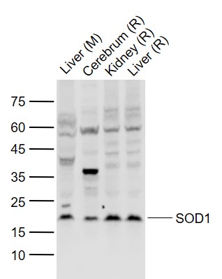
Sample:
Lane 1: Liver (Mouse) Lysate at 40 ug
Lane 2: Cerebrum (Rat) Lysate at 40 ug
Lane 3: Kidney (Rat) Lysate at 40 ug
Lane 4: Liver (Rat) Lysate at 40 ug
Primary: Anti-SOD1 (bs-10216R) at 1/1000 dilution
Secondary: IRDye800CW Goat Anti-Rabbit IgG at 1/20000 dilution
Predicted band size: 17 kD
Observed band size: 19 kD
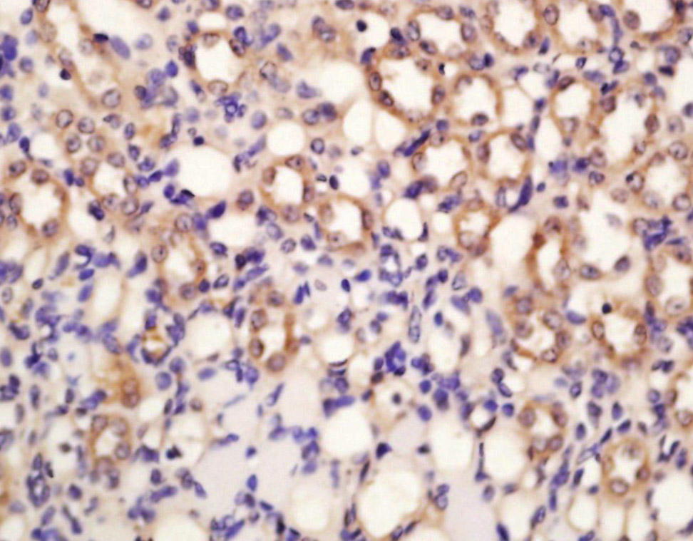
Tissue/cell: rat kidney tissue; 4% Paraformaldehyde-fixed and paraffin-embedded;
Antigen retrieval: citrate buffer ( 0.01M, pH 6.0 ), Boiling bathing for 15min; Block endogenous peroxidase by 3% Hydrogen peroxide for 30min; Blocking buffer (normal goat serum,C-0005) at 37℃ for 20 min;
Incubation: Anti-SOD1 Polyclonal Antibody, Unconjugated(bs-10216R) 1:200, overnight at 4°C, followed by conjugation to the secondary antibody(SP-0023) and DAB(C-0010) staining
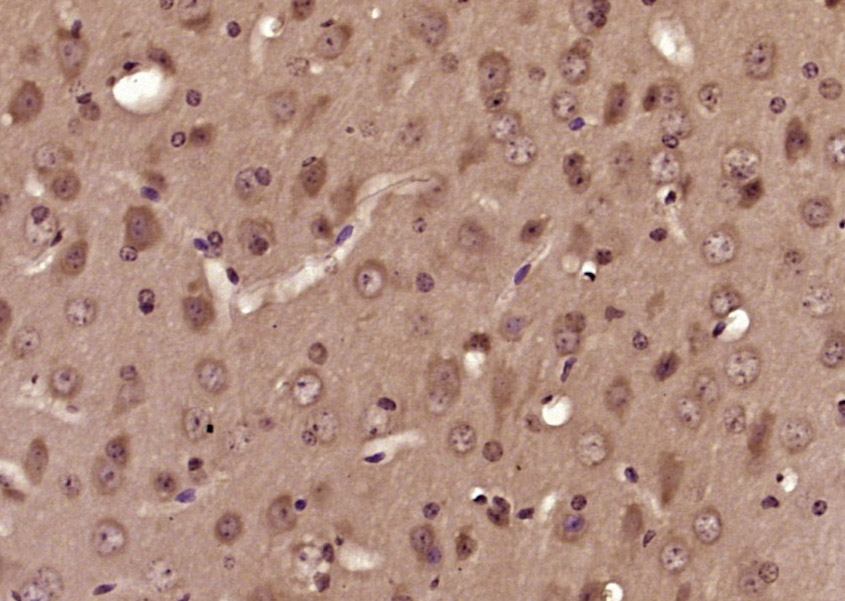
Paraformaldehyde-fixed, paraffin embedded (mouse brain tissue); Antigen retrieval by boiling in sodium citrate buffer (pH6.0) for 15min; Block endogenous peroxidase by 3% hydrogen peroxide for 20 minutes; Blocking buffer (normal goat serum) at 37°C for 30min; Antibody incubation with (SOD1) Polyclonal Antibody, Unconjugated (bs-10216R) at 1:200 overnight at 4°C, followed by operating according to SP Kit(Rabbit) (sp-0023) instructionsand DAB staining.
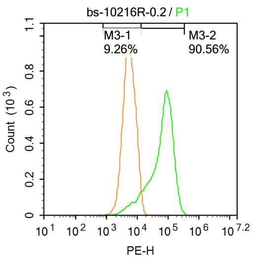
Blank control: Raji.
Primary Antibody (green line): Rabbit Anti-SOD1 antibody (bs-10216R)
Dilution: 1μg /10^6 cells;
Isotype Control Antibody (orange line): Rabbit IgG .
Secondary Antibody : Goat anti-rabbit IgG-PE
Dilution: 1μg /test.
Protocol
The cells were fixed with 4% PFA (10min at room temperature)and then permeabilized with PBST for 20 min at room temperature. The cells were then incubated in 5%BSA to block non-specific protein-protein interactions for 30 min at at room temperature .Cells stained with Primary Antibody for 30 min at room temperature. The secondary antibody used for 40 min at room temperature. Acquisition of 20,000 events was performed.
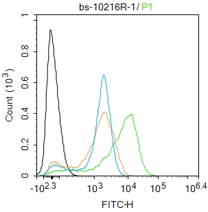
Blank control: Jurkat.
Primary Antibody (green line): Rabbit Anti-SOD1 antibody (bs-10216R)
Dilution: 1ug/Test;
Secondary Antibody : Goat anti-rabbit IgG-FITC
Dilution: 0.5ug/Test.
Protocol
The cells were fixed with 4% PFA (10min at room temperature)and then permeabilized with 0.1% PBST for 20 min at room temperature.The cells were then incubated in 5%BSA to block non-specific protein-protein interactions for 30 min at room temperature .Cells stained with Primary Antibody for 30 min at room temperature. The secondary antibody used for 40 min at room temperature. Acquisition of 20,000 events was performed.
|
| 1、抗体溶解方法 | |
| 2、抗体修复方式 | |
| 3、常用试剂的配制 | |
| 4、免疫组化操作步骤 | |
| 5、免疫组化问题解答 | |
| 6、Western Blotting 操作步骤 | |
| 7、Western Blotting 问题解答 | |
| 8、关于肽链的设计 | |
| 9、多肽的溶解与保存 | |
| 10、酶标抗体效价测定程序 | |


