

| 产品编号 | bs-8853R |
| 英文名称 | phospho-Smad2/Smad3 (Thr8) Rabbit pAb |
| 中文名称 | 磷酸化细胞信号转导分子SMAD2/SMAD3抗体 |
| 别 名 | SMAD2 & SMAD3 (phospho-T8); p-SMAD2 & SMAD3; phospho-SMAD2 & SMAD3; SMAD2 & SMAD3 (phospho-Thr8); CHTD8; JV18; JV18-1; LDS6; MADH2; MADR2; hMAD-2; hSMAD2; 7120426M23Rik; Smad-2; mMad2; SMAD2_HUMAN; SMAD2; MAD homolog 2; Mothers against DPP homolog 2; Mad- |
 |
Specific References (8) | bs-8853R has been referenced in 8 publications.
|
| 产品类型 | 磷酸化抗体 |
| 研究领域 | 肿瘤 细胞生物 免疫学 信号转导 细胞凋亡 转录调节因子 表观遗传学 |
| 抗体来源 | Rabbit |
| 克隆类型 | Polyclonal |
| 克 隆 号 | |
| 交叉反应 | Human,Mouse (predicted: Rat,Pig,Cow,Chicken,Dog,Horse) |
| 产品应用 | WB=1:500-2000,Flow-Cyt=1μg/Test
not yet tested in other applications. optimal dilutions/concentrations should be determined by the end user. |
| 理论分子量 | 52 kDa |
| 检测分子量 | 48,52 |
| 细胞定位 | 细胞核 细胞浆 |
| 性 状 | Liquid |
| 浓 度 | 1mg/ml |
| 免 疫 原 | KLH conjugated synthesised phosphopeptide derived from human Smad2/Smad3 around the phosphorylation site of Thr8: PF(p-T)PP |
| 亚 型 | IgG |
| 纯化方法 | affinity purified by Protein A |
| 缓 冲 液 | 0.01M TBS (pH7.4) with 1% BSA, 0.02% Proclin300 and 50% Glycerol. |
| 保存条件 | Shipped at 4℃. Store at -20℃ for one year. Avoid repeated freeze/thaw cycles. |
| 注意事项 | This product as supplied is intended for research use only, not for use in human, therapeutic or diagnostic applications. |
| PubMed | PubMed |
| 产品介绍 |
The protein encoded by this gene belongs to the SMAD, a family of proteins similar to the gene products of the Drosophila gene 'mothers against decapentaplegic' (Mad) and the C. elegans gene Sma. SMAD proteins are signal transducers and transcriptional modulators that mediate multiple signaling pathways. This protein mediates the signal of the transforming growth factor (TGF)-beta, and thus regulates multiple cellular processes, such as cell proliferation, apoptosis, and differentiation. This protein is recruited to the TGF-beta receptors through its interaction with the SMAD anchor for receptor activation (SARA) protein. In response to TGF-beta signal, this protein is phosphorylated by the TGF-beta receptors. The phosphorylation induces the dissociation of this protein with SARA and the association with the family member SMAD4. The association with SMAD4 is important for the translocation of this protein into the nucleus, where it binds to target promoters and forms a transcription repressor complex with other cofactors. This protein can also be phosphorylated by activin type 1 receptor kinase, and mediates the signal from the activin. Alternatively spliced transcript variants have been observed for this gene. [provided by RefSeq, May 2012] Function: SMAD is a family of proteins similar to the gene products of the Drosophila gene 'mothers against decapentaplegic' (Mad) and the C. elegans gene Sma. SMAD proteins are signal transducers and transcriptional modulators that mediate multiple signaling pathways. They mediate the signal of the transforming growth factor (TGF)-beta, and thus regulate multiple cellular processes, such as cell proliferation, apoptosis, and differentiation. Subcellular Location: Cytoplasm. Nucleus. Note: Cytoplasmic in the absence of ligand. Migrates to the nucleus when complexed with SMAD4. SWISS: Q15796 Gene ID: 4087 Database links: Entrez Gene: 4087 Human Entrez Gene: 4088 Human Entrez Gene: 17126 Mouse Entrez Gene: 17127 Mouse Omim: 601366 Human Omim: 603109 Human SwissProt: P84022 Human SwissProt: Q15796 Human SwissProt: Q62432 Mouse SwissProt: Q8BUN5 Mouse Unigene: 12253 Human Unigene: 714621 Human Unigene: 10636 Rat |
| 产品图片 |
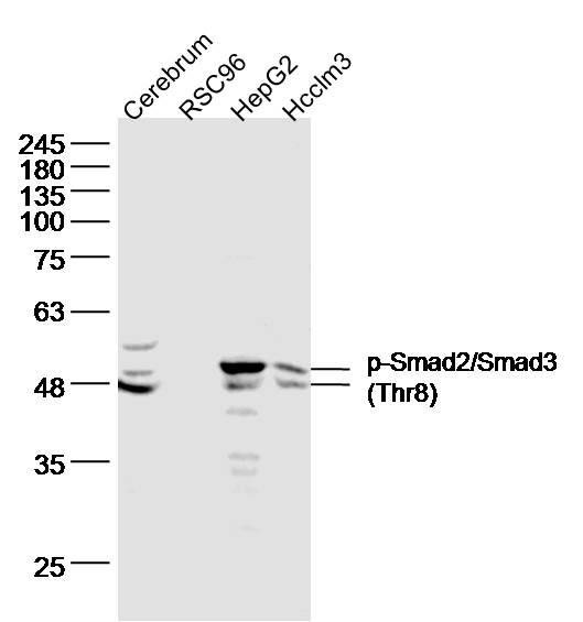
Sample:
cerebrum(mouse) Lysate at 40 ug
RSC96 cell(rat) Lysate at 30 ug
hepG2 cell(human) Lysate at 30 ug
Hcclm3 cell(human) Lysate at 30 ug
Primary: Anti- p-Smad2/Smad3 (Thr8) (bs-8853R) at 1/500 dilution
Secondary: IRDye800CW Goat Anti-Rabbit IgG at 1/20000 dilution
Predicted band size: 52kD
Observed band size: 48,52 kD
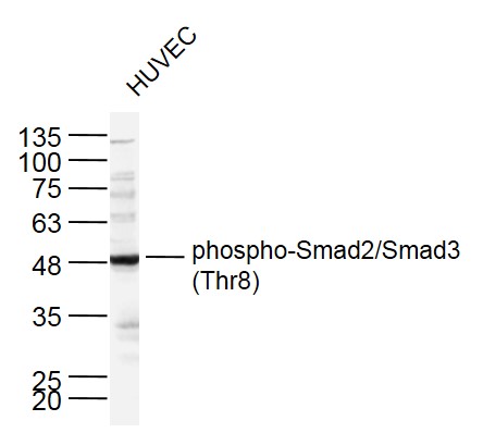
Sample:
HUVEC cell(human) Lysate at 30 ug
Primary: Anti- p-Smad2/Smad3 (Thr8) (bs-8853R) at 1/500 dilution
Secondary: IRDye800CW Goat Anti-Rabbit IgG at 1/20000 dilution
Predicted band size: 52kD
Observed band size: 52 kD
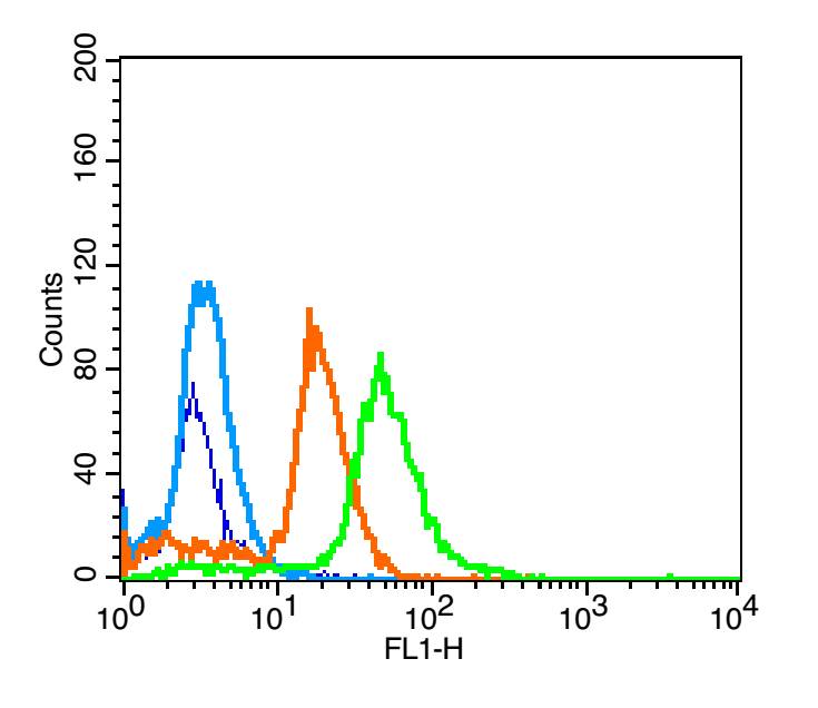
blank: A549 cells (blue line)
isotype control: rabbit IgG (orange line)
second antibody: goat anti-rabbit IgG (white blue line)
primary antibody: rabbit Anti-phospho-Smad2/Smad3 (Thr8) (green line); contration: 3μg /10^6 cells
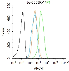
Blank control: Hela.
Primary Antibody (green line): Rabbit Anti-phospho-Smad2/Smad3 (Thr8) antibody (bs-8853R)
Dilution: 1μg /10^6 cells;
Isotype Control Antibody (orange line): Rabbit IgG .
Secondary Antibody : Goat anti-rabbit IgG-AF647
Dilution: 1μg /test.
Protocol
The cells were fixed with 4% PFA (10min at room temperature)and then permeabilized with 90% ice-cold methanol for 20 min at -20℃. The cells were then incubated in 5%BSA to block non-specific protein-protein interactions for 30 min at room temperature .Cells stained with Primary Antibody for 30 min at room temperature. The secondary antibody used for 40 min at room temperature. Acquisition of 20,000 events was performed.
|
| 1、抗体溶解方法 | |
| 2、抗体修复方式 | |
| 3、常用试剂的配制 | |
| 4、免疫组化操作步骤 | |
| 5、免疫组化问题解答 | |
| 6、Western Blotting 操作步骤 | |
| 7、Western Blotting 问题解答 | |
| 8、关于肽链的设计 | |
| 9、多肽的溶解与保存 | |
| 10、酶标抗体效价测定程序 | |


