

| 产品编号 | bs-10726R |
| 英文名称 | CDK2 Rabbit pAb |
| 中文名称 | 周期素依赖性激酶2抗体 |
| 别 名 | CDKN2; p33(CDK2); A630093N05Rik; CDK2_HUMAN; CDK2; Cell division protein kinase 2; p33 protein kinase; 2.7.11.22; CDK2_MOUSE; CDK2_RAT; |
 |
Specific References (6) | bs-10726R has been referenced in 6 publications.
[IF=6.1] Cuifang Chang. et al. The orphan GPR50 receptor interacting with TβRI induces G1/S-phase cell cycle arrest via Smad3-p27/p21 in BRL-3A cells. BIOCHEM PHARMACOL. 2022 Aug;202:115117 WB ; Rat.
[IF=5.075] Tianjie Wang. et al. Effect of Fumonisin B1 on Proliferation and Apoptosis of Intestinal Porcine Epithelial Cells. TOXINS. 2022 Jul;14(7):471 WB ; Pig.
[IF=4.221] Jingtao Zhang. et al. Ze-Qi decoction inhibits non-small cell lung cancer growth and metastasis by modulating the PI3K/Akt/p53 signaling pathway. Journal of Traditional and Complementary Medicine. 2023 Mar;: WB ; Mouse.
[IF=2.65] Ruoyang Lin. et al. Inhibitory Effects of Rabdosia rubescens in Esophageal Squamous Cell Carcinoma: Network Pharmacology and Experimental Validation. EVID-BASED COMPL ALT. 2022 Nov 10;2022:2696347 WB ; Human.
[IF=2.28] Wang, Haihe, et al. "RBP-J-interacting and tubulin-associated protein induces apoptosis and cell cycle arrest in human hepatocellular carcinoma by activating the p53-Fbxw7 pathway." Biochemical and Biophysical Research Communications (2014). WB ; Human.
[IF=1.99] He, Xiangming, et al. "CDK2-AP1 inhibits growth of breast cancer cells by regulating cell cycle and increasing docetaxel sensitivity in vivo and in vitro." Cancer Cell International 14.1 (2014): 130. WB ; Human.
|
| 研究领域 | 肿瘤 细胞生物 染色质和核信号 信号转导 细胞周期蛋白 激酶和磷酸酶 |
| 抗体来源 | Rabbit |
| 克隆类型 | Polyclonal |
| 克 隆 号 | |
| 交叉反应 | Human,Mouse (predicted: Rat,Rabbit,Pig,Sheep,Cow,Horse) |
| 产品应用 | WB=1:500-2000,Flow-Cyt=3μg/Test
not yet tested in other applications. optimal dilutions/concentrations should be determined by the end user. |
| 理论分子量 | 33 kDa |
| 检测分子量 | 30 |
| 细胞定位 | 细胞核 细胞浆 |
| 性 状 | Liquid |
| 浓 度 | 1mg/ml |
| 免 疫 原 | KLH conjugated synthetic peptide derived from human CDK2: 201-298/298 |
| 亚 型 | IgG |
| 纯化方法 | affinity purified by Protein A |
| 缓 冲 液 | 0.01M TBS (pH7.4) with 1% BSA, 0.02% Proclin300 and 50% Glycerol. |
| 保存条件 | Shipped at 4℃. Store at -20℃ for one year. Avoid repeated freeze/thaw cycles. |
| 注意事项 | This product as supplied is intended for research use only, not for use in human, therapeutic or diagnostic applications. |
| PubMed | PubMed |
| 产品介绍 |
The protein encoded by this gene is a member of the Ser/Thr protein kinase family. This protein kinase is highly similar to the gene products of S. cerevisiae cdc28, and S. pombe cdc2. It is a catalytic subunit of the cyclin-dependent protein kinase complex, whose activity is restricted to the G1-S phase, and essential for cell cycle G1/S phase transition. This protein associates with and regulated by the regulatory subunits of the complex including cyclin A or E, CDK inhibitor p21Cip1 (CDKN1A) and p27Kip1 (CDKN1B). Its activity is also regulated by its protein phosphorylation. Two alternatively spliced variants and multiple transcription initiation sites of this gene have been reported. [provided by RefSeq, Jul 2008]. Function: Serine/threonine-protein kinase involved in the control of the cell cycle; essential for meiosis, but dispensable for mitosis. Phosphorylates CTNNB1, USP37, p53/TP53, NPM1, CDK7, RB1, BRCA2, MYC, NPAT, EZH2. Interacts with cyclins A, B1, B3, D, or E. Triggers duplication of centrosomes and DNA. Acts at the G1-S transition to promote the E2F transcriptional program and the initiation of DNA synthesis, and modulates G2 progression; controls the timing of entry into mitosis/meiosis by controlling the subsequent activation of cyclin B/CDK1 by phosphorylation, and coordinates the activation of cyclin B/CDK1 at the centrosome and in the nucleus. Crucial role in orchestrating a fine balance between cellular proliferation, cell death, and DNA repair in human embryonic stem cells (hESCs). Activity of CDK2 is maximal during S phase and G2; activated by interaction with cyclin E during the early stages of DNA synthesis to permit G1-S transition, and subsequently activated by cyclin A2 (cyclin A1 in germ cells) during the late stages of DNA replication to drive the transition from S phase to mitosis, the G2 phase. EZH2 phosphorylation promotes H3K27me3 maintenance and epigenetic gene silencing. Phosphorylates CABLES1 (By similarity). Cyclin E/CDK2 prevents oxidative stress-mediated Ras-induced senescence by phosphorylating MYC. Involved in G1-S phase DNA damage checkpoint that prevents cells with damaged DNA from initiating mitosis; regulates homologous recombination-dependent repair by phosphorylating BRCA2, this phosphorylation is low in S phase when recombination is active, but increases as cells progress towards mitosis. In response to DNA damage, double-strand break repair by homologous recombination a reduction of CDK2-mediated BRCA2 phosphorylation. Phosphorylation of RB1 disturbs its interaction with E2F1. NPM1 phosphorylation by cyclin E/CDK2 promotes its dissociates from unduplicated centrosomes, thus initiating centrosome duplication. Cyclin E/CDK2-mediated phosphorylation of NPAT at G1-S transition and until prophase stimulates the NPAT-mediated activation of histone gene transcription during S phase. Required for vitamin D-mediated growth inhibition by being itself inactivated. Involved in the nitric oxide- (NO) mediated signaling in a nitrosylation/activation-dependent manner. USP37 is activated by phosphorylation and thus triggers G1-S transition. CTNNB1 phosphorylation regulates insulin internalization. Subunit: Found in a complex with CABLES1, CCNA1 and CCNE1. Interacts with CABLES1. Interacts with UHRF2. Part of a complex consisting of UHRF2, CDK2 and CCNE1. Interacts with the Speedy/Ringo proteins SPDYA and SPDYC. Found in a complex with both SPDYA and CDKN1B/KIP1. Binds to RB1 and CDK7. Binding to CDKN1A (p21) leads to CDK2/cyclin E inactivation at the G1-S phase DNA damage checkpoint, thereby arresting cells at the G1-S transition during DNA repair. Associated with PTPN6 and beta-catenin/CTNNB1. Interacts with CACUL1. May interact with CEP63. Subcellular Location: Cytoplasm, cytoskeleton, centrosome. Nucleus, Cajal body. Cytoplasm. Endosome. Note=Localized at the centrosomes in late G2 phase after separation of the centrosomes but before the start of prophase. Nuclear-cytoplasmic trafficking is mediated during the inhibition by 1,25-(OH)(2)D(3). Post-translational modifications: Phosphorylated at Thr-160 by CDK7 in a CAK complex. Phosphorylation at Thr-160 promotes kinase activity, whereas phosphorylation at Tyr-15 by WEE1 reduces slightly kinase activity. Phosphorylated on Thr-14 and Tyr-15 during S and G2 phases before being dephosphorylated by CDC25A. Nitrosylated after treatment with nitric oxide (DETA-NO). Similarity: Belongs to the protein kinase superfamily. CMGC Ser/Thr protein kinase family. CDC2/CDKX subfamily. Contains 1 protein kinase domain. SWISS: P24941 Gene ID: 1017 Database links: Entrez Gene: 1017 Human Entrez Gene: 12566 Mouse Omim: 116953 Human SwissProt: P24941 Human SwissProt: P97377 Mouse Unigene: 19192 Human Unigene: 689624 Human Unigene: 111326 Mouse Unigene: 104460 Rat |
| 产品图片 |
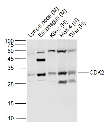
Sample:
Lane 1: Lymph node (Mouse) Lysate at 40 ug
Lane 2: Esophagus (Mouse) Lysate at 40 ug
Lane 3: K562 (Human) Cell Lysate at 30 ug
Lane 4: Molt-4 (Human) Cell Lysate at 30 ug
Lane 5: Siha (Human) Cell Lysate at 30 ug
Primary:
Anti-CDK2 (bs-10726R) at 1/1000 dilution
Secondary: IRDye800CW Goat Anti-Rabbit IgG at 1/20000 dilution
Predicted band size: 30 kD
Observed band size: 30 kD
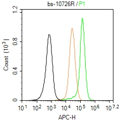
Blank control (Black line): U87MG(Black).
Primary Antibody (green line): Rabbit Anti-CDK2 antibody (bs-10726R)
Dilution: 3μg /10^6 cells;
Isotype Control Antibody (orange line): Rabbit IgG .
Secondary Antibody (white blue line): Goat anti-rabbit IgG-AF647
Dilution: 1μg /test.
Protocol
The cells were fixed with 4% PFA (10min at room temperature)and then permeabilized with 90% ice-cold methanol for 20 min at room temperature. The cells were then incubated in 5%BSA to block non-specific protein-protein interactions for 30 min at room temperature .Cells stained with Primary Antibody for 30 min at room temperature. The secondary antibody used for 40 min at room temperature. Acquisition of 20,000 events was performed.
|
| 1、抗体溶解方法 | |
| 2、抗体修复方式 | |
| 3、常用试剂的配制 | |
| 4、免疫组化操作步骤 | |
| 5、免疫组化问题解答 | |
| 6、Western Blotting 操作步骤 | |
| 7、Western Blotting 问题解答 | |
| 8、关于肽链的设计 | |
| 9、多肽的溶解与保存 | |
| 10、酶标抗体效价测定程序 | |


