
| 产品编号 | bs-20364R |
| 英文名称 | caspase-3 p17 subunit |
| 中文名称 | 活化半胱胺酸蛋白酶蛋白-3抗体 |
| 别 名 | Caspase-3 subunit p17; cleaved Caspase 3; cleaved Caspase-3; APOPAIN; CASP3; Caspase 3 apoptosis related cysteine protease; Caspase3; CPP32; CPP32B; Cysteine protease CPP32; Human cysteine protease CPP32 isoform alpha mRNA complete cds; PARP cleavage protease; SCA 1; SCA1; SREBP cleavage activity 1; Yama; CASP3_HUMAN; Caspase-3; CASP-3; Apopain; Protein Yama; SREBP cleavage activity 1; SCA-1. |
 |
Specific References (3) | bs-20364R has been referenced in 3 publications.
[IF=4.932] Yi Ren. et al. Pravastatin attenuates sepsis-induced acute lung injury through decreasing pulmonary microvascular permeability via inhibition of Cav-1/eNOS pathway. Int Immunopharmacol. 2021 Nov;100:108077 WB ; Mouse.
[IF=4.599] Juan Tan. et al. Circ_0124644 Serves as a ceRNA for miR-590-3p to Promote Hypoxia-Induced Cardiomyocytes Injury via Regulating SOX4. Front Genet. 2021; 12: 667724 WB ; Human.
[IF=2.523] Zhi Chen. et al. Klotho deficiency aggravates diabetes-induced podocyte injury due to DNA damage caused by mitochondrial dysfunction. Int J Med Sci. 2020; 17(17): 2763–2772 WB ; Mouse.
|
| 研究领域 | 细胞生物 免疫学 信号转导 细胞凋亡 |
| 抗体来源 | Rabbit |
| 克隆类型 | Polyclonal |
| 交叉反应 | Human,Mouse |
| 产品应用 | WB=1:500-2000, IHC-P=1:100-500, IHC-F=1:100-500, ICC=1:100-500, IF=1:100-500, Flow-Cyt=1μg/Test, ELISA=1:5000-10000
not yet tested in other applications. optimal dilutions/concentrations should be determined by the end user. |
| 理论分子量 | 17/32kDa |
| 细胞定位 | 细胞浆 |
| 性 状 | Liquid |
| 浓 度 | 1mg/ml |
| 免 疫 原 | KLH conjugated synthetic peptide derived from human caspase-3 p17 subunit : 1-80/277 |
| 亚 型 | IgG |
| 纯化方法 | affinity purified by Protein A |
| 缓 冲 液 | Preservative: 15mM Sodium Azide, Constituents: 1% BSA, 0.01M PBS, pH 7.4 |
| 保存条件 | Shipped at 4℃. Store at -20 °C for one year. Avoid repeated freeze/thaw cycles. |
| 注意事项 | This product as supplied is intended for research use only, not for use in human, therapeutic or diagnostic applications. |
| PubMed | PubMed |
| 产品介绍 |
The caspase family of cysteine proteases play a key role in apoptosis. Caspase 3 is the most extensively studied apoptotic protein among caspase family members. Caspase 3 is synthesized as inactive pro enzyme that is processed in cells undergoing apoptosis by self proteolysis and/or cleavage by other upstream proteases (e.g. Caspases 8, 9 and 10). The processed form of Caspase 3 consists of large (17kDa) and small (12kDa) subunits which associate to form an active enzyme. Caspase 3 is cleaved at Asp28 Ser29 and Asp175 Ser176. The active Caspase 3 proteolytically cleaves and activates other caspases (e.g. Caspases 6, 7 and 9), as well as relevant targets in the cells (e.g. PARP and DFF). Alternative splicing of this gene results in two transcript variants which encode the same protein. In immunohistochemical studies Caspase 3 expression has been shown to be widespread but not present in all cell types (e.g. commonly reported in epithelial cells of skin, renal proximal tubules and collecting ducts). Differences in the level of Caspase 3 have been reported in cells of short lived nature (eg germinal centre B cells) and those that are long lived (eg mantle zone B cells). Caspase 3 is the predominant caspase involved in the cleavage of amyloid beta 4A precursor protein, which is associated with neuronal death in Alzheimer's disease. Reacts with Caspase-3 subunit p17 and precursor. Function: Involved in the activation cascade of caspases responsible for apoptosis execution. At the onset of apoptosis it proteolytically cleaves poly(ADP-ribose) polymerase (PARP) at a '216-Asp-|-Gly-217' bond. Cleaves and activates sterol regulatory element binding proteins (SREBPs) between the basic helix-loop-helix leucine zipper domain and the membrane attachment domain. Cleaves and activates caspase-6, -7 and -9. Involved in the cleavage of huntingtin. Triggers cell adhesion in sympathetic neurons through RET cleavage. Subunit: Heterotetramer that consists of two anti-parallel arranged heterodimers, each one formed by a 17 kDa (p17) and a 12 kDa (p12) subunit. Interacts with BIRC6/bruce. Subcellular Location: Cytoplasm. Tissue Specificity: Highly expressed in lung, spleen, heart, liver and kidney. Moderate levels in brain and skeletal muscle, and low in testis. Also found in many cell lines, highest expression in cells of the immune system. Post-translational modifications: Cleavage by granzyme B, caspase-6, caspase-8 and caspase-10 generates the two active subunits. Additional processing of the propeptides is likely due to the autocatalytic activity of the activated protease. Active heterodimers between the small subunit of caspase-7 protease and the large subunit of caspase-3 also occur and vice versa. S-nitrosylated on its catalytic site cysteine in unstimulated human cell lines and denitrosylated upon activation of the Fas apoptotic pathway, associated with an increase in intracellular caspase activity. Fas therefore activates caspase-3 not only by inducing the cleavage of the caspase zymogen to its active subunits, but also by stimulating the denitrosylation of its active site thiol. Similarity: Belongs to the peptidase C14A family. SWISS: P42574 Gene ID: 836 Database links: Entrez Gene: 836 Human Entrez Gene: 12367 Mouse Entrez Gene: 100008840 Rabbit Omim: 600636 Human SwissProt: P42574 Human SwissProt: P70677 Mouse SwissProt: Q8MJC3 Rabbit Unigene: 141125 Human Unigene: 34405 Mouse Unigene: 10562 Rat
Caspase3广泛分布于各种不同类型的细胞中,是Caspase家族中最重要的凋亡执行者之一,激活的Caspase-3能使许多与细胞结构、细胞周期及DNA修复等相关蛋白或激酶失活,从而使细胞凋亡. |
| 产品图片 |
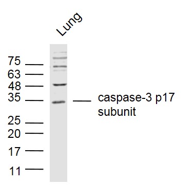
Sample:
Lung (Mouse) Lysate at 40 ug Primary: Anti- caspase-3 p17 subunit (bs-20364R) at 1/300 dilution Secondary: IRDye800CW Goat Anti-Rabbit IgG at 1/20000 dilution Predicted band size: 17/32 kD Observed band size: 17/32 kD 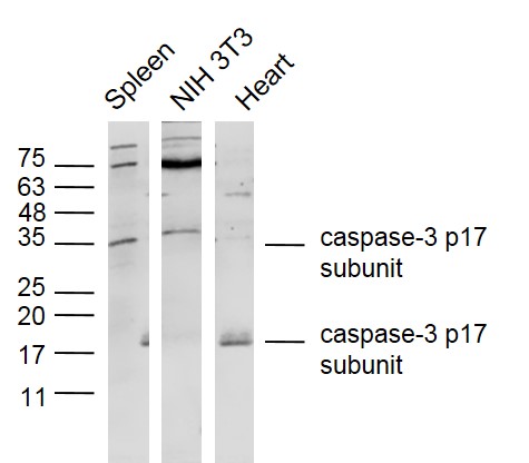
Sample:
Spleen (Mouse) Lysate at 40 ug NIH/3T3 (Mouse) Cell Lysate at 30 ug Heart (Mouse) Lysate at 40 ug Primary: Anti- caspase-3 p17 subunit (bs-20364R) at 1/300 dilution Secondary: IRDye800CW Goat Anti-Rabbit IgG at 1/20000 dilution Predicted band size: 17/32 kD Observed band size: 17/32 kD 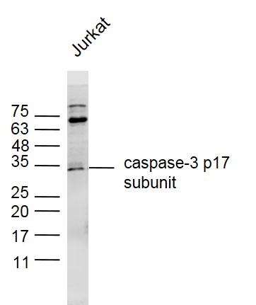
Sample:
Jurkat (human) Cell Lysate at 30 ug Primary: Anti- caspase-3 p17 subunit (bs-20364R) at 1/300 dilution Secondary: IRDye800CW Goat Anti-Rabbit IgG at 1/20000 dilution Predicted band size: 17/32 kD Observed band size: 17/32 kD 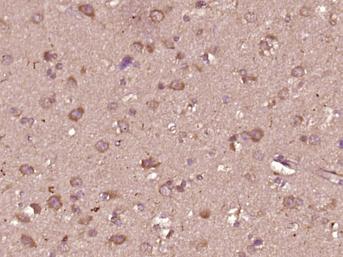
Paraformaldehyde-fixed, paraffin embedded (Human glioma); Antigen retrieval by boiling in sodium citrate buffer (pH6.0) for 15min; Block endogenous peroxidase by 3% hydrogen peroxide for 20 minutes; Blocking buffer (normal goat serum) at 37°C for 30min; Antibody incubation with (caspase-3 p17 subunit) Polyclonal Antibody, Unconjugated (bs-20364R) at 1:400 overnight at 4°C, followed by operating according to SP Kit(Rabbit) (sp-0023) instructionsand DAB staining.

Blank control (blue line): Hela (fixed with 80% methanol (5 min at -20℃) and then permeabilized with 0.1% PBS-Tween for 20 min at room temperature).
Primary Antibody (green line): Rabbit Anti-caspase-3 p12 subunit antibody (bs-20364R),Dilution: 1μg /10^6 cells; Isotype Control Antibody (orange line): Rabbit IgG . Secondary Antibody (white blue line): Goat anti-rabbit IgG-FITC,Dilution: 1μg /test. 
Overlay histogram showing HL 60 cells stained with bs-20364R (Green line).
The cells were fixed with 90% methanol (5 min) and then permeabilized with 0.01M PBS-Tween for 20 min. The cells were then incubated in 1x PBS / 10% normal goat serum to block non-specific protein-protein interactions followed by the antibody (bs-20364R,1μg/1x10^6 cells) for 30 min at 22℃. The secondary antibody used was fluorescein isothiocyanate goat anti-rabbit IgG (H+L) (bs- 0295G-FITC , Brillant blue line) at 1/200 dilution for 30 min at 22℃. Isotype control antibody was rabbit IgG (polyclonal,bs-0295P,Orange line) (1μg/1x10^6 cells) used under the same conditions. Unlabelled sample (blue line) was also used as a control. Acquisition of 20,000 events were collected using a 20mW Argon ion laser (488nm) and 525/30 bandpass filter. 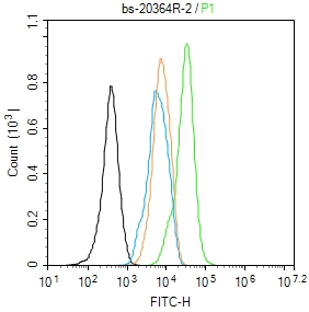
Blank control: NIH/3T3.
Primary Antibody (green line): Rabbit Anti-caspase-3 p17 subunit antibody (bs-20364R) Dilution: 1μg /10^6 cells; Isotype Control Antibody (orange line): Rabbit IgG . Secondary Antibody : Goat anti-rabbit IgG-AF488 Dilution: 1μg /test. Protocol The cells were fixed with 4% PFA (10min at room temperature)and then permeabilized with 90% ice-cold methanol for 20 min at -20℃. The cells were then incubated in 5%BSA to block non-specific protein-protein interactions for 30 min at room temperature .Cells stained with Primary Antibody for 30 min at room temperature. The secondary antibody used for 40 min at room temperature. Acquisition of 20,000 events was performed. |
| 1、抗体溶解方法 | |
| 2、抗体修复方式 | |
| 3、常用试剂的配制 | |
| 4、免疫组化操作步骤 | |
| 5、免疫组化问题解答 | |
| 6、Western Blotting 操作步骤 | |
| 7、Western Blotting 问题解答 | |
| 8、关于肽链的设计 | |
| 9、多肽的溶解与保存 | |
| 10、酶标抗体效价测定程序 | |


