

| 产品编号 | bs-23103R |
| 英文名称 | Ki-67 Rabbit pAb |
| 中文名称 | Ki67抗体 |
| 别 名 | KIA; MIB-; MIB-1; PPP1R105; KI67_HUMAN; MKI67; Antigen identified by monoclonal antibody Ki-67 (Antigen KI-67 | Antigen Ki67); marker of proliferation Ki-67; antigen identified by monoclonal antibody Ki-67; protein phosphatase 1, regulatory subunit 105; Molecular Immunology Borstel antibody 1 |
 |
Specific References (24) | bs-23103R has been referenced in 24 publications.
|
| 研究领域 | 肿瘤 细胞生物 神经生物学 细胞周期蛋白 细胞分化 细胞类型标志物 表观遗传学 |
| 抗体来源 | Rabbit |
| 克隆类型 | Polyclonal |
| 交叉反应 | Human |
| 产品应用 | Flow-Cyt=1:50-100,ICC/IF=1:100-500
not yet tested in other applications. optimal dilutions/concentrations should be determined by the end user. |
| 理论分子量 | 358kDa |
| 细胞定位 | 细胞核 |
| 性 状 | Liquid |
| 浓 度 | 1mg/ml |
| 免 疫 原 | KLH conjugated synthetic peptide derived from human Ki-67: 1231-1330/3256 |
| 亚 型 | IgG |
| 纯化方法 | affinity purified by Protein A |
| 缓 冲 液 | 0.01M TBS (pH7.4) with 1% BSA, 0.02% Proclin300 and 50% Glycerol. |
| 保存条件 | Shipped at 4℃. Store at -20℃ for one year. Avoid repeated freeze/thaw cycles. |
| 注意事项 | This product as supplied is intended for research use only, not for use in human, therapeutic or diagnostic applications. |
| PubMed | PubMed |
| 产品介绍 |
Ki67 antigen is the prototypic cell cycle related nuclear protein, expressed by proliferating cells in all phases of the active cell cycle (G1, S, G2 and M phase). It is absent in resting (G0) cells. Ki67 antibodies are useful in establishing the cell growing fraction in neoplasms (immunohistochemically quantified by determining the number of Ki67 positive cells among the total number of resting cells = Ki67 index). In neoplastic tissues the prognostic value is comparable to the tritiated thymidine labelling index. The correlation between low Ki67 index and histologically low grade tumours is strong. Ki67 is routinely used as a neuronal marker of cell cycling and proliferation. Function: Thought to be required for maintaining cell proliferation. Subcellular Location: Nucleus. Chromosome. Predominantly localized in the G1 phase in the perinucleolar region, in the later phases it is also detected throughout the nuclear interior, being predominantly localized in the nuclear matrix. In mitosis, it is present on all chromosomes. Similarity: Contains 1 FHA domain. SWISS: P46013 Gene ID: 4288 Database links: Entrez Gene: 4288 Human Entrez Gene: 17345 Mouse Omim: 176741 Human SwissProt: P46013 Human SwissProt: Q91VE6 Mouse Unigene: 689823 Human Unigene: 80976 Human Unigene: 4078 Mouse Unigene: 233802 Rat 细胞增殖标志物(Proliferation Marker) Ki67与PCNA一样,为细胞增殖的一种标记,在细胞凋亡中S、G2 、M期均有表达,G0期缺如。 Ki-67增殖指数高低与许多肿瘤的分化程度、浸润、转移以及预后密切相关,因此被广泛作为各种恶性肿瘤的必检项目之一。 |
| 产品图片 |
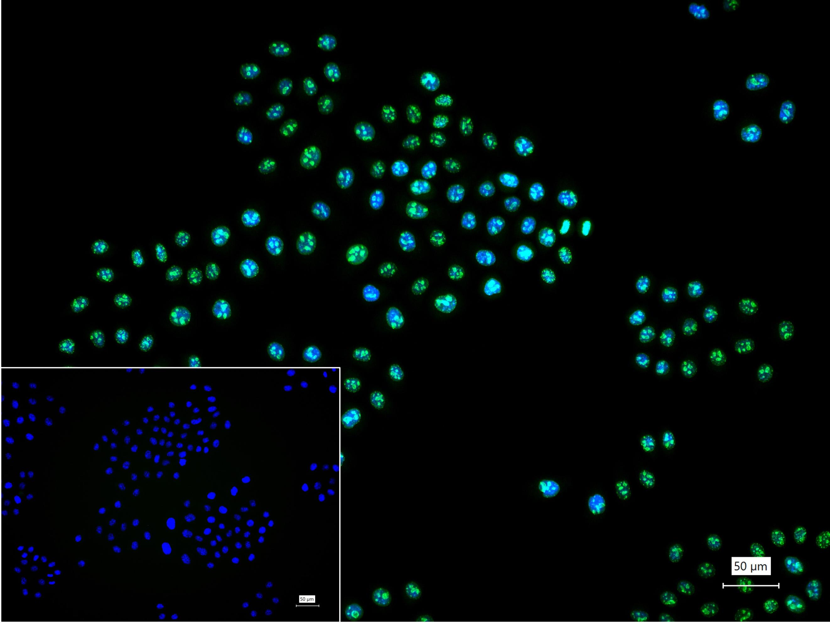
4% Paraformaldehyde-fixed Hela (H) cell; Triton X-100 at r.t. for 20 min; Antibody incubation with (Ki-67) polyclonal Antibody, unconjugated (bs-23103R) 1:100, 90 min at 37°C; followed by conjugated Goat Anti-Rabbit IgG antibody (green, bs-60295G-BF488) at 37°C for 90 min, DAPI (blue, C02-04002) was used to stain the cell nuclei. PBS instead of the primary antibody was used as the blank control.
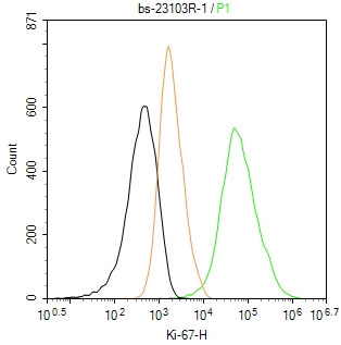
The HepG2 (H) cells were fixed with 4% PFA (10 min at r.t.) and then permeabilized with 90% ice-cold methanol for 20 min at -20℃,the cells then were incubated in 5%BSA to block non-specific protein-protein interactions (30 min at r.t.), followed by secondary antibody incubation for 40 min at room temperature. Primary Antibody (green):Rabbit Anti-Ki-67 antibody (bs-23103R,1:100); Isotype Control (orange): Rabbit IgG (bs-0295P). Blank control (black): PBS. Acquisition of 20,000 events was performed.
|
| 1、抗体溶解方法 | |
| 2、抗体修复方式 | |
| 3、常用试剂的配制 | |
| 4、免疫组化操作步骤 | |
| 5、免疫组化问题解答 | |
| 6、Western Blotting 操作步骤 | |
| 7、Western Blotting 问题解答 | |
| 8、关于肽链的设计 | |
| 9、多肽的溶解与保存 | |
| 10、酶标抗体效价测定程序 | |


