

| 产品编号 | bs-41192R |
| 英文名称 | TNF alpha Rabbit pAb |
| 中文名称 | 人肿瘤坏死因子 |
| 别 名 | Tumor necrosis factor-α; TNFalpha; APC1; Cachectin; DIF; Differentiation inducing factor; Macrophage cytotoxic factor; MCF; Necrosin; TNF a; TNF-alpha; TNF; TNF Macrophage Derived; TNF Monocyte Derived; TNF Superfamily Member 2; TNFA; TNFSF2; Tumor necros |
 |
Specific References (1) | bs-41192R has been referenced in 1 publications.
[IF=2.464] Zhou, Taocheng. et al. Leonurine Alleviates Alcoholic Steatohepatitis Through the TLR4/NF-κB Signalling Pathway. Revista Brasileira de Farmacognosia-Brazilian Journal of Pharmacognosy. 2022 Aug;:1-15 WB ; Mouse.
|
| 研究领域 | 肿瘤 细胞生物 免疫学 神经生物学 信号转导 细胞凋亡 生长因子和激素 |
| 抗体来源 | Rabbit |
| 克隆类型 | Polyclonal |
| 交叉反应 | Human,Mouse |
| 产品应用 | WB=1:500-2000,Flow-Cyt=1ug/Test
not yet tested in other applications. optimal dilutions/concentrations should be determined by the end user. |
| 理论分子量 | 17/26 kDa |
| 检测分子量 | |
| 细胞定位 | 分泌型蛋白 |
| 性 状 | Liquid |
| 浓 度 | 1mg/ml |
| 免 疫 原 | Recombinant human TNF-Alpha Protein: 77-233/233aa <Extracellular> |
| 亚 型 | IgG |
| 纯化方法 | affinity purified by Protein A |
| 缓 冲 液 | 0.01M TBS (pH7.4) with 1% BSA, 0.02% Proclin300 and 50% Glycerol. |
| 保存条件 | Shipped at 4℃. Store at -20℃ for one year. Avoid repeated freeze/thaw cycles. |
| 注意事项 | This product as supplied is intended for research use only, not for use in human, therapeutic or diagnostic applications. |
| PubMed | PubMed |
| 产品介绍 |
This gene encodes a multifunctional proinflammatory cytokine that belongs to the tumor necrosis factor (TNF) superfamily. This cytokine is mainly secreted by macrophages. It can bind to, and thus functions through its receptors TNFRSF1A/TNFR1 and TNFRSF1B/TNFBR. This cytokine is involved in the regulation of a wide spectrum of biological processes including cell proliferation, differentiation, apoptosis, lipid metabolism, and coagulation. This cytokine has been implicated in a variety of diseases, including autoimmune diseases, insulin resistance, psoriasis, rheumatoid arthritis ankylosing spondylitis, tuberculosis, autosomal dominant polycystic kidney disease, and cancer. Mutations in this gene affect susceptibility to cerebral malaria, septic shock, and Alzheimer disease. Knockout studies in mice also suggested the neuroprotective function of this cytokine. [provided by RefSeq, Aug 2020] Function: Cytokine that binds to TNFRSF1A/TNFR1 and TNFRSF1B/TNFBR. It is mainly secreted by macrophages and can induce cell death of certain tumor cell lines. It is potent pyrogen causing fever by direct action or by stimulation of interleukin-1 secretion and is implicated in the induction of cachexia, Under certain conditions it can stimulate cell proliferation and induce cell differentiation. The TNF intracellular domain (ICD) form induces IL12 production in dendritic cells. Subunit: Homotrimer. Interacts with SPPL2B. Subcellular Location: Cell membrane; Single-pass type II membrane protein. Tumor necrosis factor, membrane form: Membrane; Single-pass type II membrane protein. Tumor necrosis factor, soluble form: Secreted. C-domain 1: Secreted. C-domain 2: Secreted. Post-translational modifications: The soluble form derives from the membrane form by proteolytic processing. The membrane-bound form is further proteolytically processed by SPPL2A or SPPL2B through regulated intramembrane proteolysis producing TNF intracellular domains (ICD1 and ICD2) released in the cytosol and TNF C-domain 1 and C-domain 2 secreted into the extracellular space. The membrane form, but not the soluble form, is phosphorylated on serine residues. Dephosphorylation of the membrane form occurs by binding to soluble TNFRSF1A/TNFR1. O-glycosylated; glycans contain galactose, N-acetylgalactosamine and N-acetylneuraminic acid. DISEASE: Genetic variations in TNF are a cause of susceptibility psoriatic arthritis (PSORAS) [MIM:607507]. PSORAS is an inflammatory, seronegative arthritis associated with psoriasis. It is a heterogeneous disorder ranging from a mild, non-destructive disease to a severe, progressive, erosive arthropathy. Five types of psoriatic arthritis have been defined: asymmetrical oligoarthritis characterized by primary involvement of the small joints of the fingers or toes; asymmetrical arthritis which involves the joints of the extremities; symmetrical polyarthritis characterized by a rheumatoidlike pattern that can involve hands, wrists, ankles, and feet; arthritis mutilans, which is a rare but deforming and destructive condition; arthritis of the sacroiliac joints and spine (psoriatic spondylitis). Similarity: Belongs to the tumor necrosis factor family. SWISS: P01375 Gene ID: 7124 Database links: Entrez Gene: 7124 Human Entrez Gene: 21926 Mouse Omim: 191160 Human SwissProt: P01375 Human SwissProt: P06804 Mouse Unigene: 241570 Human Unigene: 1293 Mouse |
| 产品图片 |

Sample:
Lane 1: A431 (Human) Cell Lysate at 30 ug
Lane 2: Hela (Human) Cell Lysate at 30 ug
Primary:
Anti-TNF alpha (bs-41192R) at 1/1000 dilution
Secondary: IRDye800CW Goat Anti-Rabbit IgG at 1/20000 dilution
Predicted band size: 36/17/26 kD
Observed band size: 36 kD
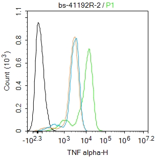
Blank control:Raw264.7.
Primary Antibody (green line): Rabbit Anti-TNF alpha antibody (bs-41192R)
Dilution: 2ug/Test;
Secondary Antibody : Goat anti-rabbit IgG-FITC
Dilution: 0.5ug/Test.
Protocol
The cells were treated with LPS (1 ug/mL, 18 hr/6 hr) and Brefeldin A (300 ng/mL, last 3 hr of stimulation).The cells were fixed with 4% PFA (10min at room temperature)and then permeabilized with 0.1% PBST for 20 min at room temperature.The cells were then incubated in 5%BSA to block non-specific protein-protein interactions for 30 min at room temperature .Cells stained with Primary Antibody for 30 min at room temperature. The secondary antibody used for 40 min at room temperature. Acquisition of 20,000 events was performed.
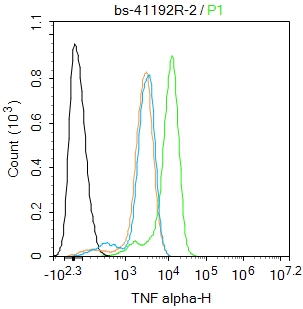
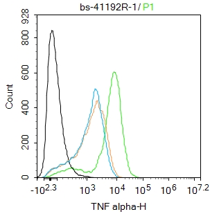
Blank control:THP-1.
Primary Antibody (green line): Rabbit Anti-TNF alpha antibody (bs-41192R)
Dilution: 1ug/Test;
Secondary Antibody : Goat anti-rabbit IgG-FITC
Dilution: 0.5ug/Test.
Protocol
The cells were treated with TPA (80 nM, overnight) and then treated with LPS (1 ug/mL, 18 hr/6 hr) and Brefeldin A (300 ng/mL, last 3 hr of stimulation) .The cells were fixed with 4% PFA (10min at room temperature)and then permeabilized with 0.1% PBST for 20 min at room temperature.The cells were then incubated in 5%BSA to block non-specific protein-protein interactions for 30 min at room temperature .Cells stained with Primary Antibody for 30 min at room temperature. The secondary antibody used for 40 min at room temperature. Acquisition of 20,000 events was performed.
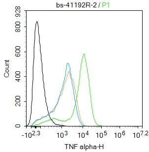
Blank control:THP-1.
Primary Antibody (green line): Rabbit Anti-TNF alpha antibody (bs-41192R)
Dilution:2ug/Test;
Secondary Antibody : Goat anti-rabbit IgG-FITC
Dilution: 0.5ug/Test.
Protocol
The cells were treated with TPA (80 nM, overnight) and then treated with LPS (1 ug/mL, 18 hr/6 hr) and Brefeldin A (300 ng/mL, last 3 hr of stimulation) .The cells were fixed with 4% PFA (10min at room temperature)and then permeabilized with 0.1% PBST for 20 min at room temperature.The cells were then incubated in 5%BSA to block non-specific protein-protein interactions for 30 min at room temperature .Cells stained with Primary Antibody for 30 min at room temperature. The secondary antibody used for 40 min at room temperature. Acquisition of 20,000 events was performed.
|
| 1、抗体溶解方法 | |
| 2、抗体修复方式 | |
| 3、常用试剂的配制 | |
| 4、免疫组化操作步骤 | |
| 5、免疫组化问题解答 | |
| 6、Western Blotting 操作步骤 | |
| 7、Western Blotting 问题解答 | |
| 8、关于肽链的设计 | |
| 9、多肽的溶解与保存 | |
| 10、酶标抗体效价测定程序 | |


