

| 产品编号 | bs-22049R |
| 英文名称 | ErbB 4 Rabbit pAb |
| 中文名称 | HER4抗体 |
| 别 名 | ALS19; HER4; p180erbB4; c-erbB-4; ERBB4_HUMAN; ERBB4; Proto-oncogene-like protein c-ErbB-4; Tyrosine kinase-type cell surface receptor HER4; 2.7.10.1; ERBB4_MOUSE; Mer4; ERBB4_RAT; Tyro-2; |
| 研究领域 | 肿瘤 免疫学 神经生物学 信号转导 生长因子和激素 转录调节因子 激酶和磷酸酶 |
| 抗体来源 | Rabbit |
| 克隆类型 | Polyclonal |
| 克 隆 号 | |
| 交叉反应 | Human,Mouse (predicted: Rat,Rabbit,Chicken,Dog,Horse) |
| 产品应用 | IHC-P=1:100-500,IHC-F=1:100-500,IF=1:100-500
not yet tested in other applications. optimal dilutions/concentrations should be determined by the end user. |
| 理论分子量 | 142 kDa |
| 细胞定位 | 细胞核 细胞浆 细胞膜 |
| 性 状 | Liquid |
| 浓 度 | 1mg/ml |
| 免 疫 原 | KLH conjugated synthetic peptide derived from human ErbB 4 : 21-120/1308 <Extracellular> |
| 亚 型 | IgG |
| 纯化方法 | affinity purified by Protein A |
| 缓 冲 液 | 0.01M TBS (pH7.4) with 1% BSA, 0.02% Proclin300 and 50% Glycerol. |
| 保存条件 | Shipped at 4℃. Store at -20℃ for one year. Avoid repeated freeze/thaw cycles. |
| 注意事项 | This product as supplied is intended for research use only, not for use in human, therapeutic or diagnostic applications. |
| PubMed | PubMed |
| 产品介绍 |
This gene is a member of the Tyr protein kinase family and the epidermal growth factor receptor subfamily. It encodes a single-pass type I membrane protein with multiple cysteine rich domains, a transmembrane domain, a tyrosine kinase domain, a phosphotidylinositol-3 kinase binding site and a PDZ domain binding motif. The protein binds to and is activated by neuregulins and other factors and induces a variety of cellular responses including mitogenesis and differentiation. Multiple proteolytic events allow for the release of a cytoplasmic fragment and an extracellular fragment. Mutations in this gene have been associated with cancer. Alternatively spliced variants which encode different protein isoforms have been described; however, not all variants have been fully characterized. [provided by RefSeq, Jul 2008]. Function: Tyrosine-protein kinase that plays an essential role as cell surface receptor for neuregulins and EGF family members and regulates development of the heart, the central nervous system and the mammary gland, gene transcription, cell proliferation, differentiation, migration and apoptosis. Required for normal cardiac muscle differentiation during embryonic development, and for postnatal cardiomyocyte proliferation. Required for normal development of the embryonic central nervous system, especially for normal neural crest cell migration and normal axon guidance. Required for mammary gland differentiation, induction of milk proteins and lactation. Acts as cell-surface receptor for the neuregulins NRG1, NRG2, NRG3 and NRG4 and the EGF family members BTC, EREG and HBEGF. Ligand binding triggers receptor dimerization and autophosphorylation at specific tyrosine residues that then serve as binding sites for scaffold proteins and effectors. Ligand specificity and signaling is modulated by alternative splicing, proteolytic processing, and by the formation of heterodimers with other ERBB family members, thereby creating multiple combinations of intracellular phosphotyrosines that trigger ligand- and context-specific cellular responses. Mediates phosphorylation of SHC1 and activation of the MAP kinases MAPK1/ERK2 and MAPK3/ERK1. Isoform JM-A CYT-1 and isoform JM-B CYT-1 phosphorylate PIK3R1, leading to the activation of phosphatidylinositol 3-kinase and AKT1 and protect cells against apoptosis. Isoform JM-A CYT-1 and isoform JM-B CYT-1 mediate reorganization of the actin cytoskeleton and promote cell migration in response to NRG1. Isoform JM-A CYT-2 and isoform JM-B CYT-2 lack the phosphotyrosine that mediates interaction with PIK3R1, and hence do not phosphorylate PIK3R1, do not protect cells against apoptosis, and do not promote reorganization of the actin cytoskeleton and cell migration. Proteolytic processing of isoform JM-A CYT-1 and isoform JM-A CYT-2 gives rise to the corresponding soluble intracellular domains (4ICD) that translocate to the nucleus, promote nuclear import of STAT5A, activation of STAT5A, mammary epithelium differentiation, cell proliferation and activation of gene expression. The ERBB4 soluble intracellular domains (4ICD) colocalize with STAT5A at the CSN2 promoter to regulate transcription of milk proteins during lactaction. The ERBB4 soluble intracellular domains can also translocate to mitochondria and promote apoptosis. Subunit: Monomer in the absence of bound ligand. Homodimer or heterodimer with another ERBB family member upon ligand binding, thus forming heterotetramers. Interacts with EGFR and ERBB2. Interacts with CBFA2T3 (By similarity). Interacts with DLG2 (via its PDZ domain), DLG3 (via its PDZ domain), DLG4 (via its PDZ domain) and SNTB2 (via its PDZ domain). Interacts with MUC1. Interacts (via its PPxy motifs) with WWOX. Interacts (via the PPxY motif 3 of isoform JM-A CYT-2) with YAP1 (via the WW domain 1 of isoform 1). Interacts (isoform JM-A CYT-1 and isoform JM-B CYT-1) with WWP1. Interacts (via its intracellular domain) with TRIM28. Interacts (via the intracellular domains of both CYT-1 and CYT-2 isoforms) with KAP1; the interaction does not phosphorylate KAP1 but represses ERBB4-mediated transcriptional activity. Interacts with PRPU, DDX23, MATR3, RBM15, ILF3, KAP1, U5S1, U2SURP, ITCH, HNRPU, AP2A1, NULC, LEO1, WWP2, IGHG1, HXK1, GRB7 AND ARS2. Interacts (phosphorylated isoform JM-A CYT-1 and isoform JM-B CYT-1) with PIK3R1. Interacts with SHC1. Interacts with GRB2. Interacts (soluble intracellular domain) with STAT5A. Interacts (soluble intracellular domain) with BCL2. Interacts (phosphorylated) with STAT1. Subcellular Location: Cell membrane; Single-pass type I membrane protein. Note=In response to NRG1 treatment, the activated receptor is internalized. ERBB4 intracellular domain: Nucleus. Mitochondrion. Note=Following proteolytical processing E4ICD (E4ICD1 or E4ICD2 generated from the respective isoforms) is translocated to the nucleus. Significantly more E4ICD2 than E4ICD1 is found in the nucleus. E4ICD2 colocalizes with YAP1 in the nucleus. Tissue Specificity: Expressed at highest levels in brain, heart, kidney, in addition to skeletal muscle, parathyroid, cerebellum, pituitary, spleen, testis and breast. Lower levels in thymus, lung, salivary gland, and pancreas. Isoform JM-A CYT-1 and isoform JM-B CYT-1 are expressed in cerebellum, but only the isoform JM-B is expressed in the heart. Post-translational modifications: Isoform JM-A CYT-1 and isoform JM-A CYT-2 are processed by ADAM17. Proteolytic processing in response to ligand or 12-O-tetradecanoylphorbol-13-acetate stimulation results in the production of 120 kDa soluble receptor forms and intermediate membrane-anchored 80 kDa fragments (m80HER4), which are further processed by a presenilin-dependent gamma-secretase to release a cytoplasmic intracellular domain (E4ICD; E4ICD1/s80Cyt1 or E4ICD2/s80Cyt2, depending on the isoform). Membrane-anchored 80 kDa fragments of the processed isoform JM-A CYT-1 are more readily degraded by the proteasome than fragments of isoform JM-A CYT-2, suggesting a prevalence of E4ICD2 over E4ICD1. Isoform JM-B CYT-1 and isoform JM-B CYT-2 lack the ADAM17 cleavage site and are not processed by ADAM17, precluding further processing by gamma-secretase. Autophosphorylated on tyrosine residues in response to ligand binding. Autophosphorylation occurs in trans, i.e. one subunit of the dimeric receptor phosphorylates tyrosine residues on the other subunit. Ligands trigger phosphorylation at specific tyrosine residues, thereby creating binding sites for scaffold proteins and effectors. Constitutively phosphorylated at a basal level when overexpressed in heterologous systems; ligand binding leads to increased phosphorylation. Phosphorylation at Tyr-1035 is important for interaction with STAT1. Phosphorylation at Tyr-1056 is important for interaction with PIK3R1. Phosphorylation at Tyr-1242 is important for interaction with SHC1. Phosphorylation at Tyr-1188 may also contribute to the interaction with SHC1. Isoform JM-A CYT-2 is constitutively phosphorylated on tyrosine residues in a ligand-independent manner. E4ICD2 but not E4ICD1 is phosphorylated on tyrosine residues. Ubiquitinated. During mitosis, the ERBB4 intracellular domain is ubiquitinated by the APC/C complex and targeted to proteasomal degradation. Isoform JM-A CYT-1 and isoform JM-B CYT-1 are ubiquitinated by WWP1. The ERBB4 intracellular domain (E4ICD1) is ubiquitinated, and this involves NEDD4. Similarity: Belongs to the protein kinase superfamily. Tyr protein kinase family. EGF receptor subfamily. Contains 1 protein kinase domain. SWISS: Q15303 Gene ID: 2066 Database links: Entrez Gene: 2066 Human Entrez Gene: 13869 Mouse Omim: 600543 Human SwissProt: Q15303 Human SwissProt: Q61527 Mouse Unigene: 390729 Human Unigene: 442420 Mouse Unigene: 163078 Rat 细胞膜受体(Membrane Receptors) c-erbB-4蛋白过去一直为乳腺癌基因研究的主要方向,研究乳腺癌组织学分级及术后生存期的关系。目前认为他与细胞增殖活性和激素受体有一定的关联,有学者将c-erbB-4蛋白研究用于神经内分泌蛋白为主。 |
| 产品图片 |
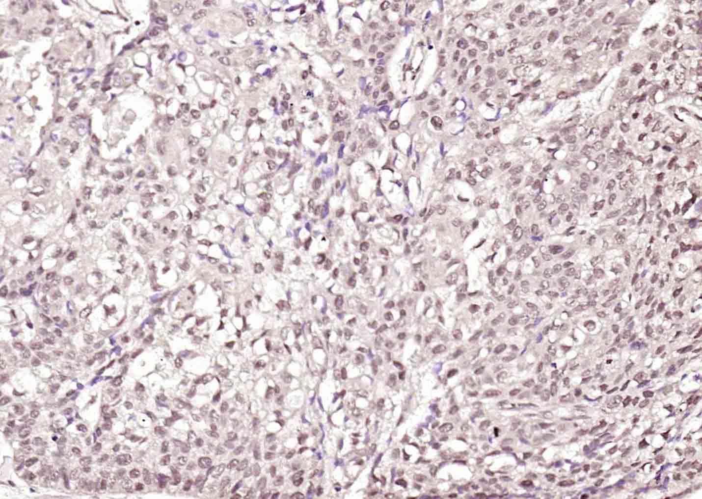
Paraformaldehyde-fixed, paraffin embedded (Human esophageal cancer); Antigen retrieval by boiling in sodium citrate buffer (pH6.0) for 15min; Block endogenous peroxidase by 3% hydrogen peroxide for 20 minutes; Blocking buffer (normal goat serum) at 37°C for 30min; Antibody incubation with (ErbB 4) Polyclonal Antibody, Unconjugated (bs-22049R) at 1:400 overnight at 4°C, followed by operating according to SP Kit(Rabbit) (sp-0023) instructionsand DAB staining.
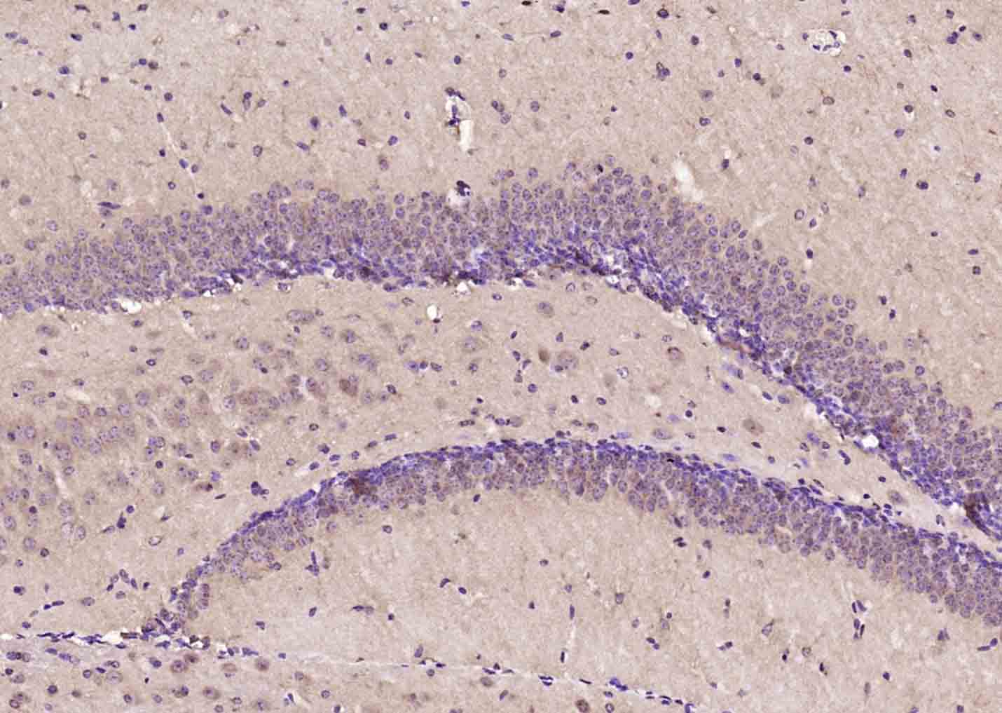
Paraformaldehyde-fixed, paraffin embedded (mouse brain); Antigen retrieval by boiling in sodium citrate buffer (pH6.0) for 15min; Block endogenous peroxidase by 3% hydrogen peroxide for 20 minutes; Blocking buffer (normal goat serum) at 37°C for 30min; Antibody incubation with (ErbB 4) Polyclonal Antibody, Unconjugated (bs-22049R) at 1:400 overnight at 4°C, followed by operating according to SP Kit(Rabbit) (sp-0023) instructionsand DAB staining.
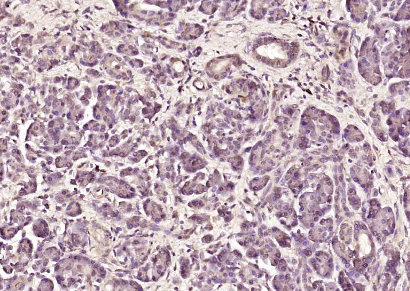
Paraformaldehyde-fixed, paraffin embedded (human pancreatic cancer); Antigen retrieval by boiling in sodium citrate buffer (pH6.0) for 15min; Block endogenous peroxidase by 3% hydrogen peroxide for 20 minutes; Blocking buffer (normal goat serum) at 37°C for 30min; Antibody incubation with (ErbB 4) Polyclonal Antibody, Unconjugated (bs-22049R) at 1:400 overnight at 4°C, followed by operating according to SP Kit(Rabbit) (sp-0023) instructionsand DAB staining.
|
| 1、抗体溶解方法 | |
| 2、抗体修复方式 | |
| 3、常用试剂的配制 | |
| 4、免疫组化操作步骤 | |
| 5、免疫组化问题解答 | |
| 6、Western Blotting 操作步骤 | |
| 7、Western Blotting 问题解答 | |
| 8、关于肽链的设计 | |
| 9、多肽的溶解与保存 | |
| 10、酶标抗体效价测定程序 | |


