
| 产品编号 | bsm-52048R |
| 英文名称 | Cyclin E1 Recombinant Rabbit mAb |
| 中文名称 | 周期素E重组兔单抗 |
| 别 名 | CCNE; pCCNE1; CycE1; CYCLE; CCNE1_HUMAN; CCNE1; CCNE1_MOUSE; CCNE1_RAT; |
 |
Specific References (1) | bsm-52048R has been referenced in 1 publications.
[IF=3.738] Qiao Zhang. et al. G6PD upregulates Cyclin E1 and MMP9 to promote clear cell renal cell carcinoma progression. Int J Med Sci. 2022; 19(1): 47–64 IHC ; Human.
|
| 研究领域 | 细胞生物 细胞周期蛋白 |
| 抗体来源 | Rabbit |
| 克隆类型 | Recombinant |
| 克 隆 号 | 4H7 |
| 交叉反应 | Human |
| 产品应用 | WB=1:500-2000,IHC-P=1:100-500,IHC-F=1:100-500,IF=1:100-500,ICC/IF=1:50-200
not yet tested in other applications. optimal dilutions/concentrations should be determined by the end user. |
| 理论分子量 | 45 kDa |
| 检测分子量 | 48 |
| 细胞定位 | 细胞核 |
| 性 状 | Liquid |
| 浓 度 | 1mg/ml |
| 免 疫 原 | A synthesized peptide derived from human Cyclin E1: 350-410 |
| 亚 型 | IgG |
| 纯化方法 | affinity purified by Protein A |
| 缓 冲 液 | 0.01M TBS (pH7.4) with 1% BSA, 0.02% Proclin300 and 50% Glycerol. |
| 保存条件 | Shipped at 4℃. Store at -20℃ for one year. Avoid repeated freeze/thaw cycles. |
| 注意事项 | This product as supplied is intended for research use only, not for use in human, therapeutic or diagnostic applications. |
| PubMed | PubMed |
| 产品介绍 |
The protein encoded by this gene belongs to the highly conserved cyclin family, whose members are characterized by a dramatic periodicity in protein abundance through the cell cycle. Cyclins function as regulators of CDK kinases. Different cyclins exhibit distinct expression and degradation patterns which contribute to the temporal coordination of each mitotic event. This cyclin forms a complex with and functions as a regulatory subunit of CDK2, whose activity is required for cell cycle G1/S transition. This protein accumulates at the G1-S phase boundary and is degraded as cells progress through S phase. Overexpression of this gene has been observed in many tumors, which results in chromosome instability, and thus may contribute to tumorigenesis. This protein was found to associate with, and be involved in, the phosphorylation of NPAT protein (nuclear protein mapped to the ATM locus), which participates in cell-cycle regulated histone gene expression and plays a critical role in promoting cell-cycle progression in the absence of pRB. [provided by RefSeq, Apr 2016] Function: Essential for the control of the cell cycle at the G1/S (start) transition. Subunit: Interacts with a member of the CDK2/CDK protein kinases to form a serine/threonine kinase holoenzyme complex. The cyclin subunit imparts substrate specificity to the complex. Found in a complex with CDK2, CABLES1 and CCNA1 (By similarity). Part of a complex consisting of UHRF2, CDK2 and CCNE1. Interacts directly with UHRF2; the interaction ubiquitinates CCNE1 and appears to occur independently of CCNE1 phosphorylation. Subcellular Location: Nucleus. Tissue Specificity: Highly expressed in testis and placenta. Low levels in bronchial epithelial cells. Post-translational modifications: Phosphorylation of Thr-395 by GSK3 and of Ser-399 by CDK2 accelerates degradation via the ubiquitin proteasome pathway. Phosphorylated upon DNA damage, probably by ATM or ATR. Similarity: Belongs to the cyclin family. Cyclin E subfamily. SWISS: P24864 Gene ID: 898 Database links: Entrez Gene: 898 Human Entrez Gene: 12447 Mouse Omim: 123837 Human SwissProt: P24864 Human SwissProt: Q61457 Mouse Unigene: 244723 Human Unigene: 16110 Mouse Unigene: 15455 Rat |
| 产品图片 |
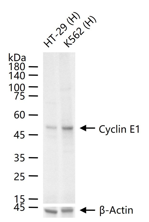
25 ug total protein per lane of various lysates (see on figure) probed with Cyclin E1 monoclonal antibody, unconjugated (bsm-52048R) at 1:1000 dilution and 4°C overnight incubation. Followed by conjugated secondary antibody incubation at r.t. for 60 min.
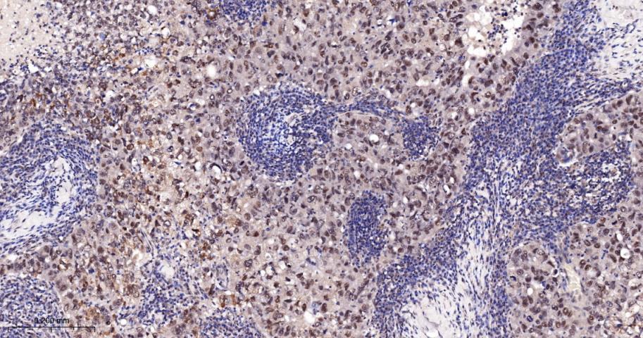
Paraformaldehyde-fixed, paraffin embedded Human Breast Cancer; Antigen retrieval by boiling in sodium citrate buffer (pH6.0) for 15 min; The section was incubated with Cyclin E1 Monoclonal Antibody, Unconjugated (bsm-52048R) at 1:200 overnight at 4°C, followed by conjugation to the bs-0295G-HRP and DAB (C-0010) staining.
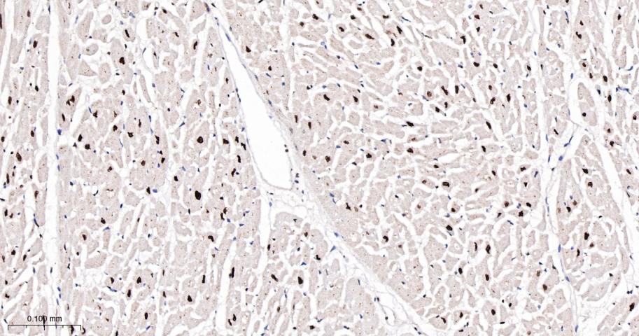
Paraformaldehyde-fixed, paraffin embedded Human Heart; Antigen retrieval by boiling in sodium citrate buffer (pH6.0) for 15 min; The section was incubated with Cyclin E1 Monoclonal Antibody, Unconjugated (bsm-52048R) at 1:200 overnight at 4°C, followed by conjugation to the bs-0295G-HRP and DAB (C-0010) staining.
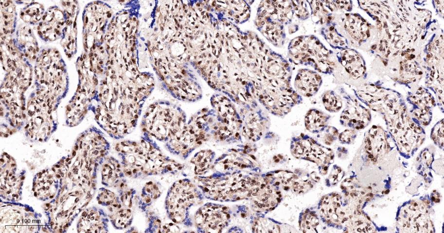
Paraformaldehyde-fixed, paraffin embedded Human Placenta; Antigen retrieval by boiling in sodium citrate buffer (pH6.0) for 15 min; The section was incubated with Cyclin E1 Monoclonal Antibody, Unconjugated (bsm-52048R) at 1:200 overnight at 4°C, followed by conjugation to the bs-0295G-HRP and DAB (C-0010) staining.
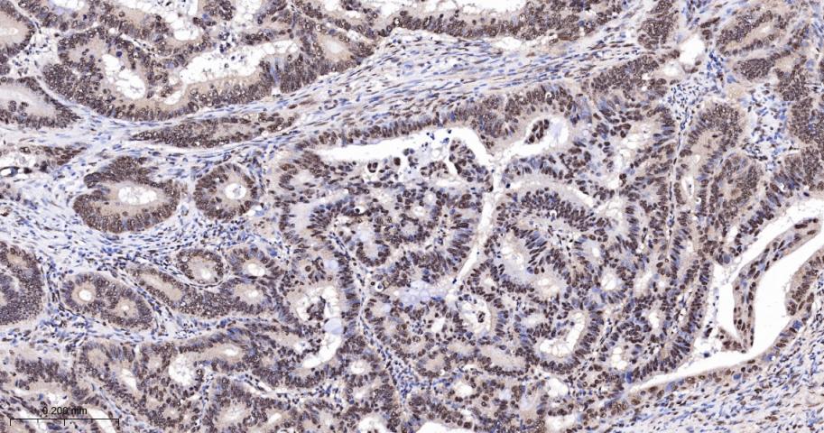
Paraformaldehyde-fixed, paraffin embedded Human Colon Cancer; Antigen retrieval by boiling in sodium citrate buffer (pH6.0) for 15 min; The section was incubated with Cyclin E1 Monoclonal Antibody, Unconjugated (bsm-52048R) at 1:200 overnight at 4°C, followed by conjugation to the bs-0295G-HRP and DAB (C-0010) staining.
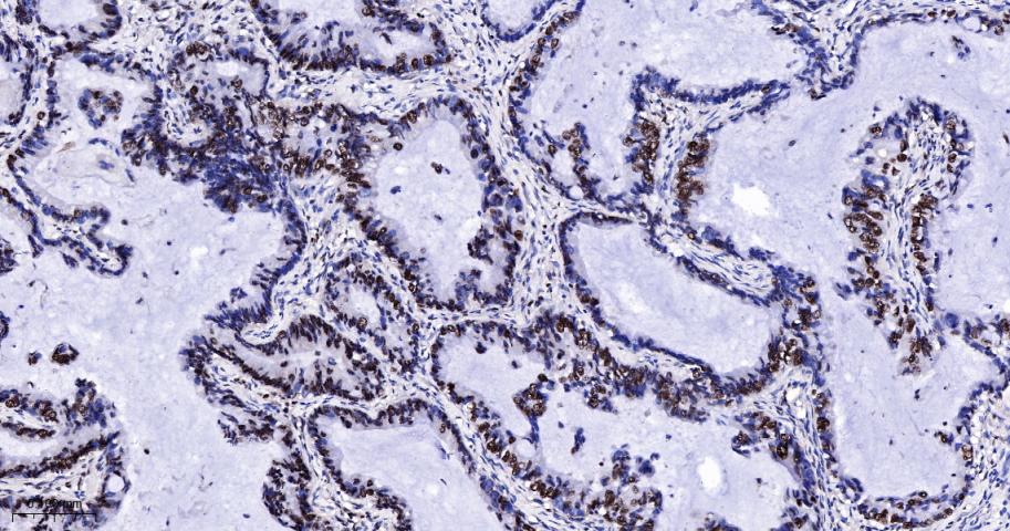
Paraformaldehyde-fixed, paraffin embedded Human Lung Cancer; Antigen retrieval by boiling in sodium citrate buffer (pH6.0) for 15 min; The section was incubated with Cyclin E1 Monoclonal Antibody, Unconjugated (bsm-52048R) at 1:200 overnight at 4°C, followed by conjugation to the bs-0295G-HRP and DAB (C-0010) staining.
|
| 1、抗体溶解方法 | |
| 2、抗体修复方式 | |
| 3、常用试剂的配制 | |
| 4、免疫组化操作步骤 | |
| 5、免疫组化问题解答 | |
| 6、Western Blotting 操作步骤 | |
| 7、Western Blotting 问题解答 | |
| 8、关于肽链的设计 | |
| 9、多肽的溶解与保存 | |
| 10、酶标抗体效价测定程序 | |


