
| 产品编号 | bsm-52223R |
| 英文名称 | Smad2 Recombinant Rabbit mAb |
| 中文名称 | 细胞信号转导分子Smad-2重组兔单抗 |
| 别 名 | CHTD8; JV18; JV18-1; LDS6; MADH2; MADR2; hMAD-2; hSMAD2; 7120426M23Rik; Smad-2; mMad2; SMAD2_HUMAN; SMAD2; MAD homolog 2; Mothers against DPP homolog 2; Mad-related protein 2 (hMAD-2); SMAD family member 2 (SMAD 2 | Smad2 | hSMAD2); SMAD2_MOUSE; Mad-relat |
 |
Specific References (1) | bsm-52223R has been referenced in 1 publications.
[IF=4.694] Qi SS et al. Protective Effects of Chromium Picolinate Against Diabetic-Induced Renal Dysfunction and Renal Fibrosis in Streptozotocin-Induced Diabetic Rats. Biomolecules. 2020 Mar 4;10(3). IHC ; rat.
|
| 研究领域 | 肿瘤 细胞生物 信号转导 细胞凋亡 生长因子和激素 转录调节因子 激酶和磷酸酶 |
| 抗体来源 | Rabbit |
| 克隆类型 | Recombinant |
| 克 隆 号 | 9A3 |
| 交叉反应 | Human,Mouse,Rat |
| 产品应用 | WB=1:500-2000,IHC-P=1:50-200,IHC-F=1:50-200,IF=1:50-200,Flow-Cyt=1ug/Test,ICC/IF=1:50-200
not yet tested in other applications. optimal dilutions/concentrations should be determined by the end user. |
| 理论分子量 | 52 kDa |
| 检测分子量 | 60 |
| 细胞定位 | 细胞核 细胞浆 |
| 性 状 | Liquid |
| 浓 度 | 1mg/ml |
| 免 疫 原 | A synthesized peptide derived from human SMAD2: 230-270 |
| 亚 型 | IgG |
| 纯化方法 | affinity purified by Protein A |
| 缓 冲 液 | 0.01M TBS (pH7.4) with 1% BSA, 0.02% Proclin300 and 50% Glycerol. |
| 保存条件 | Shipped at 4℃. Store at -20℃ for one year. Avoid repeated freeze/thaw cycles. |
| 注意事项 | This product as supplied is intended for research use only, not for use in human, therapeutic or diagnostic applications. |
| PubMed | PubMed |
| 产品介绍 |
SMAD2 or Mothers against decapentaplegic homolog 2 is a polypeptide that, as its name describes, is a homolog of the Drosophila gene: "Mothers against decepentaplegic". It belongs to the SMAD family of proteins, which belong to the TGF-Beta superfamily of modulators. Like many other TGF-Beta family members SMAD2 is involved in cell signalling. SMAD2 modulates signals of activin and TGF-Beta's. It interacts with SMAD anchor for receptor activation (SARA). The binding of ligands causes the phosphorylation of the SMAD2 protein and the dissociation from SARA and the association with SMAD4. It is subsequently transferred to the nucleus where it forms complexes with other proteins and acts as a transcription factor. SMAD2 is a receptor regulated SMAD (R-SMAD) and is activated by bone morphogenetic protein type 1 receptor kinase. This antibody is cross reactive with Smad3. Function: Receptor-regulated SMAD (R-SMAD) that is an intracellular signal transducer and transcriptional modulator activated by TGF-beta (transforming growth factor) and activin type 1 receptor kinases. Binds the TRE element in the promoter region of many genes that are regulated by TGF-beta and, on formation of the SMAD2/SMAD4 complex, activates transcription. May act as a tumor suppressor in colorectal carcinoma. Positively regulates PDPK1 kinase activity by stimulating its dissociation from the 14-3-3 protein YWHAQ which acts as a negative regulator. Subunit: Momomer; the absence of TGF-beta. Heterodimer; in the presence of TGF-beta. Forms a heterodimer with co-SMAD, SMAD4, in the nucleus to form the transactivation complex SMAD2/SMAD4. Interacts with AIP1, HGS, PML and WWP1. Interacts with NEDD4L in response to TGF-beta. Found in a complex with SMAD3 and TRIM33 upon addition of TGF-beta. Interacts with ACVR1B, SMAD3 and TRIM33. Interacts (via the MH2 domain) with ZFYVE9; may form trimers with the SMAD4 co-SMAD. Interacts with FOXH1, homeobox protein TGIF, PEBP2-alpha subunit, CREB-binding protein (CBP), EP300 and SKI. Interacts with SNON; when phosphorylated at Ser-465/467. Interacts with SKOR1 and SKOR2. Interacts with PRDM16. Interacts (via MH2 domain) with LEMD3. Interacts with RBPMS. Interacts with WWP1. Interacts (dephosphorylated form, via the MH1 and MH2 domains) with RANBP3 (via its C-terminal R domain); the interaction results in the export of dephosphorylated SMAD3 out of the nucleus and termination ot the TGF-beta signaling. Interacts with PDPK1 (via PH domain). Subcellular Location: Cytoplasm. Nucleus. Note=Cytoplasmic and nuclear in the absence of TGF-beta. On TGF-beta stimulation, migrates to the nucleus when complexed with SMAD4. On dephosphorylation by phosphatase PPM1A, released from the SMAD2/SMAD4 complex, and exported out of the nucleus by interaction with RANBP1. Tissue Specificity: Expressed at high levels in skeletal muscle, heart and placenta. Post-translational modifications: Phosphorylated on one or several of Thr-220, Ser-245, Ser-250, and Ser-255. In response to TGF-beta, phosphorylated on Ser-465/467 by TGF-beta and activin type 1 receptor kinases. Able to interact with SMURF2 when phosphorylated on Ser-465/467, recruiting other proteins, such as SNON, for degradation. In response to decorin, the naturally occurring inhibitor of TGF-beta signaling, phosphorylated on Ser-240 by CaMK2. Phosphorylated by MAPK3 upon EGF stimulation; which increases transcriptional activity and stability, and is blocked by calmodulin. Phosphorylated by PDPK1. In response to TGF-beta, ubiquitinated by NEDD4L; which promotes its degradation. Acetylated on Lys-19 by coactivators in response to TGF-beta signaling, which increases transcriptional activity. Isoform short: Acetylation increases DNA binding activity in vitro and enhances its association with target promoters in vivo. Acetylation in the nucleus by EP300 is enhanced by TGF-beta. Similarity: Belongs to the dwarfin/SMAD family. Contains 1 MH1 (MAD homology 1) domain. Contains 1 MH2 (MAD homology 2) domain. SWISS: Q15796 Gene ID: 4087 Database links: Entrez Gene: 4087 Human Entrez Gene: 17126 Mouse Omim: 601366 Human SwissProt: Q15796 Human SwissProt: Q62432 Mouse Unigene: 12253 Human Unigene: 705764 Human Unigene: 391091 Mouse Unigene: 2755 Rat |
| 产品图片 |
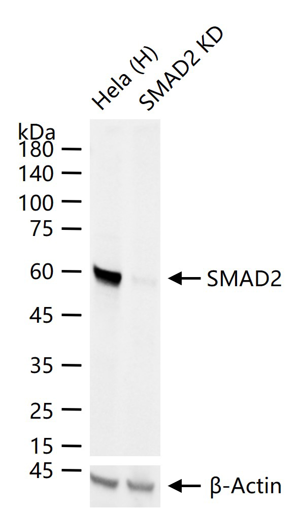
25 ug total protein per lane of various lysates (see on figure) probed with SMAD2 monoclonal antibody, unconjugated (bsm-52223R) at 1:1000 dilution and 4°C overnight incubation. Followed by conjugated secondary antibody incubation at r.t. for 60 min.
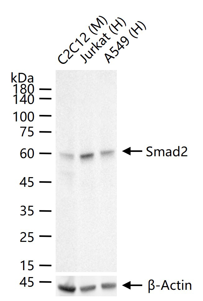
25 ug total protein per lane of various lysates (see on figure) probed with Smad2 monoclonal antibody, unconjugated (bsm-52223R) at 1:1000 dilution and 4°C overnight incubation. Followed by conjugated secondary antibody incubation at r.t. for 60 min.
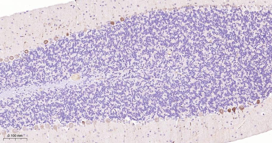
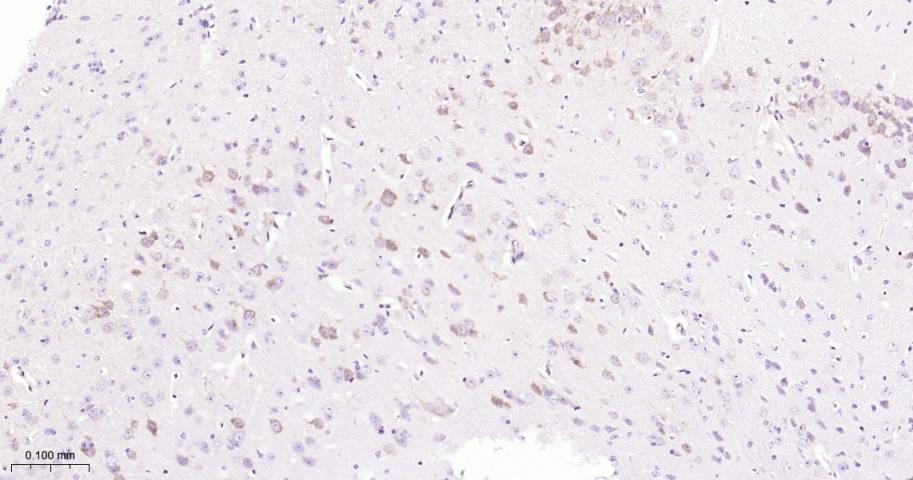
Paraformaldehyde-fixed, paraffin embedded Rat Cerebellum; Antigen retrieval by boiling in sodium citrate buffer (pH6.0) for 15 min; Antibody incubation with Smad2 Monoclonal Antibody, Unconjugated(bsm-52223R) at 1:200 overnight at 4°C, followed by conjugation to the bs-0295G-HRP and DAB (C-0010) staining.
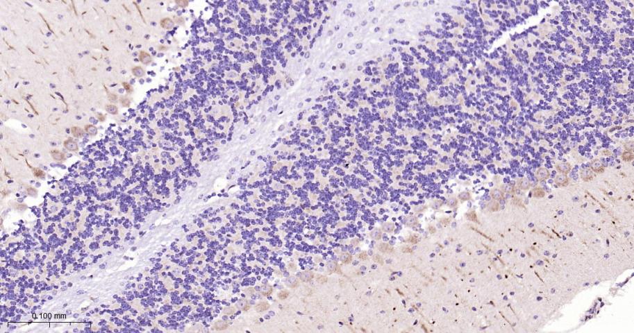
Paraformaldehyde-fixed, paraffin embedded Mouse Cerebellum; Antigen retrieval by boiling in sodium citrate buffer (pH6.0) for 15 min; Antibody incubation with Smad2 Monoclonal Antibody, Unconjugated(bsm-52223R) at 1:200 overnight at 4°C, followed by conjugation to the bs-0295G-HRP and DAB (C-0010) staining.

Paraformaldehyde-fixed, paraffin embedded Human Cerebrum; Antigen retrieval by boiling in sodium citrate buffer (pH6.0) for 15 min; Antibody incubation with Smad2 Monoclonal Antibody, Unconjugated(bsm-52223R) at 1:200 overnight at 4°C, followed by conjugation to the bs-0295G-HRP and DAB (C-0010) staining.
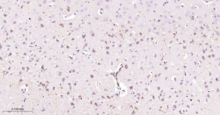
Paraformaldehyde-fixed, paraffin embedded Rat Cerebrum; Antigen retrieval by boiling in sodium citrate buffer (pH6.0) for 15 min; Antibody incubation with Smad2 Monoclonal Antibody, Unconjugated(bsm-52223R) at 1:200 overnight at 4°C, followed by conjugation to the bs-0295G-HRP and DAB (C-0010) staining.
Paraformaldehyde-fixed, paraffin embedded Mouse Cerebrum; Antigen retrieval by boiling in sodium citrate buffer (pH6.0) for 15 min; Antibody incubation with Smad2 Monoclonal Antibody, Unconjugated(bsm-52223R) at 1:200 overnight at 4°C, followed by conjugation to the bs-0295G-HRP and DAB (C-0010) staining.
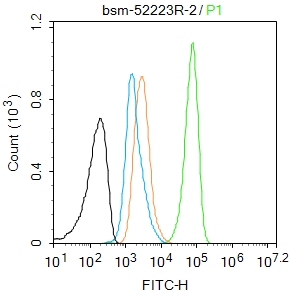
Blank control: A431.
Primary Antibody (green line): Rabbit Anti-Smad2 antibody (bsm-52223R)
Dilution: 1μg /10^6 cells;
Isotype Control Antibody (orange line): Rabbit IgG .
Secondary Antibody : Goat anti-rabbit IgG-AF488
Dilution: 1μg /test.
Protocol
The cells were fixed with 4% PFA (10min at room temperature)and then permeabilized with 90% ice-cold methanol for 20 min at-20℃. The cells were then incubated in 5%BSA to block non-specific protein-protein interactions for 30 min at at room temperature .Cells stained with Primary Antibody for 30 min at room temperature. The secondary antibody used for 40 min at room temperature. Acquisition of 20,000 events was performed.
|
| 1、抗体溶解方法 | |
| 2、抗体修复方式 | |
| 3、常用试剂的配制 | |
| 4、免疫组化操作步骤 | |
| 5、免疫组化问题解答 | |
| 6、Western Blotting 操作步骤 | |
| 7、Western Blotting 问题解答 | |
| 8、关于肽链的设计 | |
| 9、多肽的溶解与保存 | |
| 10、酶标抗体效价测定程序 | |


