
| 产品编号 | bsm-52018R |
| 英文名称 | Aurora A Recombinant Rabbit mAb |
| 中文名称 | 有丝分裂激酶A与重组兔单抗 |
| 别 名 | AIK; ARK1; AURA; BTAK; PPP1R47; STK15; STK6; STK7; AIRK1; ARK-1; Aurora-A; Ayk1; IAK; IAK1; AURKA_HUMAN; AURKA; Aurora 2; Aurora/IPL1-related kinase 1 (ARK-1 | Aurora-related kinase 1); Breast tumor-amplified kinase; Ipl1- and aurora-related kinase 1; Ser |
 |
Specific References (1) | bsm-52018R has been referenced in 1 publications.
[IF=2.426] Yuanyuan Wang. et al. Role of AURKA in the hypothalamus–pituitary–testicular axis in Tibetan sheep from Tianzhu. Gen Comp Endocr. 2021 Jan;300:113617 IF,IHC ; Sheep.
|
| 研究领域 | 肿瘤 细胞生物 细胞周期蛋白 激酶和磷酸酶 |
| 抗体来源 | Rabbit |
| 克隆类型 | Recombinant |
| 克 隆 号 | 1A7 |
| 交叉反应 | Human,Mouse,Rat |
| 产品应用 | IHC-P=1:50-200,IHC-F=1:50-200,IF=1:50-200,ICC/IF=1:50-200
not yet tested in other applications. optimal dilutions/concentrations should be determined by the end user. |
| 理论分子量 | 48 kDa |
| 细胞定位 | 细胞浆 |
| 性 状 | Liquid |
| 浓 度 | 1mg/ml |
| 免 疫 原 | A synthesized peptide derived from human Aurora kinase A: 1-48/403 |
| 亚 型 | IgG |
| 纯化方法 | affinity purified by Protein A |
| 缓 冲 液 | 0.01M TBS (pH7.4) with 1% BSA, 0.02% Proclin300 and 50% Glycerol. |
| 保存条件 | Shipped at 4℃. Store at -20℃ for one year. Avoid repeated freeze/thaw cycles. |
| 注意事项 | This product as supplied is intended for research use only, not for use in human, therapeutic or diagnostic applications. |
| PubMed | PubMed |
| 产品介绍 |
The protein encoded by this gene is a cell cycle-regulated kinase that appears to be involved in microtubule formation and/or stabilization at the spindle pole during chromosome segregation. The encoded protein is found at the centrosome in interphase cells and at the spindle poles in mitosis. This gene may play a role in tumor development and progression. A processed pseudogene of this gene has been found on chromosome 1, and an unprocessed pseudogene has been found on chromosome 10. Multiple transcript variants encoding the same protein have been found for this gene. [provided by RefSeq, Jul 2008] Function: Contributes to the regulation of cell cycle progression. Required for normal mitosis. Associates with the centrosome and the spindle microtubules during mitosis and functions in centrosome maturation, spindle assembly, maintenance of spindle bipolarity, centrosome separation and mitotic checkpoint control. Phosphorylates numerous target proteins, including ARHGEF2, BRCA1, KIF2A, NDEL1, PARD3, PLK1 and BORA. Regulates KIF2A tubulin depolymerase activity. Required for normal axon formation. Plays a role in microtubule remodeling during neurite extension. Important for microtubule formation and/or stabilization. Subunit: Interacts with FBXL7. Interacts with CPEB1, JTB, TACC1, TPX2, PPP2CA, as well as with the protein phosphatase type 1 (PP1) isoforms PPP1CA, PPP1CB and PPP1CC. Interacts also with its substrates ARHGEF2, BORA, BRCA1, KIF2A, PARD3, and p53/TP53. Interaction with BORA promotes phosphorylation of PLK1. Interacts with PIFO. Interacts with GADD45A, competing with its oligomerization. Interacts (via C-terminus) with AUNIP (via C-terminus). Identified in a complex with AUNIP and NIN. Interacts with FRY; this interaction facilitates AURKA-mediated PLK1 phosphorylation. Subcellular Location: Cytoplasm. Tissue Specificity: Highly expressed in testis and weakly in skeletal muscle, thymus and spleen. Also highly expressed in colon, ovarian, prostate, neuroblastoma, breast and cervical cancer cell lines. Post-translational modifications: Activated by phosphorylation at Thr-288; this brings about a change in the conformation of the activation segment. Phosphorylation at Thr-288 varies during the cell cycle and is highest during M phase. Autophosphorylated at Thr-288 upon TPX2 binding. Phosphorylated upon DNA damage, probably by ATM or ATR. Ubiquitinated by CHFR, leading to its degradation by the proteasome. Ubiquitinated by the anaphase-promoting complex (APC), leading to its degradation by the proteasome. Similarity: Belongs to the protein kinase superfamily. Ser/Thr protein kinase family. Aurora subfamily. Contains 1 protein kinase domain. SWISS: O14965 Gene ID: 6790 Database links: Entrez Gene: 6790 Human Entrez Gene: 20878 Mouse Omim: 603072 Human SwissProt: O14965 Human SwissProt: P97477 Mouse Unigene: 250822 Human Unigene: 249363 Mouse |
| 产品图片 |
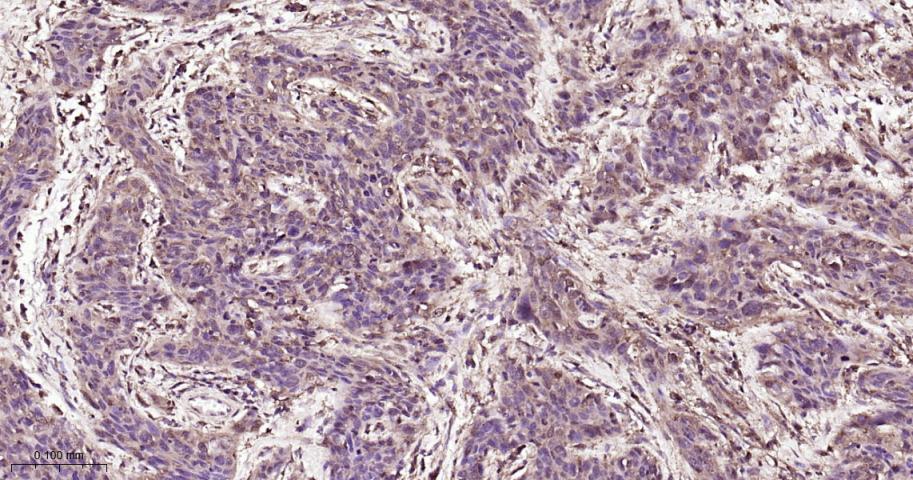
Paraformaldehyde-fixed, paraffin embedded Human Cervical Cancer; Antigen retrieval by boiling in sodium citrate buffer (pH6.0) for 15 min; Antibody incubation with Aurora A Monoclonal Antibody, Unconjugated(bsm-52018R) at 1:200 overnight at 4°C, followed by conjugation to the bs-0295G-HRP and DAB (C-0010) staining.
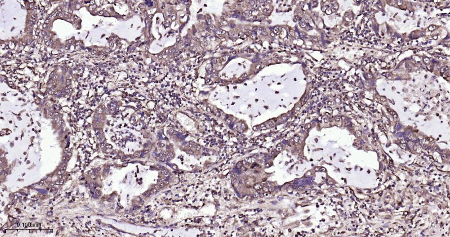
Paraformaldehyde-fixed, paraffin embedded Human Lung Cancer; Antigen retrieval by boiling in sodium citrate buffer (pH6.0) for 15 min; Antibody incubation with Aurora A Monoclonal Antibody, Unconjugated(bsm-52018R) at 1:200 overnight at 4°C, followed by conjugation to the bs-0295G-HRP and DAB (C-0010) staining.
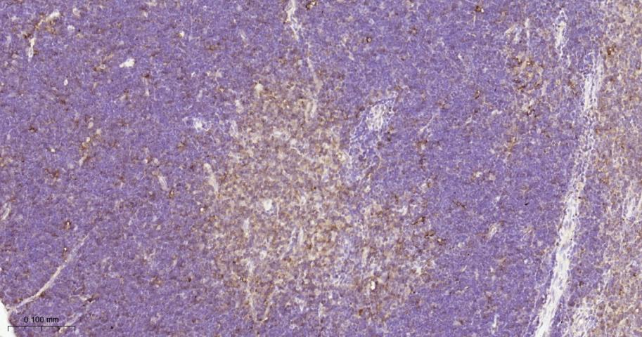
Paraformaldehyde-fixed, paraffin embedded Rat Thymus; Antigen retrieval by boiling in sodium citrate buffer (pH6.0) for 15 min; Antibody incubation with Aurora A Monoclonal Antibody, Unconjugated(bsm-52018R) at 1:200 overnight at 4°C, followed by conjugation to the bs-0295G-HRP and DAB (C-0010) staining.
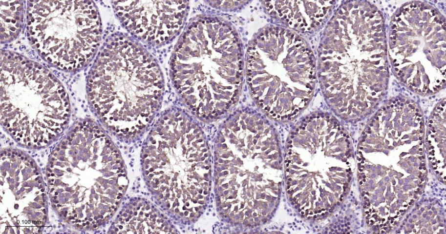
Paraformaldehyde-fixed, paraffin embedded Rat Testicles; Antigen retrieval by boiling in sodium citrate buffer (pH6.0) for 15 min; Antibody incubation with Aurora A Monoclonal Antibody, Unconjugated(bsm-52018R) at 1:200 overnight at 4°C, followed by conjugation to the bs-0295G-HRP and DAB (C-0010) staining.
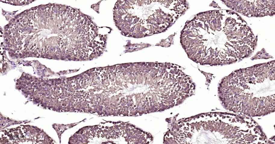
Paraformaldehyde-fixed, paraffin embedded Mouse Testicles; Antigen retrieval by boiling in sodium citrate buffer (pH6.0) for 15 min; Antibody incubation with Aurora A Monoclonal Antibody, Unconjugated(bsm-52018R) at 1:200 overnight at 4°C, followed by conjugation to the bs-0295G-HRP and DAB (C-0010) staining.
|
| 1、抗体溶解方法 | |
| 2、抗体修复方式 | |
| 3、常用试剂的配制 | |
| 4、免疫组化操作步骤 | |
| 5、免疫组化问题解答 | |
| 6、Western Blotting 操作步骤 | |
| 7、Western Blotting 问题解答 | |
| 8、关于肽链的设计 | |
| 9、多肽的溶解与保存 | |
| 10、酶标抗体效价测定程序 | |


