
| 产品编号 | bsm-52051R |
| 英文名称 | Cytokeratin 1 Recombinant Rabbit mAb |
| 中文名称 | 细胞角蛋白1重组兔单抗 |
| 别 名 | AEI2; CK1; EHK; EHK1; EPPK; K1; KRT1A; NEPPK; Krt-2.1; Krt2-1; Krt86; Kb1; K2C1_HUMAN; KRT1; 67 kDa cytokeratin; Cytokeratin-1 (CK-1); Hair alpha protein; Keratin-1 (K1); Type-II keratin Kb1; KRTA; K2C1_MOUSE; K2C1_RAT; |
| 研究领域 | 肿瘤 信号转导 干细胞 激酶和磷酸酶 |
| 抗体来源 | Rabbit |
| 克隆类型 | Recombinant |
| 克 隆 号 | 1C2 |
| 交叉反应 | Human,Mouse,Rat |
| 产品应用 | WB=1:500-2000,IHC-P=1:100-500,IHC-F=1:100-500,IF=1:100-500,ICC/IF=1:50-200
not yet tested in other applications. optimal dilutions/concentrations should be determined by the end user. |
| 理论分子量 | 70 kDa |
| 检测分子量 | 65 |
| 细胞定位 | 细胞浆 细胞膜 |
| 性 状 | Liquid |
| 浓 度 | 1mg/ml |
| 免 疫 原 | A synthesized peptide derived from human Cytokeratin 1: 600-644 |
| 亚 型 | IgG |
| 纯化方法 | affinity purified by Protein A |
| 缓 冲 液 | 0.01M TBS (pH7.4) with 1% BSA, 0.02% Proclin300 and 50% Glycerol. |
| 保存条件 | Shipped at 4℃. Store at -20℃ for one year. Avoid repeated freeze/thaw cycles. |
| 注意事项 | This product as supplied is intended for research use only, not for use in human, therapeutic or diagnostic applications. |
| PubMed | PubMed |
| 产品介绍 |
The protein encoded by this gene is a member of the keratin gene family. The type II cytokeratins consist of basic or neutral proteins which are arranged in pairs of heterotypic keratin chains coexpressed during differentiation of simple and stratified epithelial tissues. This type II cytokeratin is specifically expressed in the spinous and granular layers of the epidermis with family member KRT10 and mutations in these genes have been associated with bullous congenital ichthyosiform erythroderma. The type II cytokeratins are clustered in a region of chromosome 12q12-q13. [provided by RefSeq]. Function: May regulate the activity of kinases such as PKC and SRC via binding to integrin beta-1 (ITB1) and the receptor of activated protein kinase C (RACK1/GNB2L1). In complex with C1QBP is a high affinty receptor for kininogen-1/HMWK. Subunit: Heterotetramer of two type I and two type II keratins. Keratin-1 is generally associated with keratin-10. Interacts with ITGB1 in the presence of GNB2L1 and SRC, and with GNB2L1. Interacts with C1QBP; the association represents a cell surface kininogen receptor. Subcellular Location: Cell membrane. Note=Located on plasma membrane of neuroblastoma NMB7 cells. Tissue Specificity: The source of this protein is neonatal foreskin. The 67-kDa type II keratins are expressed in terminally differentiating epidermis. Post-translational modifications: Undergoes deimination of some arginine residues (citrullination). DISEASE: Defects in KRT1 are a cause of epidermolytic hyperkeratosis (EHK) [MIM:113800]. An autosomal dominant skin disorder characterized by widespread blistering and an ichthyotic erythroderma at birth that persist into adulthood. Histologically there is a diffuse epidermolytic degeneration in the lower spinous layer of the epidermis. Within a few weeks from birth, erythroderma and blister formation diminish and hyperkeratoses develop. Defects in KRT1 are the cause of ichthyosis hystrix Curth-Macklin type (IHCM) [MIM:146590]. IHCM is a genodermatosis with severe verrucous hyperkeratosis. Affected individuals manifest congenital verrucous black scale on the scalp, neck, and limbs with truncal erythema, palmoplantar keratoderma and keratoses on the lips, ears, nipples and buttocks. Defects in KRT1 are a cause of palmoplantar keratoderma non-epidermolytic (NEPPK) [MIM:600962]. NEPKK is a dermatological disorder characterized by focal palmoplantar keratoderma with oral, genital, and follicular lesions. Defects in KRT1 are a cause of ichthyosis annular epidermolytic (AEI) [MIM:607602]; also known as cyclic ichthyosis with epidermolytic hyperkeratosis. AEI is a skin disorder resembling bullous congenital ichthyosiform erythroderma. Affected individuals present with bullous ichthyosis in early childhood and hyperkeratotic lichenified plaques in the flexural areas and extensor surfaces at later ages. The feature that distinguishes AEI from BCIE is dramatic episodes of flares of annular polycyclic plaques with scale, which coalesce to involve most of the body surface and can persist for several weeks or even months. Defects in KRT1 are the cause of palmoplantar keratoderma striate type 3 (SPPK3) [MIM:607654]; also known as keratosis palmoplantaris striata III. SPPK3 is a dermatological disorder affecting palm and sole skin where stratum corneum and epidermal layers are thickened. There is no involvement of non-palmoplantar skin, and both hair and nails are normal. Similarity: Belongs to the intermediate filament family. SWISS: P04264 Gene ID: 3848 Database links: Entrez Gene: 3848 Human Entrez Gene: 16678 Mouse Omim: 139350 Human SwissProt: P04264 Human SwissProt: P04104 Mouse Unigene: 80828 Human Unigene: 183137 Mouse Unigene: 31789 Rat |
| 产品图片 |
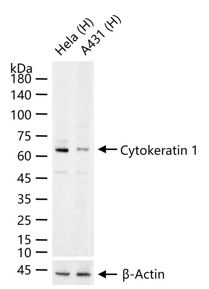
25 ug total protein per lane of various lysates (see on figure) probed with Cytokeratin 1 monoclonal antibody, unconjugated (bsm-52051R) at 1:1000 dilution and 4°C overnight incubation. Followed by conjugated secondary antibody incubation at r.t. for 60 min.
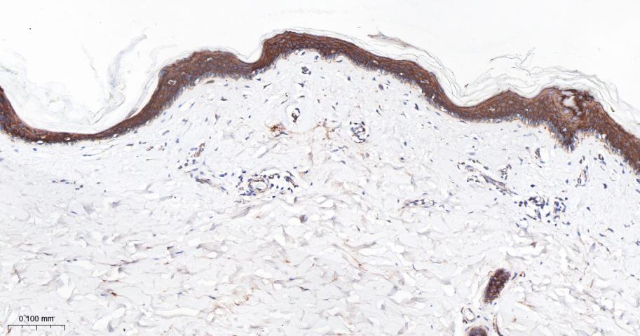
Paraformaldehyde-fixed, paraffin embedded Human Skin; Antigen retrieval by boiling in sodium citrate buffer (pH6.0) for 15 min; Antibody incubation with Cytokeratin 1 Monoclonal Antibody, Unconjugated(bsm-52051R) at 1:200 overnight at 4°C, followed by conjugation to the bs-0295G-HRP and DAB (C-0010) staining.
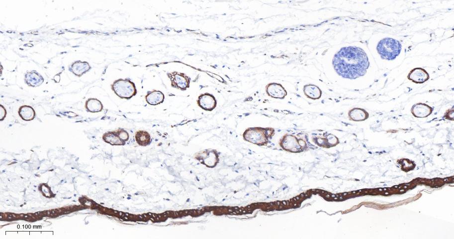
Paraformaldehyde-fixed, paraffin embedded Rat Skin; Antigen retrieval by boiling in sodium citrate buffer (pH6.0) for 15 min; Antibody incubation with Cytokeratin 1 Monoclonal Antibody, Unconjugated(bsm-52051R) at 1:200 overnight at 4°C, followed by conjugation to the bs-0295G-HRP and DAB (C-0010) staining.
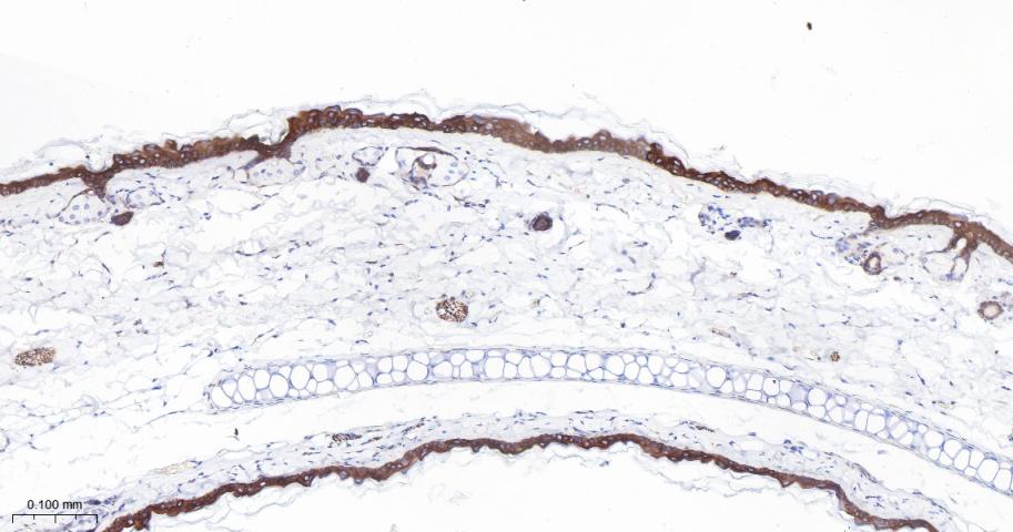
Paraformaldehyde-fixed, paraffin embedded Mouse Skin; Antigen retrieval by boiling in sodium citrate buffer (pH6.0) for 15 min; Antibody incubation with Cytokeratin 1 Monoclonal Antibody, Unconjugated(bsm-52051R) at 1:200 overnight at 4°C, followed by conjugation to the bs-0295G-HRP and DAB (C-0010) staining.
|
| 1、抗体溶解方法 | |
| 2、抗体修复方式 | |
| 3、常用试剂的配制 | |
| 4、免疫组化操作步骤 | |
| 5、免疫组化问题解答 | |
| 6、Western Blotting 操作步骤 | |
| 7、Western Blotting 问题解答 | |
| 8、关于肽链的设计 | |
| 9、多肽的溶解与保存 | |
| 10、酶标抗体效价测定程序 | |


